Obstructive Sleep Apnea Diagnosis and Treatment - part 4 doc
Bạn đang xem bản rút gọn của tài liệu. Xem và tải ngay bản đầy đủ của tài liệu tại đây (942.08 KB, 47 trang )
Bilevel Pressure and Adaptive Servo-Ventilation 127
is more effective in splinting the pharynx open than BPAP in patients with OSA.
Another study of 10 selected patients with OSA requiring ≥ 10 cm H
2
O of CPAP failed
to show optimal resolution of SDB at any level of IPAP until a critical level of EPAP
was achieved indicating a clear need to select an adequate EPAP level (11).
An alternative approach to BPAP titration has been to optimize all SDB events
with CPAP and then to reduce both the EPAP and IPAP as allowed taking advantage
of the natural hysteresis that occurs during a typical CPAP titration episode. In one
study, a small but significant reduction in the optimal CPAP level occurred when a
downward titration followed an initial titration procedure (12).
SELECTION OF PATIENTS
Severe Obesity
The importance of recognizing and addressing the differences in respiratory system
loading that occurs during the inspiratory phase in specific subtypes of patients has
helped guide which patients might best be considered for BPAP as well. The impact
of obesity on the mechanical loading of the respiratory system, neuromuscular
control, and the pathogenesis of apnea and related hypoventilation has been known
for many years. This issue was more precisely studied in three groups of severely
obese patients, who were eucapnic obese without OSA (O), eucapnic obese with
OSA (OSA), or hypercapnic obese with sleep apnea (OH), and were compared to
nonobese volunteers (NO) who were subjected to abdominal mass loading (13).
This was assessed with diaphragmatic electromyogram (EMGdi), a measure of muscle
activation, and mouth occlusion pressure (P0.15), an assessment of mechanical drive,
during CO
2
stimulation. The authors showed that P0.15 responses were decreased
in OSA and OH but the EMGdi responses did not differ from the control subjects.
However, when the NO control subjects were subjected to mass loading, the EMGdi
and P0.15 responses increased. Their findings showed that both OSA and OH
patients did not develop the expected increase in respiratory muscle response for a
given level of activation and the impaired mass load compensation predisposes
obese patients to develop hypercapnia. These kinds of observations may help
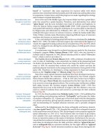
FIGURE 2 Algorithm for adjustment of inspiratory positive airway pressure and expiratory positive
airway pressure during the trial of nasal bilevel positive airway pressure (BiPAP
®
). Source: From
Ref. 9.
128 Gay
explain why the early BiPAP study focused on patients with a mean body mass
index (BMI) of 57.4 kg/m
2
even though they were normocapnic and why severe
obesity has emerged as an independent predictor of failed CPAP therapy (9,14).
The unloading of inspiratory muscle activity with BPAP measured as dia-
phragmatic pressure time product was studied in 18 obese subjects with a BMI ≥ 40
kg/m
2
including five healthy controls with simple obesity (SO), seven patients with
OSA, and six with obesity-hypoventilation syndrome (OHS) (15). Although the
overall ventilation as measured by end-tidal carbon dioxide with BPAP was not
changed in SO and OSA, it was decreased in OHS, while the inspiratory muscle
activity was reduced by at least 40% in all groups. The authors concluded that BPAP
may be particularly effective for improving ventilation in patients with OHS by
unloading the inspiratory muscles.
Hypercapnia and Other Factors
Other studies have specifically targeted the presence of hypercapnia in patients
with severe OSA as a reason to justify short- or long-term treatment with modes
enabled to deliver more aggressive ventilatory capability (13,16,17). In most cases
these studies showed marked improvement in daytime PaCO
2
levels with treat-
ment of accompanying nocturnal hypoventilation with BPAP, which in some cases
allowed resumption of CPAP treatment alone for satisfactory response to the under-
lying OSA. Patient adherence remained high in these patients (18).
In order to assess the reasons why a BPAP device was provided to a group of
moderate-to-severe OSA patients, a study was done to investigate the frequency of
BPAP prescription when CPAP was ineffective or not tolerated during titration (19).
Of 286 consecutive adult patients referred to two sleep labs, 130 patients were
enrolled and 105 (84% males) completed the study. A split-night (diagnostic and
therapeutic) polysomnogram (PSG) was done, followed by another PSG with BPAP
if CPAP was not tolerated, or failed to correct sleep-related breathing (SRB) abnor-
malities. There were 24 patients (23% overall) that received BPAP with the highest
prevalence (11 of 17) in patients with OSA associated with OHS. The BPAP treated
patients were more obese, hypercapnic, had severe SRB desaturations, and also had
more obstruction, restriction, and hypoxia.
The prevalence and mechanism of hypercapnia in morbidly obese patients
were investigated in a selected group of 285 patients presented to a sleep laboratory
without other significant comorbid diseases. There were 89 morbidly obese patients
(31.2%) who had a BMI ≥ 40 kg/m
2
and surprisingly 59.6% were predominately
females (20). This group was further divided into three subgroups who were nor-
mocapnic without OSA, normocapnic with OSA, and lastly hypercapnic (PaCO
2
≥
45 mmHg) with OSA. Their results showed that hypercapnia was found in 27% of
the morbidly obese subjects (who were predominately males) but only in 11% of the
nonmorbidly obese patients (P < 0.01). Several characteristics were more common in
the patients with hypercapnia and OSA than patients with or without OSA including
significantly more restriction based on a mean total lung capacity (% predicted) of
63.8% ± 16.4%, a higher respiratory disturbance index of 46.3 ± 26.9 events/hour, a
longer total sleep time with SpO
2
< 90% (TSTSpO
2
< 90%) of 63.4 ± 33.9 minutes, and
a lower rapid eye movement (REM) sleep at 9.5 ± 1.2%. Their conclusion about
important factors associated with hypercapnia and OSA allowed them to construct a
predictive model for diurnal hypercapnia: PaCO
2
= −0.03 FVC %predicted − 0.05
FEV1 % predicted + 0.036 TSTSpO
2
< 90% − PaO
2
+ 57.13 (r
2
= 0.44).
Bilevel Pressure and Adaptive Servo-Ventilation 129
Another small study investigated whether a variety of factors were associated
with failure of CPAP therapy to resolve the apnea–hypopnea index (AHI) to < 5 or
mean SaO2 > 90% (14). This study of 13 patients with OSA (Group A) over a
15-month period compared to an age- and AHI-matched control group (Group B)
successfully treated by CPAP used logistic regression analysis to identify factors
associated with initial failure to CPAP. The Group A versus Group B patients were
significantly more obese (mean BMI = 44.2 kg/m
2
vs. 31.2 kg/m
2
; P < 0.001), hypoxic
at rest (P < 0.001), and at exercise (P < 0.005). Hypercapnia at rest (P < 0.001) and
worsening during exercise was also more likely, and Group A patients also spent
significantly (P < 0.0001) more time with oxygen saturation < 90%, which was the
only factor independently associated with the initial failure of CPAP [odds ratio
(OR) 1.13; 95% confidence interval (CI) 1.0–1.2]. The patients’ awake blood gases
proved that both daytime hypoxemia and hypercapnia improved significantly
(P < 0.05) after three months of treatment with BPAP.
ADHERENCE
CPAP can produce objective and subjective improvements in patients with OSA and
other types of SDB (21–23). Despite its demonstrable efficacy, many OSA patients
have difficulty with long-term acceptance of CPAP (24,25). Several intervention
strategies have been suggested to improve adherence to CPAP, including use of
specialized education and follow-up programs (26,27) and added airway humidifi-
cation (28). Others have suggested that modifications of the airflow delivery pattern,
as with continuously auto-adjusting CPAP (29) or bilevel devices capable of varying
inspiratory and expiratory levels (9), may also boost adherence in more difficult-
to-treat OSA patients.
As these earlier reports indicated, there was a general reduction in the mean
effective PAP level using BPAP compared to CPAP (3,9). It was therefore thought
that bilevel devices might prove to be a benefit for improvement in adherence to
PAP in OSA patients. There have been two high-level evidence randomized trials
that compared the use of BPAP versus CPAP for OSA patients who were first time
users without complicating comorbid medical problems. The first study random-
ized 83 OSA patients to receive either CPAP or BPAP with a primary endpoint of
adherence based on mean
machine timer hours of CPAP (30). A total of 62 patients
were evaluated and
followed for one year and of these, 26 received BPAP and 36
CPAP pressures. The groups did not differ for BPAP versus CPAP by age (48 ±
1 years vs. 46 ± 1 years) or BMI (40 ± 1 kg/m
2
vs. 39 ± 1 kg/m
2
), but were different
by gender (65.5% vs. 52.8% males). Over the 12-month period, the mean machine
timer hours of CPAP versus bilevel therapy were not different at 5.0 ± 0.19 (SEM)
versus 4.9 ± 0.23 hours per night. There was also no difference between high and
low
hourly users for the CPAP or BPAP pressures required during therapy. These
patients had similar percentages of time that the machine was
running at the pre-
scribed effective pressure at 80% in the CPAP group
and 82% in the BPAP users with
both groups reporting an equal number of complaints with
respect to mask discom-
fort, machine noise, and nasal stuffiness.
The second high-level evidence trial studied newly diagnosed OSA patients
without coexisting daytime respiratory disease and compared CPAP with a bilevel
device that also employed a prototype flow feature of the presently available
Bi-Flex
®
device (Respironics, Inc., Murrysville, Pennsylvania, U.S.) (31). The primary
endpoint was the percentage of nights with at least four hours of use and hours of
130 Gay
use per night after 30 days treatment. This was based on objective machine-
determined measurement of time at effective pressure beginning after diagnosis
and titration by split-night polysomnography (PSG) in a sleep laboratory. There
were no significant baseline group differences for the 27 adults (22 men) in age,
BMI, AHI (mean ± SD, CPAP vs. BPAP group of 46.1 ± 23.1 events/hour vs. 41.8 ±
25.8 events/hour, respectively), CPAP requirement, or scores on the Epworth sleepi-
ness scale and Functional Outcomes of Sleep Questionnaire. The percentage of
nights with ≥ 4 hours/night use was high in both groups but was not significantly
different (CPAP vs. BPAP = 80.5 ± 24% vs. 77.6 ± 24.8%). In both of these studies, the
BPAP appeared to be as effective as CPAP for the treatment of OSA but offered no
advantages in patients receiving first-time therapy for OSA.
A randomized controlled trial published in 2005 investigated whether BPAP
with Bi-Flex could prove valuable in patients who were considered nonadherent to
CPAP therapy (32). The unique design had two phases that first attempted to
improve adherence to a treatment threshold of four hours per night of CPAP. There
were 204 adult patients diagnosed with OSA (AHI ≥ 10) by PSG within 24 months
who had been titrated to an optimal CPAP level but were not able to adhere to regular
CPAP use (nonresponders) at a mean treatment time of 254 ± 333 (SD) days. Patients
were questioned about various complaints and these were addressed with a
systematic conventional intervention program including further education, CPAP
desensitization as needed, alternative mask or resizing, and heated humidification.
After two more weeks of CPAP therapy there were 24% (49 patients) who became
responders (≥ 4 hours/night), 76% (155 patients) who were still nonresponders or
who withdrew. The nonresponders who agreed to proceed on to phase 2 then had a
full-night PSG retitration and blinded randomization to either CPAP or Bi-Flex for
three months. In the 104 patients initiating the trial, there were no significant differ-
ences in baseline characteristics of the Bi-Flex versus CPAP groups for age (51.9 ±
11.3 years vs. 52.5 ± 11.3 years), sex (65% vs. 72% males), BMI (33.4 ± 7.9 kg/m
2
vs.
32.5 ± 6.3 kg/m
2
), and AHI (40.4 ± 23.4 events/hour vs. 44.0 ± 26.1 events/hour).
At three months, there was a trend to a higher success rate with Bi-Flex versus CPAP
treatment (49% vs. 28.3% of patients using PAP therapy > 4 hours/night; p < 0.05) but the
overall success rate for either PAP treatment was low at near 40%. The authors
found no strong clinical predictors to distinguish patients in either arm who become
adherent (responders) after conventional intervention techniques and concluded
that sleep specialists must be very aggressive at achieving initial CPAP adherence as
subsequent efforts to achieve optimal adherence to PAP therapy are less fruitful but
consideration could be given to repeat PSG and use of alternative modes of flow
delivery to further encourage patients to use PAP treatment.
COMPLEX SLEEP APNEA
Central and obstructive apneas may occur in the same individual, either simultane-
ously within a single breath as a mixed apnea, or as sequential breathing events (33).
The majority of OSA patients can be expected to respond favorably to CPAP but
CPAP often initially exaggerates central sleep apnea (CSA) and some patients
identified as having OSA, develop frequent central apneas and/or Cheyne-Stokes
respiratory (CSR) pattern after application of CPAP. This is an increasingly recog-
nized but not new clinical problem encountered when patients with significant OSA
develop CSA when exposed to CPAP (34,35). These patients that develop new or
very prominent CSA during CPAP titration are now referred to as complex sleep
Bilevel Pressure and Adaptive Servo-Ventilation 131
apnea (CompSA) and from a consecutive series of 133 patients referred to a sleep lab
for OSA, 34 (25.6%) proved to have CompSA (36).
In this study, the mean age (near 55 years) and total diagnostic AHI (near
30 events/hour) were similar between the groups but there were a few distinguishing
features between the patients with OSA versus CompSA. The CompSA patients were
more likely to be males (82.4% vs. 63.9%; p = 0.03) and the OSA patients tended to be
slightly heavier (36 ± 10.3 kg/m
2
vs. 33 ± 5.9 kg/m
2
; p < 0.03). By definition, CompSA
patients had a significantly higher AHI during a standard CPAP titration (CompSA vs.
OSA AHI on CPAP = 24.6 ± 21.6 events/hour vs. 2.1 ± 2.7 events/hour; p < 0.0001) with
most or all the residual difference related to the central apneas that emerged on CPAP
(19.4 ± 19.0 vs. 2.1 ± 2.7 central events/hour; p < 0.0001). In a different investigation
selecting patients who were chosen to undergo BPAP for a variety of reasons, many
patients showed a CompSA like response to BPAP (rarely was a backup rate added)
but it was not possible to discern an incidence estimate from the data provided (37).
Treatment of Complex Sleep Apnea with Bilevel Positive Airway
Pressure Vs. Adaptive Servo-Ventilation
BPAP and ASV have been shown to improve SDB in patients with CSA (38). The best
approach for treatment of CompSA patients remains unclear but one study pursued
this issue comparing the response for both BPAP and the newly approved VPAP
Adapt™, Adapt Servo Ventilator or ASV (ResMed, Poway, CA) (39). This study investi-
gated 21 adult patients (95% male) with CSA/CSR mixed apnea or CompSA who had
previously undergone diagnostic PSG and titration with PAP in a randomized cross-
over design. Patients with a diagnostic AHI = 54.7 ± 23.8 events/hour, mean age and
BMI of 65 ± 12.4 (SD) years and 31.0 ± 4.9 kg/m
2
, respectively, were randomly assigned
initially to either BPAP or ASV during two full-night PSG studies. Following previously
attempted optimal titration with CPAP, the CompSA patients (n = 15) had a mean AHI
> 30 but with either BPAP or ASV the AHI improved markedly from baseline to 6.2 ±
7.6 events/hour or 0.8 ± 2.4 events/hour, respectively. The treatment arms were
different; however based on the preselected endpoints of the AHI and respiratory
arousal index proving to be significantly superior for ASV (p < 0.02). The authors
concluded that both BPAP and ASV seem to be effective in the acute setting for treat-
ment of SDB in patients with CompSA and far more efficacious than CPAP alone but
the CompSA patients seemed to respond optimally to ASV. A classic example of a
CompSA patient response at baseline, treated with CPAP, and then the improvement
with ASV can been seen in Figures 3 to 5, respectively.
REIMBURSEMENT CRITERIA
The delivery of appropriate treatment for any condition may at times become prob-
lematic if coverage criteria are not met or are not well-documented for the individual
patient. This is especially true in the world of SDB for Medicare patients with respect
to PAP therapy. There are separate criteria specifically for BPAP or in Center for
Medicare and Medicaid (CMS) vernacular, respiratory assist devices. The coverage
criteria are divided into four categories but two of these related to patients with
either neuromuscular or chronic obstructive lung disease are not pertinent for this
chapter. The specifics can be obtained from any regional durable medical equipment
regional carrier (DMERC) web site (40). The new regulations released in March 2006
and retroactive to January 1, 2006 recognize two basic pathways for BPAP treatment
132 Gay
coverage. The simplest avenue to BPAP is for OSA patients who have a “facility-based”
diagnostic PSG and meet criteria for CPAP treatment; yet, CPAP “has been tried and
proven ineffective.” Although this is nonspecific, it can be utilized if the patient has
tried CPAP and is intolerant, nonadherent to CPAP, or it does not satisfy the thera-
peutic goals. A BPAP device without a backup rate (E0470) can be prescribed and
covered under these circumstances. The patients with CompSA can obtain BPAP
with a backup rate (E0471 and ASV is considered an equivalent device) through the
same pathway as the patients with CSA. This approach also requires a diagnostic
PSG and failure with CPAP titration with a primary diagnosis of CSA or CompSA
which the DMERC web sites specifically define as:
“Complex sleep apnea (CompSA) is a form of central apnea specifically iden-
tified by the persistence or emergence of central apneas or hypopneas upon exposure
to CPAP (E0601) or an E0470 device once obstructive events have disappeared.
These patients have predominately obstructive or mixed apneas during the diag-
nostic sleep study occurring at greater than or equal to five times per hour. With use
of a CPAP or E0470, they show a pattern of apneas and hypopneas that meets the
definition of CSA described earlier.”
Central apnea is precisely defined as:
1. An AHI greater than five,
2. Central apneas/hypopneas greater than 50% of the total apneas/hypopneas,
3. Central apneas or hypopneas greater than or equal to 5 times per hour, and
4. Symptoms of either excessive sleepiness or disrupted sleep.
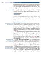
FIGURE 3 A two-minute epoch during Stage 2 sleep with frequent episodes of obstructive apnea and
oxygen desaturation. Incidental periodic limb movements are also noted in leg electromyogram channel.
Abbreviations: ABD, abdominal plethysmogram; C4-A1, right central-left reference EEG; CZ-OZ,
midline central-occipital EEG; ECG, electrocardiogram; EEG, electroencephalogram; EMG, electro-
myogram; FZ-CZ, midline frontal-central EEG; HR, heart rate; LOC and ROC, left and right outer canthi
(eye movements), respectively; Nasal P, nasal pressure via transducer; RC, rib cage plethysmogram;
Sono, sonogram (snoring intensity); SpO
2
, oxygen saturation; Sum, plethysmogram summed signal.
Bilevel Pressure and Adaptive Servo-Ventilation 133
The last complicating feature regarding reimbursement is that patients must
be followed up and proven adherent with the PAP treatment at least four hours per
night between 61 and 90 days after initiation. The whole process demands that
formal documentation be readily available for patients and although exact follow-up
guidelines are not otherwise provided in any literature, some formal and regular
follow-up is generally accepted as good clinical practice (23).
CONCLUSIONS
This chapter reviewed the rationale for the design of BPAP to treat patients with
SDB primarily due to OSA and additional clinical recommendations from evidence
review and practice parameter paper were published in 2006 (23,41). Even though
there have been no comparative studies to determine the optimal titration protocol,
a few titration techniques were discussed above. There was no evidence found that
BPAP improves efficacy or adherence to therapy in OSA patients who are first-time
PAP users but studies did at least support equivalency of CPAP and BPAP. There
was also no evidence available to support the practice of BPAP use at an arbitrary
higher level of CPAP pressure during a routine titration of OSA patients. There may
be some role for BPAP treatment in patients who have struggled with CPAP but
there is limited data. Special considerations are also needed for patients with mixed
apnea or CompSA who respond poorly to CPAP and it seems most appropriate to
FIGURE 4 Another two-minute epoch during non-rapid eye movement stage 2 sleep now with
10 cm H
2
O of continuous positive airway pressure applied. Note the exaggerated periodic breathing
with central apnea pattern now predominating. Abbreviations: ABD, abdominal plethysmogram;
C4-A1, right central-left reference EEG; CZ-OZ, midline central-occipital EEG; ECG, electrocardio-
gram; EEG, electroencephalogram; EMG, electromyogram; FZ-CZ, midline frontal-central EEG; HR,
heart rate; LOC and ROC, left and right outer canthi (eye movements), respectively; Nasal P, nasal
pressure via transducer; RC, rib cage plethysmogram; Sono, sonogram (snoring intensity); SpO
2
,
oxygen saturation; Sum, plethysmogram summed signal; VEST, estimated flow from device.
134 Gay
consider treatment with BPAP with a backup rate or an ASV device. It is important
to know and document coverage criteria so that the information is available to
enable proper reimbursement and some formal follow-up program is encouraged.
Lastly, future research should be performed to pursue ways to verify the efficacy
and treatment adherence benefits with BPAP or ASV in patients with CSA or
CompSA especially over the long term.
REFERENCES
1. Gastaut H, Tassinari A, Duron B. Polygraphic study of the episodic diurnal and
nocturnal (hypnic and respiratory) manifestations of the Pickwickian syndrome. Brain
Res 1966; 2:167.
2. Martin RJ, Pennock BE, Orr WC, et al. Respiratory mechanics and timing during sleep in
occlusive sleep apnea. J Appl Physiol 1980; 48(3):432–437.
3. Sanders MH, Moore SE. Inspiratory and expiratory partitioning of airway resistance
during sleep in patients with sleep apnea. Am Rev Respir Dis 1983; 127(5):554–558.
FIGURE 5 This 180-second epoch shows the patient stabilized in non-rapid eye movement
stage 2 sleep now using the Adaptive Servo Ventilator (ASV) device. Note the subtle alterations
in the ASV pressure (P) as the abdominal excursion is reduced or increased. The ASV P falls
during increased abdominal effort and vice versa to create a more uniform breathing pattern.
Abbreviations: ABD, abdominal plethysmogram; ASV P, mask pressure from ASV device; C4-A1,
right central-left reference EEG; CZ-OZ, midline central-occipital EEG; ECG, electrocardiogram;
EEG, electroencephalogram; EMG, electromyogram; Fpz-Cz, midline fronto-parietal-central EEG;
HR, heart rate; LOC and ROC, left and right outer canthi (eye movements), respectively; Nasal P,
nasal pressure via transducer; RC, rib cage plethysmogram; Sono, sonogram (snoring intensity);
SaO
2
, oxygen saturation; SUM, plethysmogram summed signal; VEST, estimated flow from
device.
Bilevel Pressure and Adaptive Servo-Ventilation 135
4. Remmers JE, Bartlett D. Reflex control of expiratory airflow and duration. J Appl Physiol
1977; 42:80–87.
5. Malhotra A, White DP. Obstructive sleep apnoea. Lancet 2002; 360:237–245.
6. Schwab RJ, Pasirstein M, Pierson R, et al. Identification of upper airway anatomic risk
factors for obstructive sleep apnea with volumetric magnetic resonance imaging. Am J
Respir Crit Care Med 2003; 168(5):522–530.
7. Juhasz J, Becker H, Cassel W, et al. Proportional positive airway pressure: a new concept
to treat obstructive sleep apnoea. Eur Respir J 2001; 17(3):467–473.
8. Farré R, Peslin R, Montserrat JM, et al. Flow-dependent positive airway pressure to
maintain airway patency in sleep apnea–hypopnea syndrome. Am J Respir Crit Care
Med 1998; 157:1855–1863.
9. Sanders MH, Kern N. Obstructive sleep apnea treated by independently adjusted inspira-
tory and expiratory positive airway pressures via nasal mask. Physiologic and clinical
implications. Chest 1990; 98(2):317–324.
10. Gugger M, Vock P. Effect of reduced expiratory pressure on pharyngeal size during nasal
positive airway pressure in patients with sleep apnoea: evaluation by continuous com-
puted tomography. Thorax 1992; 47(10):809–813.
11. Resta O, Guido P, Picca V, et al. The role of the expiratory phase in obstructive sleep
apnoea. Respir Med 1999; 93(3):190–195.
12. Bureau MP, Series F. Comparison of two in-laboratory titration methods to determine
effective pressure levels in patients with obstructive sleep apnoea. Thorax 2000;
55:741–745.
13. Lopata M, Onal E. Mass loading, sleep apnea, and the pathogenesis of obesity hypo-
ventilation. Am Rev Respir Dis 1982; 126(4):640–645.
14. Schafer H, Ewig S, Hasper E, et al. Failure of CPAP therapy in obstructive sleep apnoea
syndrome: predictive factors and treatment with bilevel-positive airway pressure. Respir
Med 1998; 92(2):208–215.
15. Pankow W, Hijjeh N, Schuttler F, et al. Influence of noninvasive positive pressure ventilation
on inspiratory muscle activity in obese subjects. Eur Respir J 1997; 10(12):2847–2852.
16. Piper AJ, Sullivan CE. Effects of short-term NIPPV in the treatment of patients with
severe obstructive sleep apnea and hypercapnia. Chest 1994; 105:434–440.
17. Waldhorn RE. Nocturnal nasal intermittent positive pressure ventilation with bi-level
positive airway pressure (BiPAP) in respiratory failure. Chest 1992; 101:516–521.
18. Laursen SB, Dreijer B, Hemmingsen C, et al. Bi-level positive airway pressure treatment
of obstructive sleep apnoea syndrome. Respiration 1998; 65:114–119.
19. Resta O, Guido P, Picca V, et al. Prescription of nCPAP and nBIPAP in obstructive sleep
apnoea syndrome: Italian experience in 105 subjects. A prospective two centre study.
Respir Med 1998; 92(6):820–827.
20. Resta O, Foschino-Barbaro MP, Bonfitto P, et al. Prevalence and mechanisms of diurnal
hypercapnia in a sample of morbidly obese subjects with obstructive sleep apnoea.
Respir Med 2000; 94(3):240–246.
21. Engelman HM, Kingshott RN, Wraith PK, et al. Randomized placebo-controlled crossover
trial of continuous positive airway pressure for mild sleep apnea/hypopnea syndrome. Am
J Respir Crit Care Med 1999; 159:461–467.
22. Jenkinson C, Davies RJ, Mullins R, et al. Comparison of therapeutic and subtherapeutic
nasal continuous positive airway pressure for obstructive sleep apnoea: a randomized
prospective parallel trial. Lancet 1999; 353:2100–2105.
23. Gay PC, Weaver T, Loube D, et al. Evaluation of positive airway pressure treatment for
sleep related breathing disorders in adults. Sleep 2006; 29(3):381–401.
24. Berry RB. Improving CPAP compliance-man more than machine. Sleep Med 2000;
1:175–178.
25. Weaver TE, Kribbs NB, Pack AI, et al. Night-to-night variability in CPAP use over the
first three months of treatment. Sleep 1997; 20:278–283.
26. Hoy CJ, Vennelle M, Kingshott RN, et al. Can intensive support improve continuous
positive airway pressure use in patients with sleep apnea/hypopnea syndrome? Am J
Respir Crit Care Med 1999; 159:1096–1100.
27. Chervin RD, Theut S, Bassetti C, et al. Compliance with nasal CPAP can be improved by
simple interventions. Sleep 1997; 20:284–289.
136 Gay
28. Massie CA, Hart RW, Peralez K, et al. Effects of humidification on nasal symptoms and
compliance in sleep apnea patients using continuous positive airway pressure. Chest
1999; 116:403–408.
29. Berry RB, Parish JM, Hartse KM. The use of auto-titrating continuous positive airway
pressure for treatment of adult obstructive sleep apnea. An American Academy of Sleep
Medicine review. Sleep 2002; 25:148–173.
30. Reeves-Hoche MK, Hudgel DW, Meck R, et al. Continuous versus bilevel positive airway
pressure for obstructive sleep apnea. Am J Respir Crit Care Med 1995; 151:443–449.
31. Gay PC, Herold DL, Olson EJ. A randomized, double-blind clinical trial comparing con-
tinuous positive airway pressure with a novel bilevel pressure system for treatment of
obstructive sleep apnea syndrome. Sleep 2003; 26:864–869.
32. Ballard R, Gay PC, Strollo PJ. Interventions to improve compliance in sleep apnea
patients previously non-compliant with continuous positive airway pressure. Accepted
Chest 2007.
33. Sleep-related breathing disorders in adults: recommendations for syndrome definition
and measurement techniques in clinical research. The Report of an American Academy
of Sleep Medicine Task Force. Sleep 1999; 22:667–689.
34. Thomas RJ, Terzano MG, Parrino L, et al. Obstructive sleep-disordered breathing with a
dominant cyclic alternating pattern—a recognizable polysomnographic variant with
practical clinical implications. Sleep 2004; 27:229–234.
35. Gilmartin GS, Daly RW, Thomas RJ. Recognition and management of complex sleep-dis-
ordered breathing. Curr Opin Pulm Med 2005; 11:485–493.
36. Pusalavidyasagar SS, Olson EJ, Gay PC, et al. Treatment of complex sleep apnea syndrome:
a retrospective comparative review. Sleep Med 2006; 7(6):474–479.
37. Johnson KG, Johnson DC. Bilevel positive airway pressure worsens central apneas
during sleep. Chest 2005; 128:2141–2150.
38. Teschler H, Dohring J, Wang YM, et al. Adaptive pressure support servo-ventilation: a
novel treatment for Cheyne-Stokes respiration in heart failure. Am J Respir Crit Care
Med 2001; 164(4):614–619.
39. Morgenthaler TI, Gay PC, Brown LK. Adaptive servo-ventilation versus noninvasive
positive pressure ventilation for central and complex sleep apnea syndromes. Sleep 2007;
in press.
40.
Docs?ReadForm&Providers/DMERC/Publications/DMEPOS+Supplier+Manual/Current
41. Standards of Practice Committee of the American Academy of Sleep Medicine. Kushida
CA, Littner MR, Hirshkowitz M, et al. Practice parameters for the use of continuous and
bilevel positive airway pressure devices to treat adult patients with sleep-related breath-
ing disorders. Sleep 2006; 29(3):375–380.
137
Auto-Positive Airway Pressure
Richard B. Berry
Division of Pulmonary, Critical Care, and Sleep Medicine, University of Florida,
Gainesville, Florida, U.S.A.
INTRODUCTION
Auto-positive airway pressure (APAP) devices provide a useful alternative for
providing positive airway pressure (PAP) treatment for patients with obstructive
sleep apnea (OSA) (1–3). One can separate the uses of these devices into two large
categories (Table 1). These include: (i) auto-titration PAP to determine an effective
fixed level of continuous positive airway pressure (CPAP) and (ii) auto-adjusting
PAP for chronic treatment. When used in the auto-titration mode the devices are
used by the patient for a period of time (one night to several weeks). Information
stored in the device is transferred to a computer and can be used to select an optimal
fixed level of CPAP for chronic treatment. When APAP devices are used for chronic
treatment they have the potential advantage of delivering the lowest effective pres-
sure in any circumstance (body position, sleep stage). The mean pressure for the
night may be lower than a single pressure that would be effective in all circum-
stances (the prescription pressure). For example, higher CPAP is usually needed in
the supine posture and during rapid eye movement (REM) sleep (4–6). A schematic
representation of the delivered pressure profile for a single night on auto-adjusting
PAP is shown in Figure 1. In this example, the patient spent only a small portion of
the night at the higher pressure required for the supine position. The average pressure
is much lower than a single pressure that would be effective throughout the night.
A substantial literature now exists evaluating the use of the devices in these modes
of operation.
DEVICE CHARACTERISTICS
Many brands and models of APAP devices are currently approved for treatment.
The devices differ in the respiratory variables that are monitored and in the
algorithms used to adjust the delivered pressure. The devices typically monitor one
or more of the following: airflow (or motor speed), airflow profile (flattening), snoring
(airway vibration), or airway impedance (forced oscillation technique). The algo-
rithms used to adjust pressure are proprietary but determine if the delivered pres-
sure should be increased or decreased. Depending on the type of respiratory event
that is detected the delivered pressure is increased by a certain amount. Typically,
pressure changes occur slowly over several minutes to prevent pressure-induced
arousals. If no respiratory events are detected within a certain time window the
delivered pressure is slowly decreased. Thus, the lowest effective pressure is delivered.
In some of the devices machine adjustment is available for various mask types and
for the type of humidifier that is being used.
Studies comparing different APAP devices provide evidence that devices from
different manufacturers will not deliver the same pressure for a given clinical
circumstance (7,8). These studies used a mechanical lung/upper airway with set
patterns of apnea or hypopnea to challenge different APAP devices. In Figure 2 the
8
138 Berry
responses of several devices differs markedly in response to apnea. Some of the
devices increased the delivered pressure in response to apnea while others did not (7).
Kessler et al. (9) compared the 95th percentile pressure of one APAP device based on
airflow to another based on the forced oscillation technique using a randomized
crossover study design. There was poor agreement between the optimal pressure
identified by the two devices. The 95th percentile pressure determined by the APAP
machine based on flow was on average 3 cm H
2
O higher than that of the other
device. Senn et al. (10) compared an APAP device responding to apnea, hypopnea,
and snoring with a second device responding to the previous variables as well as
airflow limitation (airflow profile flattening). The adherence and clinical outcomes
were similar although the median applied pressure was slightly higher with the
device that responded to airflow limitation (10). Thus, differences in the devices do
not always translate into differences in outcomes.
The problems of mask/mouth leak and central apnea have provided a chal-
lenge for the designers of APAP algorithms. However, these two problems are familiar
to technologists manually titrating CPAP. Mask/mouth leaks tend to raise the base-
line flow delivered by blower units and diminish the variations in flow during
inspiration and expiration. The resulting airflow signal may be interpreted as an
apnea or hypopnea and prompt an increase in pressure that may further increase
leak. Teschler and Berthon-Jones (3) reported on their clinical experience in 1000
patients using the AutoSet T
™
(ResMed, North Ryde, Australia) APAP device and
estimated that leak exceeds 0.4 L/s (considered high leak) on average for 10% of an
attended APAP night and 15% on an unattended APAP night. To handle the leak
problem many APAP units have algorithms that limit pressure increases when leak
exceeds certain values or when increases in blower speed no longer result in increases
TABLE 1 Potential Uses of Auto-Positive Airway Pressure
Auto-titrating mode
Attended auto-titration in CPAP naïve patient (technologist extender)
Unattended auto-titration in CPAP naïve patient
Check prescription pressure after weight gain/loss
Salvage a failed manual CPAP titration
Auto-adjusting mode
Initial chronic treatment of OSA (no titration needed)
Chronic treatment in patients not tolerating CPAP
Chronic treatment in patients with difficult mask/mouth leak
Abbreviations: CPAP, continuous positive airway pressure; OSA, obstructive
sleep apnea.
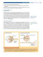
FIGURE 1 This schematic tracing of delivered pressure over an entire night illustrates that the
patient slept at a lower pressure for most of the night than a single fixed pressure that would be
effective in all body positions. The APAP device increased pressure when the patient was supine.
Abbreviation: APAP, auto-positive airway pressure.
Auto-Positive Airway Pressure 139
in mask pressure. Other units have leak alarms that can prompt the patient to adjust
the mask. Mouth leaks can be approached by using a chin strap or full-face mask.
Central apnea during APAP treatment/titration is another difficult problem in
some patients (1). Central apneas of the Cheyne-Stokes type are common in patients
in congestive heart failure. Other patients with OSA may have central apneas during
CPAP titration (treatment-emergent central apneas). Algorithms often include limits
on upward titration of pressure for apnea to avoid the delivery of high pressure for
central apneas. For example, pressure is not increased above 10 cm H
2
O unless
apnea is associated with snoring or airflow profile flattening. Of note, many
published studies of APAP excluded patients with congestive heart failure or
frequent central apneas on the preceding diagnostic sleep study.
FIGURE 2 Response of the auto-positive airway pressure (APAP) devices (D1–D5) when
subjected to a disturbed flow (V′) breathing pattern consisting of repetitive apneas. P and V′ are the
actual pressure and flow, respectively, measured at the entrance of the APAP device (V′ > 0: inspi-
ration). Source: From Ref. 7.
140 Berry
Recently, expiratory pressure relief (C-Flex
™
, Respironics, Inc., Murrysville,
Pennsylvania, U.S.) is now available for one brand of APAP devices. This mode
allows a reduction in pressure during early expiration with a return to the current
set pressure at end expiration. This feature could improve patient tolerance to pres-
sure. However, only one published study has demonstrated an advantage for CPAP
with C-Flex compared to traditional CPAP treatment (11). Another recent develop-
ment is the availability of APAP machines providing automatic adjustment of bilevel
PAP (BPAP) (Fig. 3). These devices vary inspiratory positive airway pressure (IPAP)
and expiratory positive airway pressure (EPAP) according to a proprietary algo-
rithm. The physician sets the minimum EPAP, maximum IPAP, and maximum
IPAP–EPAP difference. The option of using inspiratory and expiratory pressure
relief (Bi-Flex
®
, Respironics, Inc., Murrysville, Pennsylvania, U.S.) is currently avail-
able for one device brand. Inspiratory pressure relief allows a drop in pressure at
end inhalation (IPAP) while expiratory pressure relief allows a pressure drop at the
start of exhalation (EPAP). The superiority of APAP devices with either C-Flex or
the ability to deliver bilevel positive pressure (with or without Bi-Flex) remains to
be demonstrated. These new features do increase treatment alternatives.
AUTO-TITRATION
The gold standard for the titration of PAP is attended polysomnography with
full-electroencephalogram (EEG) monitoring to detect the presence and stage of
sleep (12). Respiratory monitoring allows classification of apneas (obstructive,
mixed, and central) and detection of hypopnea or drop in arterial oxygen
saturation. Snoring and evidence of airflow limitation or leak can also be detected
with the proper monitoring equipment. Body position is identified by technologist
documentation or by position sensors. Manual PAP titration is labor intensive
and usually a single technologist can titrate only two patients at a time. Patients
in some geographical areas may have limited or delayed access to a sleep
laboratory offering polysomnography. In addition, the gold standard PAP titration
method may result in suboptimal titrations due to a number of problems including
poor sleep, lack of supine REM sleep, high mask leak, or uncorrected mouth leak.
Patient characteristics such as weight gain may also render previously selected
pressures inadequate. Auto-titrating PAP devices can be used to address some of
these problems.
FIGURE 3 A single night profile showing changes in inspiratory positive airway pressure and
expiratory positive airway pressure over the night with auto-bilevel positive airway pressure.
Auto-Positive Airway Pressure 141
Evidence of Efficacy of Auto-Titrating Positive Airway Pressure
One important use of APAP devices is selection of a fixed CPAP pressure as an alter-
native to traditional manual (attended) PAP titration (1,2,10–19). Information stored
in the device memory can be analyzed and a pressure can be chosen for fixed CPAP
treatment. A common method is to choose the 90th or 95th percentile pressure (pressure
exceeded only 10% or 5% of the time, respectively) as the prescription pressure. This
assumes periods of high leak have been eliminated from the analysis.
There is considerable evidence that APAP devices can be successfully used to
establish a fixed CPAP pressure. However, in reviewing the literature it is important
to note whether the studies were attended or unattended and the proportion of
patients excluded. There are really two methods with which to judge APAP selection
of a fixed CPAP pressure. First, will APAP titration allow selection of a fixed level of
CPAP resulting in acceptable treatment [apnea–hypopnea index (AHI) < 10/hour]
and/or good clinical outcome? A second approach is to determine if APAP titration
can identify an optimal pressure for fixed CPAP treatment that is similar to the pressure
chosen by manual titration.
Several studies found that APAP titration could select a fixed CPAP pressure
that was effective. For example, Gagnadoux et al. (13) performed an attended APAP
titration followed by treatment with fixed CPAP chosen as the P95 (i.e., 95th percentile
pressure). After three months of treatment a sleep study on the fixed pressure found
the AHI < 10/hour in 21 of 24 patients. Two other attended studies (14,15) reached
similar conclusions. Unattended APAP titration also appears to be effective. Berkani
et al. (16) found that unattended APAP was successful at identifying an effective
CPAP level. Similarly, Series (17) found that treatment with fixed CPAP using a
pressure identified by unattended APAP titration reduced the AHI to < 10/hour in
38/40 subjects.
Studies have also compared the CPAP pressure chosen by attended manual
titration and APAP titration. Lloberes et al. (18) compared a partially attended
APAP titration with a manual CPAP titration on another night. In 15 of 20 patients
the difference between the APAP pressure (taken as P95) and the CPAP chosen by
manual titration was equal to or less than 1 cm H
2
O. Stradling et al. (19) used a
randomized parallel-group design and found the APAP and manual titration
pressures to be very similar in two well-matched groups of OSA patients (APAP:
8.2 ± 2.1, manual CPAP titration: 8.7 ± 2.1 cm H
2
O). The selection of the effective
pressure on the APAP night was performed after visualization of raw data. The
pressure effective “most” of the time during APAP titration was chosen.
Fletcher et al. (20) studied the feasibility of using ambulatory monitoring for
diagnosis followed by ambulatory APAP titration/treatment as therapy for a large
group with OSA. Exclusion criteria included a suspicion of other sleep disorders
(narcolepsy, restless legs syndrome), complicating medical illnesses, acute decom-
pensation requiring hospitalization, or a prior diagnosis of OSA. Of the 45 patients
that underwent APAP titration, it was deemed adequate in 35 (78%).
Based on a literature search pre-2002 the Standards of Practice Committee of
the American Academy of Sleep Medicine did not recommend the routine use of
unattended APAP for the choice of a fixed CPAP prescription pressure (1). Since this
literature review, further evidence of the ability of APAP devices to function in the
unattended auto-titration mode has been published. In a large multicenter study,
Masa et al. (21) randomized patients newly diagnosed with OSA to one of the three
treatment arms using a parallel-group design. The study groups included: (i) standard
attended titration with polysomnography, (ii) auto-titration at home, or (iii) use of a
142 Berry
level of CPAP chosen by a prediction formula (22) followed by pressure adjustment
based on spouse observations. The randomization was performed after a 20-minute
trial of CPAP during the day. Of note, 23% of the original study population was
excluded because of severe nasal congestion, prior palate surgery, refusal to partici-
pate, psychiatric incapacity, alcohol addiction, inability to place the CPAP mask,
and absence of a partner at home. In the auto-titrating arm patients were seen after
one night of auto-titration. The CPAP prescription pressure was chosen as the 90th
percentile pressure. If the patient slept poorly (patient report) or the machine infor-
mation revealed high leak, the auto-titration was repeated on a second or third
night. About 5% of patients in the auto-adjusting arm were considered titration fail-
ures. One night of titration was successful in 98 of 119 patients. In the predicted
formula group, CPAP was chosen based on neck size, body mass index, and the AHI
(22). Pressure was increased for observed apnea or snoring by fixed protocol. The
patients were treated with CPAP in all three groups and adherence and outcomes
measures were compared between the groups. There was no difference in the
improvement in the Epworth sleepiness scale (subjective sleepiness), adherence
(objective hours per night), drop out rates, or quality-of-life measures. The mean
CPAP levels in all three groups were similar (around 9 cm H
2
O). The mean nightly
use was approximately five hours. This large study suggests that ambulatory APAP
titration is as effective as manual titration (provided similar methods and exclusions
are used). However, the large number of excluded patients and the relatively low
mean pressure may mean that this finding may not apply to all populations of OSA
patients. The 20-minute CPAP trial during the day is a very appealing technique for
early identification of patients likely to need attended titration or who will be unable
to tolerate positive pressure at all.
In summary, there is convincing evidence that auto-titrating PAP devices can
be useful for identifying an appropriate fixed CPAP level in appropriate patients.
Recent studies have provided evidence that unattended as well as attended titration
can be effective. However, it is likely that unattended APAP titration will not be
effective in a significant proportion of OSA patients. In general, patients with a sig-
nificant proportion of central apneas, those with low baseline SaO
2
values or who
have difficulty applying the mask and/or operating an APAP device are probably
best studied using attended manual titration with polysomnography. A brief practice
trial with APAP may be useful to identify mask leak or nasal congestion problems
requiring intervention before the patient takes the APAP device home. If APAP titra-
tion is performed, close review of the data is needed to ensure that the titration was
adequate. Those patients not having an adequate APAP titration should be referred
for a traditional CPAP titration.
Technique of Auto-Titration
As with manual attended PAP titrations with polysomnography, patient education,
and mask fitting are essential for successful auto-titration. Patients must feel com-
fortable applying the mask interface and operating the APAP device if an unat-
tended titration is planned. One study found a short 20-minute trial of APAP to be
very useful in identifying patient problems including claustrophobia, mask leak or
an inability to operate the device (21). The physician ordering the APAP titration
designates the lower and upper pressure limits (for example, 4 and 20 cm H
2
O). The
APAP device then titrates between these limits. Depending on the type of APAP
device utilized, information on applied pressure, leak, snoring, flattening, and a
Auto-Positive Airway Pressure 143
moving time average of the AHI is stored in the device memory. After transfer to a
computer the information is available for review. It is possible to determine statistics
for all or a portion of the data. Most devices allow the ability to look at one or more
single nights of data in detail (pressure, leak, residual events vs. time) (Fig. 4).
Periods of high leak and the frequently associated increase in pressure can be appre-
ciated. High leak can result in many devices promptly increasing pressure until the
upper pressure limit is reached. In addition to detailed time profiles, summary
statistics can be displayed for a single night or multiple nights. Typically available
information includes: 90th or 95th percentile pressure, median pressure, maximum
pressure, maximum leak, median leak, and residual AHI.
Usually either the 90th or 95th percentile pressure is chosen for the prescrip-
tion pressure. However, simply noting one number can be very misleading. The cli-
nician must first determine if the titration duration (amount of sleep on the device)
was adequate and if the residual AHI is reasonably low (AHI < 5–10/hour). Patients
with suboptimal or inconclusive APAP titrations should have a repeat APAP titration
or be referred for an attended lab PAP titration. High-residual AHI could be secondary
to frequent central apneas, high leak, or too low maximum pressure limit. High leak
could be secondary to inadequate mask seal or mouth leak if a nasal mask is being
utilized. Patients with a high AHI and leak may undergo a repeat APAP titration
after mask adjustment or change to a full-face mask (or addition of a chin strap) as
indicated. A persistently high-residual AHI despite repeated attempts at APAP
titration would be an indication of the need for a traditional manual PAP titration.
In Figure 4 an example of an ideal titration is shown. Leak is relatively low as
is the residual AHI. The lower and upper pressure limits in this example were 4 and
15 cm H
2
O. A prescription pressure of 10 cm H
2
O was chosen based on the 95th per-
centile pressure of 9.4 cm H
2
O. Figure 5 shows a less ideal titration. Here there is a
FIGURE 4 This is a single night profile of pressure, leak, and residual apnea-hypopnea index
(AHI) of an ideal titration. The leak and residual AHI are low. The 95th percentile pressure was 9.4 cm
H
2
O. The patient was treated with a prescription pressure of 10 cm H
2
O. Source: From Ref. 42.
144 Berry
FIGURE 5 This is a single night profile of pressure, leak and residual apnea-hypopnea index (AHI) of
a fair titration. The leak is higher than ideal (> 0.4 L/s) at times. However, the residual AHI remained low.
The 95th percentile pressure was 7.8 cm H
2
O. The prescription pressure was chosen to be 8 cm H
2
O.
high leak but the residual AHI remains low. In Figure 6 an unacceptable auto-titration
is shown. Very high leak is obvious and the machine pressure climbed to the upper
pressure limit and stayed there for most of the night. In Table 2 examples of results
for two patients undergoing auto-titration are illustrated. In patient one the statistics
from five days of monitoring showed a low-residual AHI. In patient two, the results
of a single night showed a very high residual apnea index. The latter patient underwent
an attended titration and was found to have a significant number of central apneas.
A number of portable monitoring units can interface with selected APAP
devices. For example, airflow, respiratory effort, oxygen saturation, body position,
and delivered pressure can be recorded for clinician analysis (23). This is especially
helpful if one night of APAP titration is utilized. For example, absence of supine
sleep could result in a lower than typically needed prescription pressure. This is also
a reason that it is often helpful to have the patient use the APAP device for several
nights. Average statistics over a week may more accurately represent a typical
night’s pressure requirements. Information from pulse oximetry during the APAP
titration can identify the need for the addition of supplemental oxygen.
CHRONIC TREATMENT WITH AUTO-POSITIVE AIRWAY PRESSURE
(AUTO-ADJUSTING POSITIVE AIRWAY PRESSURE)
When originally developed one anticipated advantage of APAP devices was that delivery
of the lowest effective pressure in any circumstance (sleep stage, body position) would
Auto-Positive Airway Pressure 145
improve acceptance and adherence to positive pressure treatment. This was based
on the premise that a lower mean nightly pressure would improve patient tolerance
to PAP treatment. Alternatively, chronic treatment with an auto-adjusting device
would obviate the need for an attended or unattended PAP titration.
Auto-Positive Airway Pressure Vs. Continuous Positive Airway
Pressure Treatment
A number of studies have examined the hypothesis that chronic treatment with
auto-adjusting PAP would increase the adherence. Some studies did find higher
adherence with APAP compared to fixed CPAP treatment (24–27). However, several
other studies did not confirm this result (10,28–32). A meta-analysis (Fig. 7) by Ayas
FIGURE 6 This is a single night profile of pressure, leak and residual apnea–hypopnea index
(AHI) of an unacceptable auto-positive airway pressure titration. The leak is very high. The pressure
increased to the upper limit (16 cm H
2
O) and remained there for most of the night. The AHI was also
elevated. This titration would need to be repeated with a better mask seal or use of a full-face mask
if mouth leak was believed to be present.
TABLE 2 Auto-Positive Airway Pressure Statistics
Patient 1 Patient 2
Days 5 1
P
max
8.2 10
P95 7.8 8
AI (/hr) 2 15
HI (/hr) 3 5
AHI (/hr) 5 20
Median leak (L/s) 0.2 0.4
Note: Patient 2 had a high residual AHI.
Abbreviations: AI, apnea index; HI, hypopnea index; AHI,
apnea-hypopnea index.
146 Berry
FIGURE 7 Effects of auto-positive airway pressure (APAP) versus continuous positive airway
pressure (CPAP) on adherence. A positive score indicated a better adherence to APAP than CPAP.
The X axis is the nightly adherence with APAP minus adherence with CPAP. Y axis: studies reporting
adherence ordered by publication year. The bottom diamond represents the pooled effect, with the
dashed line drawn though the mean of this estimate. The composite data are consistent with no
increase in adherence on APAP versus CPAP treatment. Source: From Ref. 33.
FIGURE 8 The number of patients with displayed differences between the manual effective con-
tinuous positive airway pressure (Pman) and the mean nightly pressure on auto-positive airway
pressure (APAP). While a few patients had a much lower mean pressure on APAP compared to the
fixed pressure (Pman), the difference was only 1 or 2 cm H
2
O in most patients. Source: From Ref. 30.
et al. (32) concluded that there was no significant increase in adherence with APAP
treatment compared to fixed CPAP in these studies. In evaluating studies of
adherence several issues must be considered. First, pressure intolerance is not the
major issue for many patients (34). Second, the difference in the mean pressure on
APAP and an adequate fixed pressure is oft en only 1 to 2 cm (30) H
2
O (Fig. 8). One
would not expect such a small pressure difference to change adherence.
Auto-Positive Airway Pressure 147
Are there subsets of OSA patients who would adhere better to APAP than
fixed CPAP? One might expect a potential advantage for APAP devices in patients
with pressure intolerance, high prescription pressures, or a large variation in the
required pressure during the night (postural or REM-related OSA) (35). However,
Noseda et al. (36) found no evidence for increased adherence in patients with a high
variability in pressure requirement. A major problem with this study is that it did
not target pressure intolerant patients. Of interest, Hukins (32) found evidence of
lower leak and fewer reported side effects on APAP compared to fixed CPAP treat-
ment. Lowering the mean pressure might be expected to minimize mask leak or the
tendency for mouth leak. Leak, especially mouth leak, often results in dryness that
may not respond to the addition of heated humidity (37–39). Thus, chronic APAP
treatment provides a useful alternative in patients with a mask or mouth leak problem
that does not respond to interventions such as a change in mask type or size or an
increase in the delivered humidity.
Although APAP treatment appears to have no advantage over CPAP with
respect to adherence in unselected patients, several studies have demonstrated a
patient preference for APAP (30,32,40). It would be useful to be able to predict which
patients might prefer treatment with APAP. Marrone et al. (40) compared APAP and
fixed CPAP treatment using a crossover design. Of 22 patients, 14 preferred APAP
treatment. However, analysis of group characteristics found no factor that would
predict a preference for APAP. Of interest is the finding that only those patients with
a preference for APAP had higher adherence to APAP than CPAP. Thus, if a patient
is having difficulty with CPAP, a trial of APAP is a reasonable intervention. If APAP
is preferred, improved adherence is likely with this mode of treatment compared to
fixed CPAP. Furthermore, information stored in the machine such as leak or residual
events may help diagnose problems with treatment.
Patients with difficulty tolerating CPAP due to pressure intolerance (“cannot
breathe out”) are often switched to BPAP. One study compared intervention with
APAP to that with BPAP in patients with difficult-to-treat sleep apnea (41). The
inclusion criteria were patients requiring CPAP > 12 cm H
2
O, those not tolerating
CPAP, and those with central apneas that worsened on CPAP. Both APAP and BPAP
improved subjective sleepiness with equivalent adherence. A majority of the patients
preferred APAP at the end of the study. As APAP devices are considerably less expen-
sive than BPAP devices, this study suggests that a trial of APAP treatment should be
given consideration when CPAP is not well-tolerated.
Although the fact that APAP treatment does not improve adherence over fixed
CPAP selected by manual titration, the equivalence of these treatments suggests
another role for APAP devices. That is, patients diagnosed with OSA could be treated
with chronic APAP without the need for either traditional laboratory titration or
auto-titration. Any additional cost of APAP devices over CPAP would usually be
less than the cost of an in-laboratory CPAP titration. In fact, one study (31) found
both a cost saving and a reduction in the time needed to adjust the PAP treatment to
a satisfactory level. Indeed it is a common experience that the optimal level of CPAP
determined by an attended in-laboratory titration often requires alteration for
patient tolerance, side effects, or control of daytime sleepiness.
Technique of Chronic Auto-Positive Airway Pressure Treatment
The physician usually orders the lower and upper limits of positive pressure. The
APAP device then delivers the lowest effective pressure between these limits.
148 Berry
The upper and lower pressure limits could be placed as wide as possible (4–18 cm
H
2
O) or narrowed based on information from a previous CPAP titration or previous
nights of APAP use. Some patients find starting at 4 cm H
2
O uncomfortable and it
may take some APAP machines several minutes to reach a pressure level that they
find comfortable. In this case the lower pressure could be increased to 6–10 cm H
2
O.
Awakening with the feeling of insufficient pressure could be another situation in
which the lower pressure limit should be increased. Bloating or evidence of exces-
sive mouth leak might be an indication to lower the upper pressure limit.
Alternatively, if the 90th percentile pressure essentially equals the upper pressure
limit, then a higher upper pressure limit is likely needed (especially if the residual
AHI is high).
Information on delivered pressure is not the only data stored in the APAP
device that is potentially useful for tailoring treatment. High leak may indicate the
need for another mask or a full-face mask (mouth leak). A high-residual AHI might
indicate a need for an increase in either the lower or upper pressure. A high number
of residual apneas might suggest that central apneas could be present.
REIMBURSEMENT CRITERIA AND ISSUES
Financial constraints may limit the use of APAP devices for chronic treatment of
OSA at least in some circumstances. In the United States durable medical equip-
ment (DME) providers receive the same reimbursement for supplying an APAP
device as a CPAP device despite the greater cost of APAP. One option is to pass the
extra cost to the patient who often pays out of pocket for the difference. Some
patients may choose to buy a unit directly from a national discount provider over
the Internet.
Unattended auto-titration is currently not reimbursed in the United States.
For this reason, it has been most widely used in circumstances where reimburse-
ment for the auto-titration is not an issue such as certain health maintenance orga-
nizations and the Veterans Health Care System. In the private sector, some DME
providers are willing to provide an APAP loaner for a few days to weeks to deter-
mine a fixed CPAP pressure (if a CPAP will be provided by them at a pressure based
on the study). In this setting, the patient is responsible for paying for the mask and
other supplies should they decline CPAP treatment.
CONCLUSIONS
This chapter has presented evidence that APAP devices can be effective in the auto-
titrating and auto-adjusting modes. They provide a useful titration or chronic treat-
ment alternative for patients with sleep apnea. Success depends on proper patient
selection, education, and detailed physician review of the stored information.
Treatment or titration results could vary between brands of APAP devices. The
devices may not work well in patients with central apneas or those with large mouth
or mask leak. Careful physician evaluation of treatment efficacy is essential. For
example, it is possible that a given patient may have better alertness or sleep with a
slightly higher or lower pressure than the 90th or 95th percentile pressure identified
by APAP titration. Familiarity with type of information provided by a given brand
of device is essential to benefit from the stored information. As with any type of PAP
treatment, close follow-up of objective adherence and outcome measures such as
subjective sleepiness is essential.
Auto-Positive Airway Pressure 149
REFERENCES
1. Berry RB, Parish JM, Hartse KM. The use of auto-titrating CPAP for treatment of adults
with obstructive sleep apnea. Sleep 2002; 25:148–173.
2. Littner M, Hirshkowitz M, Davila D, et al. Standards of Practice Committee of the
American Academy of Sleep Medicine Practice parameters for the use of auto-titrating
continuous airway pressure devices for titrating pressures and treating adult patients
with obstructive sleep apnea syndrome. An American Academy of Sleep Medicine
Report. Sleep 2002; 15(25):143–147.
3. Teschler H, Berthon-Jones M. Intelligent CPAP systems: clinical experience. Thorax 1998;
53:S49–S54.
4. Pevernagie DA, Sheard JW Jr. Relations between sleep stage, posture and effective nasal
CPAP levels in OSA. Sleep 1992; 15:162–167.
5. Oksenberg A, Silverberg DS, Arons E, et al. The sleep supine position has a major effect
on optimal nasal continuous positive airway pressure. Chest 1999; 116:1000–1006.
6. Neill AM, Angus SM, Sajkov D, McEvoy RD. Effects of sleep posture on upper airway stability
in patients with obstructive sleep apnea. Am J Respir Crit Care Med 1997; 155:199–204.
7. Farré R, Montserrat JM, Rigau J, Trepat X, Pinto P, Navajas D. Response of automatic
continuous positive pressure devices to different sleep breathing patterns. Am J Respir
Crit Care Med 2002; 166:469–473.
8. Abdenbi F, Chambille B, Escourrou P. Bench testing of auto-adjusting positive airway
pressure devices. Eur Respir J 2004; 24:649–658.
9. Kessler R, Weitzenblum E, Chaouat A, Iamandt C, Alliotte T. Evaluation of unattended
automated titration to determine therapeutic continuous positive airway pressure in
patients with obstructive sleep apnea. Chest 2003; 123:704–710.
10. Senn O, Brack T, Matthews F, Russi EW, Bloch KE. Randomized short-term trial of two
AutoCPAP devices versus fixed continuous positive airway pressure for treatment of
sleep apnea. Am J Respir Crit Care Med 2003; 168:1506–1511.
11. Aloia MS, Stanchina M, Arnedt JT, Malhotra A, Millman RP. Treatment adherence and outcomes
in flexible versus standard continuous positive airway pressure. Chest 2005; 127:2085–2093.
12. American Sleep Disorders Standard of Practice Committee, Chesson A. Chairman. Practice
Parameters for the Indications for Polysomnography and Related procedures. Sleep 1997;
20:406–422.
13. Gagnadoux F, Rakotonanahary D, Martins de Araujo MT, et al. Evaluation of an auto-
CPAP device for treatment of obstructive sleep apnea. Sleep 1999; 22:1095–1097.
14. Teschler H, Berthon-Jones M, Thompson AB, et al. Automated continuous positive airway pressure
titration for obstructive sleep apnea syndrome. Am J Respir Crit Care Med 1996; 154:734–740.
15. Teschler H, Farhat AA, Exner V, et al. AutoSet nasal CPAP titration: Constancy of pressure,
compliance, and effectiveness at 8 months follow-up. Eur Respir J 1997; 10:2073–2078.
16. Berkani M, Lofaso F, Chouaid C, et al. Eur Respir J 1998; 12:759–763.
17. Series F. Accuracy of unattended home CPAP titration in the treatment of obstructive
sleep apnea. Am J Respir Crit Care Med 2000; 162:94–97.
18. Lloberes P, Ballester E, Montserrat JM, et al. Comparison of manual and automatic CPAP
titration in patients with sleep apnea/hypopnea syndrome. Am J Respir Crit Care Med
1996; 154:1755–1758.
19. Stradling J, Barbour C, Pitson DJ, Davies RJO. Automatic nasal continuous positive
airway pressure titration in the laboratory: patient outcomes. Thorax 1997; 52:72–75.
20. Fletcher EC, Stich J, Yang KL. Unattended home diagnosis and treatment of obstructive
sleep apnea without polysomnography. Arch Fam Med 2000; 9:168–174.
21. Masa JF, Jimenez A, Duran J, et al. Alternative methods of titrating continuous positive
airway pressure. Am J Respir Crit Care Med 2004; 170:1218–1224.
22. Miljeteig H, Hoffstein V. Determinants of continuous positive airway pressure for treat-
ment of obstructive sleep apnea. Am Rev Respir Dis 1993; 147:1526–1530.
23. Marrone O, Insalaco, Salvaggio A, Bonsignore G. Role of different nocturnal monitorings
in the evaluation of CPAP titration by autoCPAP devices. Respir Med 2005; 99:313–320.
24. Konermann M, Sanner BM, Vyleta M, et al. Use of conventional and self-adjusting nasal
continuous positive airway pressure for treatment of severe obstructive sleep apnea
syndrome. Chest 1998; 113:714–718.
150 Berry
25. Meurice JC, Marc I, Series F. Efficacy of auto-CPAP in the treatment of obstructive sleep
apnea/hypopnea syndrome. Am J Respir Crit Care Med 1996; 153:794–798.
26. Hudgel DW, Fung C. A long-term randomized, cross-over comparison of auto-titrating
and standard nasal continuous airway pressure. Sleep 2000; 23:645–648.
27. Massie CA, McArdle N, Hart RW, et al. Comparison between automatic and fixed positive
airway pressure therapy in the home. Am J Respir Crit Care Med 2003; 167:20–23.
28. d’Ortho PM, Grillier-Lanoir V, Levy P, et al. Constant vs automatic continuous positive
airway pressure therapy. Chest 2000; 118:1010–1017.
29. Teschler H, Wessendorf TE, Farhat AA, et al. Two months auto-adjusting versus conven-
tional nCPAP for obstructive sleep apnoea syndrome. Eur Respir J 2000; 15:990–995.
30. Randerath WJ, Schraeder, Galetke W, Feldmeyer F, Rühle K-H. Autoadjusting CPAP
based on impedance efficacy, compliance, and acceptance. Am J Respir Crit Care Med
2001; 163:652–657.
31. Planès C, d’Ortho M, Foucher A, et al. Efficacy and cost of home-initiated auto-nCPAP
versus conventional nCPAP. Sleep 2003; 26:156–160.
32. Hukins C. Comparative study of autotitrating and fixed-pressure CPAP in the home: a
randomized, single-blind crossover trial. Sleep 2004; 27:1512–1517.
33. Ayas NT, Patel SR, Malhotra A, et al. Auto-titrating versus standard continuous positive
airway pressure for the treatment of obstructive sleep apnea: results of a meta-analysis:
Sleep 2004; 27:249–253.
34. Rolfe I, Olson LG, Sanders NA. Long-term acceptance of continuous positive airway
pressure in obstructive sleep apnea. Am Rev Respir Dis 1991; 144:1130–1133.
35. Series F, Marc I. Importance of sleep stage and body position dependence of sleep apnea
in determining benefits to auto-CPAP therapy. Eur Respir J 2001; 18:170–175.
36. Noseda A, Kekmpenaers C, Kerkhofs M, Braun S, Linkowski P, Jann E. Constant vs auto-
continuous positive airway pressure in patients with sleep apnea hypopnea syndrome
with a high variability in pressure requirement. Chest 2004; 126:31–37.
37. Martins de Araujo MT, Vieira SB, Vasquez EC, Fleury B. Heated humidification or face
mask to prevent upper airway dryness during continuous positive airway pressure therapy.
Chest 2000; 117:142–147.
38. Richards GN, Cistulli PA, Ungar RG, Berthon-Jones M, Sullivan CE. Mouth leak with
nasal continuous positive airway pressure increases nasal airway resistance. Am J Respir
Crit Care Med 1996; 154:182–186.
39. Massie CA, Hart RW, Peralez K, Richards G. Effects of humidification on nasal symptoms
and compliance in sleep apnea patients using continuous positive airway pressure.
Chest 1999; 116:403–408.
40. Marrone O, Resta O, Salvaggio A, Giliberti T, Stefan A, Insalaco G. Preference for fixed or
automatic CPAP in patients with obstructive sleep apnea. Sleep Med 2004; 5:247–251.
41. Randerath W, Galetke W, Ruehle KH. Auto-adjusting CPAP based on impedance versus
bilevel pressure in difficult to treat sleep apnea syndrome: a prospective randomized
crossover study. Med Sci Monit 2003; 9:CR353–358.
42. Berry, RB. Sleep Medicine Pearls. Philadelphia: Hanley and Belfus, 2003.
151
Critical Factors in Positive
Pressure Therapy
Scott M. Leibowitz
The Sleep Disorders Center of Cardiac Disease Specialists,
Atlanta, Georgia, U.S.A.
Mark S. Aloia
Butler Hospital, Providence, Rhode Island, U.S.A.
INTRODUCTION
Obstructive sleep apnea (OSA) is a condition of cyclical or repetitive obstructions of
the upper airway during sleep, with micro-arousals occurring at the termination
of a respiratory event (1,2). In adults, micro-arousal activity has been postulated
to disrupt the normal restorative processes of sleep and has been demonstrated to
produce sleepiness and/or daytime performance deficits when induced by various
sensory stimuli in normal subjects (3,4). In addition to nonrestorative sleep, OSA
has been found to have a strong association with cardiovascular disease (5), including
hypertension (6), congestive heart failure (7), cardiac arrhythmias (8), coronary
artery disease (9), and stroke (10).
There are several different modalities that have been found to be effective for
the treatment of OSA. The first-line treatment however, has become the use of positive
airway pressure (PAP) therapy, especially continuous positive airway pressure
(CPAP) therapy (11). The first reported use of nasal CPAP for OSA in adults was
by Sullivan et al. (12) in 1981. The therapeutic mechanism of successful treatment is
the creation of a “pneumatic splint” by an effective positive pressure applied to
the pharynx, providing immediate relief from obstruction (13), thus allowing sleep
continuity and preservation of sleep architecture (14,15).
BENEFITS OF CONTINUOUS POSITIVE PRESSURE THERAPY
Studies have shown that positive pressure therapy, if successfully implemented, can
have significant impact on a variety of parameters of health and daily living. A
number of studies have shown that PAP therapy significantly improves subjective
and objective measures of daytime sleepiness (16,17), improves quality of life (18–20),
and has a positive impact on neurocognitive function in patients with OSA (18,19).
Further studies have shown that significant reductions in adverse
cardiovascular disease outcomes may be obtained with successful treatment of OSA
(5,10). CPAP reduces blood pressure in hypertensive patients (21,22), reverses
hemodynamic changes in the cerebral circulation (23), and may reduce pulmonary
pressures in OSA patients (24). Moreover, CPAP has been shown to prevent OSA-
associated bradyarrhythmias (25,26), decrease recurrence of atrial fibrillation after
cardioversion (27), and abolish ventricular arrhythmias (28,29). Furthermore,
patients with congestive heart failure (CHF) and OSA have shown a marked
improvement in left ventricular ejection fraction and functional class after initiation
of CPAP therapy (30,31).
9

