Obstructive Sleep Apnea Diagnosis and Treatment - part 7 pot
Bạn đang xem bản rút gọn của tài liệu. Xem và tải ngay bản đầy đủ của tài liệu tại đây (662.22 KB, 47 trang )
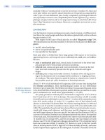
268 Pelayo and Li
A study from Israel found that children with SDB had lower scores on neuro-
cognitive testing compared to controls but the scores improve after treatment (23).
This prospective study of 39 children aged five to nine years underwent a battery of
neurocognitive tests containing process-oriented intelligence scales. Children with
SDB had lower scores compared with healthy children in some Kaufman Assessment
Battery for Children (K-ABC) subtests and in the general scale Mental Processing
Composite, indicating impaired neurocognitive function. Six to 10 months after
adenotonsillectomy, the children with OSAs demonstrated significant improvement
in sleep characteristics, as well as in daytime behavior. Their neurocognitive perfor-
mance improved considerably, reaching the level of the control group in the sub-
tests Gestalt Closure, Triangles, Word Order, and the Matrix analogies, as well as in
the K-ABC general scales, Sequential and Simultaneous Processing scales, and the
Mental Processing Composite scale. The authors concluded neurocognitive func-
tion is impaired in otherwise healthy children with SDB. Most functions improve to
the level of the control group, indicating that the impaired neurocognitive functions
are mostly reversible, at least 3 to 10 months following adenotonsillectomy (23). An
abrupt and persistent deterioration in grades must also raise the question of abnormal
sleep and SDB (20,21,50,51).
In schools the tiredness and sleepiness may be labeled as “inattentiveness in
class,” “daydreaming,” or “not being there” (22,52). Concerns about school perfor-
mance were raised in the original description of OSA syndrome in children (3).
More recently, the possible association between SDB, learning problems, and atten-
tion-deficit disorder has been studied (8,18,19,21,22,52–56). A study by Gozal et al.
examined the hypothesis that domains of neurobehavioral function would be selec-
tively affected by SDB. They study children with reported symptoms of attention-
deficit/hyperactivity disorder (ADHD) and also determined the incidence of
snoring and other sleep problems in 5- to 7-year-old children in a public school
system. Children with reported symptoms of ADHD and control children were ran-
domly selected for an overnight polysomnographic assessment and a battery of
neurocognitive tests. Frequent and loud snoring was reported for 673 children
(11.7%). Similarly, 418 (7.3%) children were reported to have hyperactivity/ADHD.
Children with reported symptoms of ADHD and control children were randomly
selected for an overnight polysomnographic assessment and a battery of neurocog-
nitive tests. Eighty-three children with parentally reported symptoms of ADHD had
sleep studies together with 34 control children. After assessment with the ADHD
subscale of the Conners Parent Rating Scale, 44 children were designated as having
“significant” symptoms of ADHD, 27 as “mild,” and 39 designated as “none” (con-
trols). Overnight polysomnography indicated that OSA was present in 5% of those
with significant ADHD symptoms, 26% of those with mild symptoms, and 5% of
those with no symptoms. The authors concluded an unusually high prevalence of
snoring was identified among a group of children designated as showing mild
symptoms of ADHD based on the Conners ADHD subscale. SDB can lead to mild
ADHD-like behaviors that can be readily misperceived and potentially delay the
diagnosis and appropriate treatment (22).
Additional clinical signs of SDB include increased respiratory efforts with
nasal flaring, suprasternal or intercostal retractions, abnormal paradoxical inward
motion of the chest occurring during inspiration, and sweating during sleep. The
sweating may be limited to only the nuchal region particularly in infants; it may be
severe enough to necessitate changing clothes during the night. The parents may
mention the child feeling warm at night or preferring to sleep without a blanket.
Obstructive Sleep Apnea in Children 269
Parents may also observe the child stop breathing, then gasping for breath. It is sur-
prising to note how often parents have observed abnormal breathing patterns
during sleep but were never questioned about it by pediatricians during regular
visits. Information regarding the sleep position is helpful. Typically, the neck is
hyper-extended and the mouth is open. Another typical sleeping position is prone
with the knee tucked under the chest with head turned to the side and hyperextended.
Rarely, the child with SDB prefers to sleep propped up on several pillows (4).
Parasomnias may be triggered or exacerbated by SDB. Ohayon (57) has found
that individuals identified with SDB have a much higher incidence of nightmares,
with reports of “drowning,” “being buried alive,” and “choking.” SDB leads to
sleep fragmentation or disruption. Any condition that disrupts slow-wave sleep
may lead to sleep terrors and sleepwalking in children (58). SDB should be included
in the evaluation of any child with parasomnias.
A physical finding that may be overlooked in a child with SDB is a narrow and
high-arched palate (4). Interestingly, the description of attention-deficit disorder in
the Diagnostic and Statistical Manual of Mental Disorders (DSM-IV) mentions that
minor physical anomalies such as high-arched palates may be present (59). Since
both conditions may have similar daytime behavior in the same age group, a child
with SDB could be misidentified as having attention-deficit disorder. The possibility
of a sleep disorder being present should be considered in any child being evaluated
for attention-deficit disorder. This is particularly important since treatment of SDB
may improve behavior and academic performance (60,61).
Diagnostic Criteria
The diagnostic criteria used for adults with OSA cannot be used reliably in children
(5,49,62,63). The diagnosis of SDB is based on the history, physical findings, and
supportive data. Laboratory testing should be, ideally, tailored to the clinical question.
For example, if there are concerns about excessive daytime sleepiness, a multiple
sleep latency test (MSLT) may be indicated (64). The MSLT is ideally performed in
subjects who are at least eight years old.
The polysomnogram in a child uses the same technology and the same type of
information as recorded in adults. Airflow, respiratory effort, and pulse oximetry
comprise the breathing measurements usually monitored. Breathing can be mea-
sured with different techniques, ranging from qualitative techniques such as nasal
thermocouples which use the temperature difference between inhaled and exhaled
air to record individual breaths, to quantitative and invasive measures such as
esophageal pressure measurements. The latter technique is less tolerable than others
but is particularly helpful to distinguish central from obstructive apneas. End-tidal
CO
2
monitoring is another technique that can help detect transient episodes of
hypoventilation. Currently, the technique that balances need for quantification with
tolerability is measuring airflow using a nasal pressure cannula (65,66). This tech-
nique allows for the identification of more subtle breathing episodes but can be harder
to interpret than earlier techniques, in particular when a child is mouth breathing.
This nasal pressure cannula has facilitated the measurement of an additional
abnormal respiratory event, RERA, which is an acronym for respiratory event-
related arousal. The term respiratory disturbance index (RDI) may now include the
total number of apneas, hypopneas, and RERAs divided by the total sleep time.
The RDI should be distinguished from the AHI. However, some sleep study reports
may equate the RDI with the AHI if the sleep study did not measure RERAs.
270 Pelayo and Li
The clinician needs to be aware that these terms may be used interchangeably,
potentially causing confusion.
The multitude of available techniques to measure breathing makes it difficult
to compare the results from different studies. Along with the absence of controlled
studies, another problem with understanding pediatric SDB is that definitions for
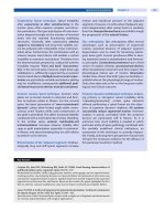
key terms vary. OSAs are defined as lasting at least 10 seconds in adults. However,
since children have faster respiratory rates clinically significant apneas can occur in
less time (Fig. 1). Apneas as brief as three or four seconds may have oxygen desatu-
rations. There is no universally accepted definition of hypopneas in children. The
clinician needs to know how apneas and hypopneas are defined and scored when
interpreting a polysomnogram report. The most recent edition of the International
Classification of Sleep Disorders (ICSD-2) defines OSA in children as having an
AHI ≥ 1 (67). In adults a higher AHI of five is required. Unfortunately it is not
uncommon for an adult cutoff value to be used in children (68). There is also contro-
versy as to when the adult cutoff value should be applied; the onset of puberty or
the age of 18 years is often debated.
FIGURE 1 Polysomnogram of a 10-year-old girl depicting several obstructive apneas and hypop-
neas during a 60-second epoch of rapid eye movement (REM) sleep, accompanied by esophageal
pressure “crescendos,” intermittent snoring (as detected by the Chin EMG and Mic), and oxygen
desaturations. Note the rapid respiration rate consistent with that of a child. Abbreviations: C3-A2,
C4-A1, O1-A2, Fp1-A2, electroencephalogram electrodes placed centrally (C3, C4), occipitally
(O1), and fronto-parietally (Fp1), and referenced to the right (A2) or left (A1) ear; Chin EMG, elec-
tromyogram recorded from chin muscles; LOC, left eye electro-oculogram; ROC, right eye electro-
oculogram; EKG, electrocardiogram; LAT and RAT, electromyogram recorded from the left and
right anterior tibialis muscles, respectively; SaO
2
, pulse oximetry; Mic, microphone to detect
snoring; Nasal, nasal pressure measured by pressure transducer; Oral, oral airflow measured by
thermistor; Chest and Abdomen, impedance bands to measure thoracic and abdominal movement,
respectively; P
es
, esophageal pressure to measure transmitted intrathoracic pressure. Source:
Courtesy of Clete A. Kushida, MD, PhD.
Obstructive Sleep Apnea in Children 271
Controversy exists over whether a diagnosis of OSA, or the larger spectrum of
SDB, should be routinely made without a formal polysomnogram. While some have
suggested that this diagnosis can be made in patients using either the history and
physical, or the history, physical, and an audio- or videotape, others have found an
inability of clinical history alone to distinguish primary snoring from OSA in children (69).
The situation is further complicated by the description of UARS in children, which
may have been missed in the studies cited above. Therefore, a sleep study is the
most definitive test for SDB (70,71). Currently, some otolaryngologists who treat
SDB in children may make the surgical recommendation based on clinical findings
of airway obstruction, sometimes reviewing an audio- or videotape (72,73).
The
clinicians must be aware of the potential pitfalls to this practice. Certainly there are
individual cases in which a diagnostic sleep studies are not available, but ideally
they should be the exception. The challenge we face in sleep medicine is providing
easily-accessible and cost-effective care working within a multidisciplinary model. We
do not know, for certain, how accurate clinical diagnosis is without objective testing.
Until we have a better answer, the diagnostic gold standard should not be disregarded
particularly in a tertiary care setting. The American Thoracic Society, American
Academy of Sleep Medicine, and AAP all support the use of sleep studies (70,74,75).
SDB is not the only sleep disorder a child may have. Clinical impression may
have both false negatives and positives resulting in possible misdiagnosis or
unnecessary surgery. For example, without confirmatory testing, a child with
symptomatic periodic limb movements might be misdiagnosed with SDB and
may have unnecessary surgery. Periodic limb movements of sleep and restless
legs syndrome may not be rare in children (76). These syndromes can have a vague
or difficult history to elicit.
Sudden Infant Death Syndrome
Sudden infant death syndrome (SIDS) remains one of the most common causes of
death among infants throughout the world. In the United States there has been a
major decrease in the incidence of SIDS since the AAP released its recommendation
in 1992 that infants be placed down for sleep in a nonprone position. A public health
initiative was developed using the slogan “back to sleep.” The recommendations
also included the need to avoid redundant soft bedding and soft objects in the
infant’s sleeping environment. The AAP further refined its position in 2000 and no
longer recognized side sleeping as a reasonable alternative to fully supine sleeping.
In 2005, the AAP again provided further recommendations to decrease the incidence
of SIDS. These included recommending that adults do not share a bed with infants.
Instead adults should share the bedroom but sleep on a different surface. The AAP
also recommended using pacifiers in the beginning of the night but replacing them in
the children’s mouths if they fell out during the night (77). Concerns have been raised
that these newer guidelines may have the unintended consequences of disrupting the
sleep of families by the infants creating an association with the need for the pacifiers
in order to return to sleep during the night. Other experts have also expressed con-
cerns that discouraging bed sharing may decrease nursing and bonding (78–81).
Sleep-Disordered Breathing in Special Populations
SDB may occur more often in special populations (82–86). Any condition or syn-
drome associated with craniofacial anomalies may be associated with SDB. Pierre
Robin (Fig. 2), Apert’s and Crouzon’s are among these syndromes. Approximately
272 Pelayo and Li
half of all children with Down syndrome have SDB. However, symptoms of day-
time sleepiness and sleep disruptions at night may be due to non-neurological
factors such as maxillofacial abnormalities, large tonsils or adenoids, micrognathia,
large tongues, or other abnormalities. Sleep disorders often occur in patients with
neuromuscular disease because of associated weakness in respiratory muscles,
which is further exacerbated by hypotonia during sleep. In disorders such as
Duchenne’s muscular dystrophy, daytime pulmonary function studies do not
predict the degree of apneic events during sleep. Rather, these patients can have
nocturnal oxygen desaturation, significant sleep fragmentation, recurrent hypoven-
tilation, and reduced REM sleep. These patients are also at increased risk for aspiration
during sleep. Diagnosis and treatment of SDB in these patients can be an important
part of comprehensive management.
Treatment
Not only are the diagnostic criteria different in children than adults but also the
treatment options. SDB in adults has four treatments options which may be
combined. The most common treatment is continuous positive airway pressure
(CPAP) to help splint open the upper airway (see also Chapter 6). When CPAP is
used correctly snoring should be absent during sleep. There are several sophisti-
cated surgical options with a wide range of success (see also Chapter 11). In adults,
oral appliances, which help reposition the mandible, have improved breathing
during sleep in selected patients (see also Chapter 12). As a conservative measure,
adults with SDB are advised to sleep off their backs, lose weight, and avoid alcohol
before sleeping (see also Chapter 13).
Unlike adults, in children surgery is the most common treatment for SDB.
Adenotonsillectomy is the most common initial treatment for SDB in children (Fig. 3).
This procedure can be extremely effective and result in dramatic improvements
(and very grateful parents). When surgery is being entertained, as a general rule, the
adenoids and tonsils should both be removed during the same surgery. It is tempting
in very small children to only remove the adenoids if the tonsils do not appear
overly enlarged since this allows for less postoperative pain and lower risk of
adverse events such as bleeding. This practice should be discouraged since even
though the tonsils do not seem enlarged the surgeon must keep in mind that they
are examining a child that is awake and sitting. The relative posterior airway space
FIGURE 2 (See color insert.) Infant with
Pierre Robin syndrome; micrognathia, spe-
cifically mandibular hypoplasia, as depicted
is characteristic of this disorder.
Obstructive Sleep Apnea in Children 273
may be obstructed when the child is supine, the tongue falling back and the airway
narrowing during REM sleep hypotonia. Also in a growing child the tonsils may
also grow larger. If only the adenoids are removed there is the risk of having to later
return for further surgery to remove the tonsils. Clinicians should be aware that
there are several different techniques used to remove tonsils and this may play a
role in the efficacy of treatment.
The anesthesiologist should be familiar with OSA since postoperative pulmo-
nary complications can occur (87). Children with OSA are often thinner than
expected. This is may be due to multiple factors including the greater caloric demand
of breathing through a narrow airway and possible disruption of growth hormone
secretion. Children after OSA surgery may unexpectedly increase their weight (88).
Surgery does not always completely cure the child’s SDB. The true cure rate of
this surgery for SDB is unknown (23,28,89,90). Most studies that have performed
postsurgical sleep studies have used older adult definitions of sleep apnea in the
children. Suen et al. designed a prospective study of 69 children aged 1 to 14 years
who were referred to an otolaryngologist. Of the 69 children 35 (51%) had a RDI > 5
on polysomnography. Thirty children with a RDI > 5 underwent adenotonsillec-
tomy. Of the 30 children 26 had follow-up polysomnography following surgery. All
26 children had a lower RDI after surgery, although four patients still had a RDI > 5.
Using a RDI cut off of 5, the cure rate of surgery would be 85%. However, three chil-
dren snored with postoperative RDI < 5. If those subjects were considered to have
residual SDB then the cure rate of surgery would only be 73%. All patients improved
with adenotonsillectomy but the true cure rate is not clear. The possibility of residual
SDB should always be considered after surgery if the child is symptomatic. Suen
et al. concluded history and physical findings were not useful in predicting outcome
(91). Different surgical techniques may improve the success of surgery in these
children (92).
Some may argue that patients with clear-cut cases of SDB may skip the post-
operative sleep study. However, the adult experience teaches us that it is precisely
these obviously more severe or “clear-cut” cases that will have residual disease.
Adenotonsillectomy will not change the relationship of tongue size and shape to the
palate. The parents may report that the child is “100% better” yet still has residual
obstruction. If the child still has trouble paying attention in school, a sleep problem
may be overlooked and no longer be considered a possibility. The child may end up
labeled as having attention-deficit disorder because there was no postoperative
sleep test done (36,93).
CPAP therapy should be considered if surgery is not a viable option for the
child (94–96) (Fig. 4). CPAP uses a small air compressor attached to a mask via a
FIGURE 3 (See color insert.) (A) Schematic diagram illustrating oral cavity before (left) and after
(right) tonsillectomy. (B) Patient’s oral cavity depicting hypertrophied tonsils. (C) Same patient’s oral
cavity following tonsillectomy.
274 Pelayo and Li
hose. The mask usually only covers the nose but masks are available that cover the
nose and mouth. By forcing positive air pressure in the airway, the negative pressure
of inspiration can be countered to avoid airway narrowing or collapse. CPAP is
effective but can be cumbersome to use. Over time the CPAP devices have become
smaller and quieter. The masks have also improved with many more styles and
sizes available. In the recent past in the United States there was no CPAP mask
certified for home use in children. Clinicians needed to obtain the mask from out-
side of the country or used the smallest available adult mask. This has now changed.
CPAP has been approved for home use in children in the United States. A wider
range of mask sizes and styles should now become available.
Despite these advances CPAP remains a second choice over surgery in most
children (70). This is due to the advantage of having a surgical option. The main
drawbacks of using CPAP are related to getting a proper-fitting CPAP mask. If the
mask is not fitted correctly the air pressure may leak out causing discomfort and
sleep disruption. If the mask is too tight it can cause facial abrasions or bruising.
In small children the possibility of the CPAP mask interfering with growth of the
maxilla should be considered. As the child grows CPAP may require adjustments
both in terms of mask size and the amount of pressure delivered to the airway. In
addition to a continuous pressure delivery mode, a bilevel mode [bilevel positive
airway pressure, (BPAP)] is available. In this mode, the pressure on expiration is
lowered from the inspiratory pressure (see also Chapter 7). This may allow the
device to be more comfortable and may be preferred in patients with neuromuscu-
lar weakness. The most recent advance in positive airway pressure has been the
development of machines, which can adjust the pressure required to keep the airway
open on a breath-by-breath basis. These so-called “smart CPAP” or auto-positive
airway pressure units (see also Chapter 8) are promising but are not part of the
mainstream treatment of children at this time (94).
The treatment of residual or persistent OSA after surgery is a difficult clinical
situation. CPAP has been the recommended option yet CPAP can be cumbersome.
FIGURE 4 (See color insert.) (A) Child awake and (B) asleep while wearing a continuous positive
airway pressure mask during polysomnographic monitoring in a sleep laboratory. Note wires con-
nected to recording electrodes that are placed on the face and on the scalp, which are hidden
beneath the head wraps used to prevent dislodgement of electrodes.
Obstructive Sleep Apnea in Children 275
If a child has clinically significant SDB after adenotonsillectomy and CPAP is not
an option or not tolerated the clinician had been forced to consider more aggres-
sive surgery such as a tracheostomy or palliative use of supplemental oxygen.
A search for better alternatives is underway. The application of more sophisticated
surgical techniques with the possible use of orthodontic treatments is being pur-
sued (97–100). In adults with persistent sleep apnea after a uvulopalatoplasty the
remaining obstruction is often at the level of the base of the tongue. This may be
due to a combination of retrognathia and a narrow hard palate. The most effective
surgical correction at this level of obstruction is bilateral maxillo-mandibular
advancement. This surgery is not advised for the growing bones of young children.
FIGURE 5 (See color insert.) Maxillary osteogenic
distraction device placed below the palate of a child’s
mouth. Source: Photograph courtesy of Kannan
Ramar, MD.
FIGURE 6 (See color insert.) Profile of child’s face (A) before and (B) after mandibular distraction
osteogenesis.
276 Pelayo and Li
The base of tongue obstruction can be minimized in some children with rapid
maxillary expansion (99,101). By widening the child’s palate the tongue can fit
into its natural position on the hard palate and be less likely to slide back into the
hypopharynx (Fig. 5). This procedure is most effective when there is a significant
narrow and high-arched palate. Such osteogenic distraction techniques are very
promising. These techniques were traditionally reserved in children with cranio-
facial anomalies to lengthen bones. These techniques are starting to be adapted for
persistent SDB to bring the mandible forward and increase the posterior airway
space in the pharynx (Fig. 6).
CONCLUSIONS
There are important similarities and differences between SRBD in adults and
children. SDB may manifest in children with daytime behavioral problems. It is
important for clinicians to be aware that snoring is unlikely to be normal in a
child. Diagnostic criteria in children recognize an AHI ≥ 1 as abnormal. Unlike
adults, surgery is the primary treatment for children. Residual SDB is possible after
surgery. Treatment options are evolving for this situation and may involve all modal-
ities of positive airway pressure, further surgery and/or orthodontic procedures.
REFERENCES
1. Lamberg L. Pediatric sleep medicine comes of age. JAMA 2005; 293(19):2327–2329.
2. Osler W. Chronic tonsillitis. The Principles and Practice of Medicine. New York: Appleton and
Co., 1892:335–339.
3. Guilleminault C, Eldridge FL, Simmons FB, Dement WC. Sleep apnea in eight children.
Pediatrics 1976; 58(1):23–30.
4. Guilleminault C, Pelayo R, Leger D, Clerk A, Bocian RC. Recognition of sleep-disor-
dered breathing in children. Pediatrics 1996; 98(5):871–882.
5. Carroll JL. Obstructive sleep-disordered breathing in children: new controversies, new
directions. Clin Chest Med 2003; 24(2):261–282.
6. Owens JA. The practice of pediatric sleep medicine: results of a community survey.
Pediatrics 2001; 108(3):E51.
7. Spruyt K, O’Brien LM, Cluydts R, Verleye GB, Ferri R. Odds, prevalence and predictors
of sleep problems in school-age normal children. J Sleep Res 2005; 14(2):163–176.
8. Mulvaney SA, Goodwin JL, Morgan WJ, Rosen GR, Quan SF, Kaemingk KL. Behavior
problems associated with sleep disordered breathing in school-aged children—the Tucson
children’s assessment of sleep apnea study. J Pediatr Psychol 2006; 31(3):322–330.
9. Wake M, Morton-Allen E, Poulakis Z, Hiscock H, Gallagher S, Oberklaid F. Prevalence,
stability, and outcomes of cry-fuss and sleep problems in the first 2 years of life: pro-
spective community-based study. Pediatrics 2006; 117(3):836–842.
10. Research NCoSD. Wake up America: a national sleep alert: report of the National
Commission on Sleep Disorders Research/submitted to the United States Congress and
to the Secretary, U.S. Department of Health and Human Services, 1993.
11. Archbold KH, Pituch KJ, Panahi P, Chervin RD. Symptoms of sleep disturbances among
children at two general pediatric clinics. J Pediatr 2002; 140(1):97–102.
12. Iglowstein I, Jenni OG, Molinari L, Largo RH. Sleep duration from infancy to adoles-
cence: reference values and generational trends. Pediatrics 2003; 111(2):302–307.
13. Weissbluth M. Naps in children: 6 months–7 years. Sleep 1995; 18(2):82–87.
14. Crosby B, LeBourgeois MK, Harsh J. Racial differences in reported napping and nocturnal
sleep in 2- to 8-year-old children. Pediatrics 2005; 115(1 suppl):225–232.
15. Acebo C, Sadeh A, Seifer R, Tzischinsky O, Hafer A, Carskadon MA. Sleep/wake patterns
derived from activity monitoring and maternal report for healthy 1- to 5-year-old children.
Sleep 2005; 28(12):1568–1577.
Obstructive Sleep Apnea in Children 277
16. Ohayon MM, Carskadon MA, Guilleminault C, Vitiello MV. Meta-analysis of quantitative
sleep parameters from childhood to old age in healthy individuals: developing normative
sleep values across the human lifespan. Sleep 2004; 27(7):1255–1273.
17. Montgomery-Downs HE, O’Brien LM, Gulliver TE, Gozal D. Polysomnographic char-
acteristics in normal preschool and early school-aged children. Pediatrics 2006;
117(3):741–753.
18. Mitchell RB, Kelly J. Behavior, neurocognition and quality-of-life in children with sleep-
disordered breathing. Int J Pediatr Otorhinolaryngol 2006; 70(3):395–406.
19. Kurnatowski P, Putynski L, Lapienis M, Kowalska B. Neurocognitive abilities in children
with adenotonsillar hypertrophy. Int J Pediatr Otorhinolaryngol 2006; 70(3):419–424.
20. O’Brien LM, Gozal D. Neurocognitive dysfunction and sleep in children: from human
to rodent. Pediatr Clin North Am 2004; 51(1):187–202.
21. Kennedy JD, Blunden S, Hirte C, et al. Reduced neurocognition in children who snore.
Pediatr Pulmonol 2004; 37(4):330–337.
22. O’Brien LM, Holbrook CR, Mervis CB, et al. Sleep and neurobehavioral characteristics
of 5- to 7-year-old children with parentally reported symptoms of attention-deficit/
hyperactivity disorder. Pediatrics 2003; 111(3):554–563.
23. Friedman BC, Hendeles-Amitai A, Kozminsky E, et al. Adenotonsillectomy improves
neurocognitive function in children with obstructive sleep apnea syndrome. Sleep 2003;
26(8):999–1005.
24. Shang CY, Gau SS, Soong WT. Association between childhood sleep problems and peri-
natal factors, parental mental distress and behavioral problems. J Sleep Res 2006 Mar;
15(1):63–73.
25. McLearn KT, Minkovitz CS, Strobino DM, Marks E, Hou W. Maternal depressive symp-
toms at 2 to 4 months post partum and early parenting practices. Arch Pediatr Adolesc
Med 2006; 160(3):279–284.
26. El-Sheikh M, Buckhalt JA, Mize J, Acebo C. Marital conflict and disruption of children’s
sleep. Child Dev 2006; 77(1):31–43.
27. Johnson EO, Roth T, Schultz L, Breslau N. Epidemiology of DSM-IV insomnia in ado-
lescence: lifetime prevalence, chronicity, and an emergent gender difference. Pediatrics
2006; 117(2):e247–256.
28. Pelayo R, Chen W, Monzon S, Guilleminault C. Pediatric sleep pharmacology: you want
to give my kid sleeping pills? Pediatr Clin North Am 2004; 51(1):117–134.
29. Owens JA, Rosen CL, Mindell JA. Medication use in the treatment of pediatric insom-
nia: results of a survey of community-based pediatricians. Pediatrics 2003; 111(5 Pt 1):
e628–635.
30. /> 31. Couturier JL, Speechley KN, Steele M, Norman R, Stringer B, Nicolson R. Parental per-
ception of sleep problems in children of normal intelligence with pervasive develop-
mental disorders: prevalence, severity, and pattern. J Am Acad Child Adolesc Psychiatry
2005; 44(8):815–822.
32. Polimeni MA, Richdale AL, Francis AJ. A survey of sleep problems in autism, Asperger’s
disorder and typically developing children. J Intellect Disabil Res 2005; 49(Pt
4):260–268.
33. Oyane NM, Bjorvatn B. Sleep disturbances in adolescents and young adults with autism
and Asperger syndrome. Autism 2005; 9(1):83–94.
34. Weiskop S, Richdale A, Matthews J. Behavioural treatment to reduce sleep problems in
children with autism or fragile X syndrome. Dev Med Child Neurol 2005; 47(2):
94–104.
35. Gozal D, O’Brien L, Row BW. Consequences of snoring and sleep disordered breathing
in children. Pediatr Pulmonol Suppl 2004; 26:166–168.
36. Gozal D, O’Brien LM. Snoring and obstructive sleep apnoea in children: why should we
treat? Paediatr Respir Rev 2004; 5(suppl A):S371–S376.
37. Montgomery-Downs HE, Gozal D. Snore-associated sleep fragmentation in infancy:
mental development effects and contribution of secondhand cigarette smoke exposure.
Pediatrics 2006; 117(3):e496–502.
38. Halbower AC, Mahone EM. Neuropsychological morbidity linked to childhood sleep-
disordered breathing. Sleep Med Rev 2006; 10(2):97–107.
278 Pelayo and Li
39. Gozal D, Kheirandish L. Oxidant stress and inflammation in the snoring child:
confluent pathways to upper airway pathogenesis and end-organ morbidity. Sleep Med
Rev 2006; 10(2):83–96.
40. Clinical practice guideline: diagnosis and management of childhood obstructive sleep
apnea syndrome. Pediatrics 2002; 109(4):704–712.
41. Montgomery-Downs HE, O’Brien LM, Holbrook CR, Gozal D. Snoring and sleep-
disordered breathing in young children: subjective and objective correlates. Sleep 2004;
27(1):87–94.
42. Tauman R, O’Brien LM, Holbrook CR, Gozal D. Sleep pressure score: a new index of
sleep disruption in snoring children. Sleep 2004; 27(2):274–278.
43. Rosen CL, Storfer-Isser A, Taylor HG, Kirchner HL, Emancipator JL, Redline S. Increased
behavioral morbidity in school-aged children with sleep-disordered breathing.
Pediatrics 2004; 114(6):1640–1648.
44. Pelayo R, Sivan Y. Increased behavioral morbidity in school-aged children with sleep-
disordered breathing. Pediatrics 2005; 116(3):797–798.
45. Ivanenko A, Crabtree VM, Gozal D. Sleep in children with psychiatric disorders. Pediatr
Clin North Am 2004; 51(1):51–68.
46. Chervin RD, Dillon JE, Archbold KH, Ruzicka DL. Conduct problems and symptoms of
sleep disorders in children. J Am Acad Child Adolesc Psychiatry 2003; 42(2):201–208.
47. Sterni LM, Tunkel DE. Obstructive sleep apnea in children: an update. Pediatr Clin
North Am 2003; 50(2):427–443.
48. Weissbach A, Leiberman A, Tarasiuk A, Goldbart A, Tal A. Adenotonsilectomy improves
enuresis in children with obstructive sleep apnea syndrome. Int J Pediatr Otorhino-
laryngol 2006; 70(8):1351–1356.
49. Guilleminault C, Li K, Khramtsov A, Palombini L, Pelayo R. Breathing patterns in pre-
pubertal children with sleep-related breathing disorders. Arch Pediatr Adolesc Med
2004; 158(2):153–161.
50. Archbold KH, Giordani B, Ruzicka DL, Chervin RD. Cognitive executive dysfunction in
children with mild sleep-disordered breathing. Biol Res Nurs 2004; 5(3):168–176.
51. Chervin RD, Clarke DF, Huffman JL, et al. School performance, race, and other corre-
lates of sleep-disordered breathing in children. Sleep Med 2003; 4(1):21–27.
52. O’Brien LM, Ivanenko A, Crabtree VM, et al. Sleep disturbances in children with attention
deficit hyperactivity disorder. Pediatr Res 2003; 54(2):237–243.
53. Kirov R, Kinkelbur J, Heipke S, et al. Is there a specific polysomnographic sleep pattern
in children with attention deficit/hyperactivity disorder? J Sleep Res 2004; 13(1):87–93.
54. Gottlieb DJ, Vezina RM, Chase C, et al. Symptoms of sleep-disordered breathing in
5-year-old children are associated with sleepiness and problem behaviors. Pediatrics
2003; 112(4):870–877.
55. Crabtree VM, Ivanenko A, Gozal D. Clinical and parental assessment of sleep in chil-
dren with attention-deficit/hyperactivity disorder referred to a pediatric sleep medi-
cine center. Clin Pediatr (Phila) 2003; 42(9):807–813.
56. Chervin RD, Archbold KH, Dillon JE, et al. Associations between symptoms of inattention,
hyperactivity, restless legs, and periodic leg movements. Sleep 2002; 25(2): 213–218.
57. Ohayon MM, Guilleminault C, Priest RG. Night terrors, sleepwalking, and confusional
arousals in the general population: their frequency and relationship to other sleep and
mental disorders. J Clin Psychiatry 1999; 60(4):268–276.
58. Guilleminault C, Palombini L, Pelayo R, Chervin RD. Sleepwalking and sleep terrors in
prepubertal children: what triggers them? Pediatrics 2003; 111(1):e17–25.
59. American Psychiatric Association. Diagnostic and Statistical Manual of Mental
Disorders: DSM-IV. Washington: American Psychiatric Press, 1994.
60. Gozal D. Sleep-disordered breathing and school performance in children. Pediatrics
1998; 102(3 Pt 1):616–620.
61. Guilleminault C, Rosekind M. The arousal threshold: sleep deprivation, sleep fragmentation,
and obstructive sleep apnea syndrome. Bull Eur Physiopathol Respir 1981; 17(3):341–349.
62. Uliel S, Tauman R, Greenfeld M, Sivan Y. Normal polysomnographic respiratory values
in children and adolescents. Chest 2004; 125(3):872–878.
63. Rosen CL. Obstructive sleep apnea syndrome in children: controversies in diagnosis
and treatment. Pediatr Clin North Am 2004; 51(1):153–167, vii.
Obstructive Sleep Apnea in Children 279
64. Carskadon MA, Dement WC, Mitler MM, Roth T, Westbrook PR, Keenan S. Guidelines
for the multiple sleep latency test (MSLT): a standard measure of sleepiness. Sleep 1986;
9(4):519–524.
65. Hosselet J, Ayappa I, Norman RG, Krieger AC, Rapoport DM. Classification of sleep-
disordered breathing. Am J Respir Crit Care Med 2001; 163(2):398–405.
66. Ayappa I, Norman RG, Krieger AC, Rosen A, O’Malley RL, Rapoport DM. Non-inva-
sive detection of respiratory effort-related arousals (REras) by a nasal cannula/pressure
transducer system. Sleep 2000; 23(6):763–771.
67. International classification of sleep disorders. 2nd ed. Westchester, Illinois: American
Academy of Sleep Medicine, 2005.
68. Chervin RD. How many children with ADHD have sleep apnea or periodic leg move-
ments on polysomnography? Sleep 2005; 28(9):1041–1042.
69. Carroll JL, McColley SA, Marcus CL, Curtis S, Loughlin GM. Inability of clinical history
to distinguish primary snoring from obstructive sleep apnea syndrome in children.
Chest 1995; 108(3):610–618.
70. Farber JM. Clinical practice guideline: diagnosis and management of childhood obstructive
sleep apnea syndrome. Pediatrics 2002; 110(6):1255–1257.
71. Schechter MS. Technical report: diagnosis and management of childhood obstructive
sleep apnea syndrome. Pediatrics 2002; 109(4):e69.
72. Guilleminault C, Pelayo R. And if the polysomnogram was faulty? Pediatr Pulmonol
1998; 26(1):1–3.
73. Messner AH. Treating pediatric patients with obstructive sleep disorders: an update.
Otolaryngol Clin North Am 2003; 36(3):519–530.
74. Standards and indications for cardiopulmonary sleep studies in children. American
Thoracic Society. Am J Respir Crit Care Med 1996; 153(2):866–878.
75. Chesson AL Jr, Ferber RA, Fry JM, et al. The indications for polysomnography and
related procedures. Sleep 1997; 20(6):423–487.
76. Allen RP, Picchietti D, Hening WA, Trenkwalder C, Walters AS, Montplaisi J. Restless
legs syndrome: diagnostic criteria, special considerations, and epidemiology. A report
from the restless legs syndrome diagnosis and epidemiology workshop at the National
Institutes of Health. Sleep Med 2003; 4(2):101–119.
77. The changing concept of sudden infant death syndrome: diagnostic coding shifts, con-
troversies regarding the sleeping environment, and new variables to consider in reduc-
ing risk. Pediatrics 2005; 116(5):1245–1255.
78. Pelayo R, Owens J, Mindell J, Sheldon S. Bed sharing with unimpaired parents is not an
important risk for sudden infant death syndrome: to the editor. Pediatrics 2006;
117(3):993–994.
79. Gessner BD, Porter TJ. Bed sharing with unimpaired parents is not an important risk for
sudden infant death syndrome. Pediatrics 2006; 117(3):990–991.
80. Eidelman AI, Gartner LM. Bed sharing with unimpaired parents is not an important risk
for sudden infant death syndrome: to the editor. Pediatrics 2006; 117(3):991–992.
81. Bartick M. Bed sharing with unimpaired parents is not an important risk for sudden infant
death syndrome: to the editor. Pediatrics 2006; 117(3):992–993.
82. Shott SR, Amin R, Chini B, Heubi C, Hotze S, Akers R. Obstructive sleep apnea: Should
all children with Down syndrome be tested? Arch Otolaryngol Head Neck Surg 2006;
132(4):432–436.
83. Pavone M, Paglietti MG, Petrone A, Crino A, De Vincentiis GC, Cutrera R. Adenotonsillectomy
for obstructive sleep apnea in children with Prader-Willi syndrome. Pediatr Pulmonol 2006;
41(1):74–79.
84. Onodera K, Niikuni N, Chigono T, Nakajima I, Sakata H, Motizuki H. Sleep disordered
breathing in children with achondroplasia. Part 2. Relationship with craniofacial and
airway morphology. Int J Pediatr Otorhinolaryngol 2006; 70(3):453–461.
85. Monfared A, Messner A. Death following tonsillectomy in a child with Williams syn-
drome. Int J Pediatr Otorhinolaryngol 2006; 70(6):1133–1135.
86. Suresh S, Wales P, Dakin C, Harris MA, Cooper DG. Sleep-related breathing disorder in
Duchenne muscular dystrophy: disease spectrum in the paediatric population. J Paediatr
Child Health 2005; 41(9–10):500–503.
280 Pelayo and Li
87. Statham MM, Elluru RG, Buncher R, Kalra M. Adenotonsillectomy for obstructive sleep
apnea syndrome in young children: prevalence of pulmonary complications. Arch
Otolaryngol Head Neck Surg 2006; 132(5):476–480.
88. Roemmich JN, Barkley JE, D’Andrea L, et al. Increases in overweight after adenotonsillec-
tomy in overweight children with obstructive sleep-disordered breathing are associated
with decreases in motor activity and hyperactivity. Pediatrics 2006; 117(2):e200–208.
89. Rosen G. Identification and evaluation of obstructive sleep apnea prior to adenotonsil-
lectomy in children: is there a problem? Sleep Med 2003; 4(4):273–274.
90. Tarasiuk A, Simon T, Tal A, Reuveni H. Adenotonsillectomy in children with obstructive
sleep apnea syndrome reduces health care utilization. Pediatrics 2004; 113(2):351–356.
91. Suen JS, Arnold JE, Brooks LJ. Adenotonsillectomy for treatment of obstructive sleep
apnea in children. Arch Otolaryngol Head Neck Surg 1995; 121(5):525–530.
92. Guilleminault C, Li K, Quo S, Inouye RN. A prospective study on the surgical outcomes
of children with sleep-disordered breathing. Sleep 2004; 27(1):95–100.
93. Pelayo R, Powell N. Evaluation of obstructive sleep apnea by polysomnography prior to pedi-
atric adenotonsillectomy. Arch Otolaryngol Head Neck Surg 1999; 125(11):1282–1283.
94. Palombini L, Pelayo R, Guilleminault C. Efficacy of automated continuous positive
airway pressure in children with sleep-related breathing disorders in an attended setting.
Pediatrics 2004; 113(5):e412–417.
95. Malow BA, Weatherwax KJ, Chervin RD, et al. Identification and treatment of obstructive
sleep apnea in adults and children with epilepsy: a prospective pilot study. Sleep Med
2003; 4(6):509–515.
96. Marcus CL, Rosen G, Ward SL, et al. Adherence to and effectiveness of positive airway pres-
sure therapy in children with obstructive sleep apnea. Pediatrics 2006; 117(3):e442–451.
97. Guilleminault C, Li KK. Maxillomandibular expansion for the treatment of sleep-disor-
dered breathing: preliminary result. Laryngoscope 2004; 114(5):893–896.
98. Li KK. Surgical therapy for obstructive sleep apnea syndrome. Semin Respir Crit Care
Med 2005; 26(1):80–88.
99. Pirelli P, Saponara M, Guilleminault C. Rapid maxillary expansion in children with
obstructive sleep apnea syndrome. Sleep 2004; 27(4):761–766.
100. Li HY, Li KK, Chen NH, Wang PC. Modified uvulopalatopharyngoplasty: the extended
uvulopalatal flap. Am J Otolaryngol 2003; 24(5):311–316.
101. Cistulli PA. Rapid maxillary expansion in obstructive sleep apnea—hope on the horizon?
Sleep 2004; 27(4):606–607.
281
Obstructive Sleep Apnea in the Elderly
Lavinia Fiorentino and Sonia Ancoli-Israel
Department of Psychiatry, University of California, San Diego and Veterans
Affairs San Diego Healthcare System, San Diego, California, U.S.A.
INTRODUCTION
Many older adults complain of poor sleep. Foley reported that sleep disruption
becomes a common problem in aging adults, with reports of 50% of adults over the
age of 65 complaining of poor sleep (1). A variety of factors contribute to sleep dis-
ruption in the elderly, including underlying medical and psychiatric illness, medi-
cation use, circadian rhythm disturbances, and specific sleep disorders (2). One type
of sleep disorder most commonly diagnosed in the elderly, with prevalence reports
of 20% to 81%, is sleep-disordered breathing (SDB) (3–5). In general, SDB encom-
passes a variety of sleep-related breathing disorders ranging from benign snoring to
obstructive sleep apnea (OSA); however, the term is often used to refer to OSA.
In this chapter, we will use the terms SDB and OSA interchangeably, except when
explicitly stated otherwise.
OSA is a condition characterized by cessation of regular breathing during
sleep. Apneas refer to complete cessation of respiration and hypopneas refer to par-
tial or reduced respiration. For the diagnosis of sleep apnea, each apneic or hypop-
neic event must last a minimum of 10 seconds and recur throughout the night. Each
respiratory event generally results in repeated arousals from sleep as well as noctur-
nal hypoxemia. The apnea index (AI) is the number of apneas per hour of sleep and
the total number of apneas plus hypopneas per hour of sleep is called the apnea–
hypopnea index (AHI) or respiratory disturbance index (RDI).
EPIDEMIOLOGY
The prevalence of SDB is higher in the elderly compared to younger adults and in
older men compared to older women. Among middle-aged adults between 30 and
60 years of age, the prevalence of SDB [defined by an AHI ≥ 5, along with the pres-
ence of excessive daytime somnolence (EDS)], has been estimated to be 4% for men
and 2% for women (6). Among older adults, as reviewed by Ancoli-Israel (3), the
prevalence of SDB (defined by different levels of AHI) was estimated to be between
19.5% to 60% for women and 28% to 62% for men. Studies that have looked at the
combined prevalence rates for men and women report prevalence rates ranging
from 5.6% to 45% (3). SDB has been reported to be more prevalent in postmeno-
pausal compared to premenopausal women, although the reason for this remains
unclear (7).
Studies using longitudinal and cross-sectional designs have shown that
the prevalence of SDB increases or stabilizes with increasing age (4,8–10). Hoch
et al. (10) in 1990 reported that the prevalence of SDB and median AHI increased
16
282 Fiorentino and Ancoli-Israel
significantly from age 60 to 90 years. The authors found an AHI ≥ 5 in
2.9% of those aged 60 to 69, 33.3% of those aged 70 to 79, and 39.5% of those aged
80 to 89 (10).
Ancoli-Israel et al. in a large study on randomly selected community-dwelling
elderly between the age of 65 and 95 years reported that 24% had an AI ≥ 5 with an
average AI of 13. In addition, 81% of the study participants had an AHI ≥ 5, with
an average AHI of 38. Using more stringent criteria, the prevalence rates reported
were 62% for an AHI ≥ 10, 44% for an AHI ≥ 20, and 24% for an AHI ≥ 40 (4). The
higher rates of SDB found in this study might be the result of objective sleep record-
ings rather than subjective measurements (such as self-reported snoring with
observed apneas), which were used in many previous studies (11).
A study of a community-based cohort of more than 6400 individuals in the
Sleep Heart Health Study reported prevalence rates of SDB by 10-year age groups
(mean age 63.5 years with an age range of 40–98 years) (12). Among those between
60 and 69 years old, 32% had an AHI of 5 to 14 and 19% had an AHI ≥ 15; between
70 and 79 years old, 33% had an AHI of 5 to 14 and 21% had an AHI ≥ 15; and
between 80 and 98 years old, 36% had an AHI of 5 to 14 and 20% had an AHI ≥ 15.
When focusing on participants with an AHI ≥ 15, it was shown that the prevalence
of SDB increased slightly for every 10-year age group except in participants between
75 and 85 years old.
Greater prevalence of SDB has been found in elderly people in nursing
homes compared to those who live independently (13–15). Ancoli-Israel et al.
studied 235 nursing home patients and found that 70% to 90% had an AHI ≥ 5
and 50% had an AHI ≥ 20 (14,15). Higher SDB rates were also found in patients
with dementia (16,17). Hoch et al. (18) reported that more than 40% of Alzheimer’s
disease (AD) patients had SDB significantly higher than age-matched depressed
or healthy elderly subjects. Ancoli-Israel (3) reviewed seven different studies
examining the prevalence of SDB in those elderly with dementia versus
without dementia and reported prevalence rates ranging from 33% to 70% in
demented subjects, compared with the reported 5.6% to 45% rate found in the
nondemented elderly.
RISK FACTORS
There are several known risk factors for SDB in the elderly, including increasing age,
male gender, obesity, and symptomatic status (19). The most predictive physical
finding of SDB in younger adults is obesity [body mass index (BMI) greater than or
equal to 28 kg/m
2
] (19), with approximately 40% of those with a BMI over 40 and
50% of those with a BMI over 50 having SDB (20). In the older adult, obesity is still
a strong predictor of SDB (4,19,21).
Other risk factors for developing SDB include: the use of sedating medica-
tions, alcohol consumption, family history, race, smoking, and upper airway config-
uration (19). While few studies have explored the association between race and
SDB, there is some evidence to suggest that SDB may be more severe but not more
prevalent in older African-Americans compared to older Caucasians (22,23).
Fiorentino et al. (24), however, found that the differences in sleep between
older African-Americans and older Caucasians at risk for SDB may be better
accounted for by health and socioeconomic status variables rather than by sleep
variables.
Obstructive Sleep Apnea in the Elderly 283
CLINICAL FEATURES
The symptoms and clinical presentations of SDB in the elderly are similar to those
of younger adults. Snoring and excessive daytime sleepiness (EDS) are the two prin-
cipal symptoms of SDB in the elderly. The snoring is caused by airway collapse or
obstruction. Snoring in patients with SDB can be extremely loud, often disrupting
the bed partner’s sleep, and resulting in the bed partner moving into another bed-
room. Enright et al. (11) in a study of 5201 older adults (age 65 and over) reported
that, for males, snoring was related to younger age, marital status, and alcohol con-
sumption, and for women snoring was related to BMI, diabetes, and arthritis.
SDB has been identified in approximately 50% of patients that habitually
snore, and snoring has been shown to be an early predictor of SDB (25). It is impor-
tant to note however that not all patients who snore have SDB and not all patients
with SDB snore. Also, because many elderly do not have a bed partner and live
alone, this symptom may at times be difficult to identify.
One of the most salient symptoms of SDB in the elderly is EDS. This symptom
is most likely a result of the recurrent night-time arousals and sleep fragmentation
due to the apneas, hypopneas, and hypoxemia. EDS can have profound and detri-
mental effects on the quality of life of elderly patients as they may often fall asleep
at inappropriate times during the day. This inadvertent napping may happen while
watching television or movies, reading, attending meetings, working, driving, and
during conversations. EDS is associated with occupational and social difficulties,
reduced vigilance, and most important in the elderly, is correlated with cognitive
deficits (26).
Morbidity and Mortality Associated with Sleep-Disordered Breathing
Cardiovascular Consequences
In younger adults, SDB has been shown to be a risk factor for hypertension (27–29).
Even minimal amounts of SDB (AHI 0.1–4.9), considered by most not to be patho-
logic, have been shown to increase the risk of developing hypertension compared to
an AHI of zero (29). A link between apnea severity and elevations in blood pressure
has also been reported. A study by Lavie et al. (27) showed that each additional
apneic event per hour of sleep increased the odds of hypertension by 1%, and each
oxygen desaturation of 10% increased the odds by 13%.
The relationship between SDB and hypertension in older adults however is
not as clear. There are studies that have reported an association between hyperten-
sion and SDB in the older adult (30,31), but more recent data from the Sleep Heart
Health Study suggested that there was no association between SDB and systolic/
diastolic hypertension in those aged ≥ 60 years (32). A recent study in middle-aged
adults found that severe SDB was associated with pulmonary hypertension and
that CPAP treatment of the SDB reduced pulmonary systolic pressure (33). Similar
studies are needed in the elderly.
There is evidence of SDB being associated with cardiac arrhythmia, myocar-
dial infarction, hypercoagulable state, and sudden death (34,35). However, the rela-
tionship between SDB and cardiovascular events in the elderly is less clear as most
studies have been performed in middle-age adults. The best data come from the
Sleep Heart Health Study, which produced strong evidence in support of the associ-
ation between SDB and ischemic heart disease (34). Results suggested a positive
association between the severity of SDB (objectively measured with polysomno-
graphy) and the risk of developing cardiovascular disease including coronary artery
284 Fiorentino and Ancoli-Israel
disease and stroke. In this study, independent of known cardiovascular risk factors,
even mild to moderate SDB was associated with the development of ischemic heart
disease.
Severity of SDB is an important factor in predicting myocardial infarction in
cardiac patients. A study by Hung et al. (36) showed that in male cardiac patients,
66 years old or younger, severe SDB was 25 times more likely to be associated with
myocardial infarction compared to mild SDB. There is also evidence that snoring by
itself increases the risk of ischemic heart disease in both men and women (37).
Studies have found a high prevalence of SDB in patients with congestive heart
failure (38,39). Some research suggests that SDB may exacerbate or even cause the
heart failure. The Sleep Heart Health Study found that the severity of SDB was posi-
tively associated with the development of congestive heart failure and, like ischemic
disease, even mild to moderate SDB was associated with its development (34).
Central sleep apnea and OSA, as well as Cheyne-Stokes respiration, are all
common in patients with heart failure. Javaheri et al. (39) reported that 40% to 50%
of outpatients, predominantly males, with stable, mild, medically treated conges-
tive heart failure had SDB. In addition, AHI has been shown to be a powerful
predictor of poor prognosis in this group of patients (40).
Studies suggest that there is a direct relationship between cerebrovascular
conditions and SDB in adults. There are reports of patients with a cerebrovascular
accident having higher prevalence of SDB compared to age- and gender-matched
controls without SDB (37). The Sleep Heart Health Study found an association
between the severity of SDB and the risk of developing cerebrovascular disease and
reported that even mild to moderate SDB increases this risk (34). In many patients
the SDB persists even after the resolution of the stroke related symptoms, strength-
ening the argument that the SDB precedes the development of cerebrovascular dis-
ease (37). For those patients who have suffered a stroke, the presence of SDB and its
severity has been found to be an independent prognostic factor related to mortality,
with a 5% increase in mortality risk for each additional unit of AHI (41). In addition,
similarly to traditional risk factors for stroke such as hypertension, smoking, and
hyperlipidemia, there is evidence of an independent association between self-
reported snoring and stroke in the elderly (42).
The nature of the relationship between SDB and cerebrovascular disease in
adults and in the elderly is still to be defined; however, as reported earlier, there is
evidence that SDB might precede the development of a stroke and may in fact be a
risk factor (37).
Cognitive Impairment and Dementia
There is evidence that SDB affects patients’ cognitive functioning. Several studies
have reported the negative effect of severe SDB (AHI ≥ 30) on cognitive dysfunction,
with specific impairments in attentional tasks, immediate and delayed recall of
verbal and visual material, executive tasks, planning and sequential thinking, and
manual dexterity (26,43,44). Studies examining the relationship between milder
SDB and cognition are less clear-cut, and have found that mild SDB (AHI 10–20)
does not cause cognitive dysfunction in the absence of sleepiness (43). However,
it is important to note that SDB might not affect all areas of cognitive functioning
equally, and therefore, it is possible that in a study that only examined a small
number of cognitive tasks, the findings could be (falsely) negative.
Researchers have proposed two explanatory theories for the cognitive deficits
found in patients with SDB. The first is that the hypoxia caused by the SDB results
Obstructive Sleep Apnea in the Elderly 285
in the cognitive impairments. Evidence for this theory comes from studies, which
found that in patients with continuous hypoxia, there is an association between the
severity of cognitive dysfunction and nocturnal oxygen saturation (45,46). In partic-
ular, as the oxygen saturation decreases, the performance on various neuropsycho-
logical testing worsens. Whether this relationship holds when the hypoxia is
intermittent is unclear. An important consideration to make is that the patient’s per-
formance on cognitive tasks might vary depending on the severity of SDB and
hypoxia experienced the night before the testing. This variability may in fact par-
tially explain some of the inconsistencies reported in the literature with regards to
the effects of SDB on cognitive functioning. It remains unclear whether these
hypoxia-related cognitive deficits are reversible with treatment.
The second theory is that the EDS contributes to the cognitive impairment
found in patients with SDB. It is well known that one of the primary symptoms of
SDB is EDS, and that EDS can impair cognitive functioning including auditory
verbal learning (47), executive functioning, and working memory (48). It is also pos-
sible that the cognitive deficits found in SDB patients are a product of multiple fac-
tors, which may include both hypoxia and EDS. In addition, there is evidence that
many of the progressive dementias involve degenerative pathologies in brainstem
regions, areas that are responsible for regulating respiration and other autonomic
functions relevant to sleep maintenance (49). Therefore, because many older adults
suffer from dementia, it is possible that sleep disorders such as SDB may be more
likely to occur in this group of patients.
There are studies showing that the severity of the dementia is associated with
the severity of the SDB (14,18). In institutionalized elderly, those patients with severe
dementia [based on the Dementia Rating Scale (DRS)] had more severe SDB com-
pared to those with mild–moderate or no dementia (14). Furthermore, there was a
positive relationship between severity of the SDB and dementia, and patients with
more severe SDB performing worse on the DRS. A study by Kim et al. (50) estimated
that an AHI = 15 is equivalent to the decrement of psychomotor efficiency associ-
ated with an additional five years of age.
There is some speculation that SDB could actually be a cause of vascular
dementia (51). Studies have shown that the hypertension, arrhythmias, decreased
cardiac output, stroke volume, and cerebral perfusion associated with SDB may
lead to an increased likelihood of cerebral ischemia and/or localized infarcts (52).
In our own laboratory, we have studied the relationship between SDB and
cognitive impairment in patients with AD that were both institutionalized and
community-dwelling (3,14,15,53). We found that SDB was highly prevalent in both
populations. In addition, in the institutionalized AD patients, as AHI increased,
cognitive functioning worsened, even when controlling for age (14). There is also
evidence to suggest that the severity of sleep disruptions in AD parallels the decline
in cognitive functioning. We are currently completing a study that examines
whether treatment of SDB in patients with AD results in improvement in cognitive
abilities (54,55).
The prevalence of SDB is also higher in patients with Parkinson’s disease (PD)
compared to age-matched controls (56,57). It is known that the majority of PD
patients experience subtle changes in cognition, and that approximately 40% will
progress to PD dementia (58). PD patients also commonly experience alterations in
respiratory function while awake; hence, there are compelling reasons to think that
patients with PD may be at risk of nocturnal hypoxemia and SDB. There is evidence
that in PD patients there is a degeneration of the neurons in the reticular activating
286 Fiorentino and Ancoli-Israel
system as well as a degeneration of the pathways arising from the dorsal raphe and
locus coeruleus, all of which are likely to contribute to sleep disturbances and day-
time sleepiness in these patients (59). The role that SDB plays in the cognitive dys-
function and eventual development of dementia experienced by the majority of PD
patients is a question that still needs to be explored.
Mortality
Researchers have suggested that patients with SDB may be at increased risk of death
compared to those without SDB. Bliwise et al. (60) followed a cohort of noninstitu-
tionalized older subjects (mean age 66) for 12 years and found that there was a
2.7 times risk of shorter survival for those with SDB.
A polysomnographic study that reviewed death certificates of patients (mostly
in their 60–70 years of age) who had died of cardiac-related death, found that those
who had died from midnight to 6 .. had a significantly higher AHI than those
who died during other time intervals during the day. This study reported that for
patients with SDB, the relative risk of sudden death from cardiac causes was 2.57
from midnight to 6 .. (35). This is particularly telling about the possible relation-
ship between SDB, heart failure and death if one considers that in general the risk of
sudden death from cardiac causes is highest from 6 to noon and lowest from
midnight to 6 .. (61).
The estimates of mortality in patients with SDB are high. It is possible that
SDB in the elderly is one of several factors which, in combination, lead to increased
mortality. There are reports of increased mortality rates in patients with heart failure
who develop SDB in combination with Cheyne-Stokes breathing (62,63). Hoch et al.
(64) reported that in elderly patients suffering from depression and cognitive impair-
ment, SDB was associated with an excess mortality rate of 450%.
Ancoli-Israel et al. (65) found that community-dwelling elderly with greater
SDB (RDI ≥ 30) had significantly shorter survival rates than those with mild–
moderate or no SDB. In other studies, however, AHI was not found to be an inde-
pendent predictor of mortality (65,66). These studies found that cardiovascular and
pulmonary conditions, including hypertension, were independent predictors of
death. Ancoli-Israel et al. reported that elderly men with congestive heart failure
(CHF) had more severe SDB than those with no heart disease. Furthermore, men
with both conditions, heart failure and SDB, had shortened life-spans compared to
those men with only CHF, only SDB or neither (Fig. 1) (67).
FIGURE 1 Survival curves
for those with congestive
heart failure (CHF), and/or
sleep-disordered breathing
(SDB), or neither. Those
with CHF plus central sleep
apnea had significantly
shorter survival ( p < 0.001)
than those with just CHF or
just SDB. Source: From
Ref. 67.
Obstructive Sleep Apnea in the Elderly 287
More research is needed to clarify the exact nature of the relationship between
SDB and mortality in the elderly. Furthermore, studies with older women are par-
ticularly necessary, since most studies completed have involved older men.
CLINICAL ASSESSMENT AND MANAGEMENT OF
SLEEP-DISORDERED BREATHING
Presentation
As discussed earlier, EDS and snoring are the primary symptoms of SDB. The EDS
manifests with high propensity to fall asleep throughout the day, sometimes inap-
propriately while talking to someone or even driving a car. In general, napping
behavior can be intentional or inadvertent. Inadvertent napping, in particular, may
be a clue that a patient has disrupted or insufficient sleep, possibly secondary to
SDB. It is known that elderly patients tend to nap more frequently than younger
adults, and that regular napping behavior is common in the elderly (68). Hence, it is
imperative that clinicians discern whether these naps are planned or unintentional,
as the latter may indicate the inability to maintain wakefulness, and thus may sug-
gest the presence of SDB or other sleep disorder. Clinicians should also keep in
mind that the EDS and the inadvertent napping may be caused by other medical
conditions, such as PD, abnormal thyroid function, malignancies, depression, noc-
turia related to benign prostatic hypertrophy, and/or sedating medications such
as long-acting hypnotics, antidepressants, antihistamines, and dopaminergics
(all commonly used by the elderly).
Insomnia may also be a presenting complaint in older patients who suffer
from SDB. The fragmented or restless sleep due to frequent nocturnal awakenings
following the apneic events may result in a subjective complaint of difficulty sleep-
ing, often labeled as “insomnia.” In addition, SDB may present with a nocturnal
confusion and/or daytime cognitive impairment, including difficulties with con-
centration, attention, and memory.
Diagnosis
Because EDS and snoring are common in the older population as well as being the
two main clinical features of SDB, it is extremely important that clinicians do not
directly assume that if an older adult has complains of snoring or EDS, that these
complaints must be due to SDB, nor should they assume that snoring or EDS are
normal signs of aging. A complete evaluation is always warranted.
A step-wise assessment process is suggested to accurately determine the pres-
ence of SDB in the elderly. First, a complete sleep history should be obtained, includ-
ing symptoms of SDB, symptoms of other sleep disorders (e.g., restless leg
syndrome), sleep-related habits and routines and, if possible, bed-partner testimo-
nials. Secondly, the patient’s medical history, including psychiatric and medical
records, should be reviewed. Particular attention should be given to associated
medical conditions and medications, the use of alcohol, and evidence of cognitive
impairment. Lastly, if from the evidence gathered there is reason to suspect SDB, an
overnight polysomnographic recording should be obtained.
The diagnosis of SDB requires an overnight polysomnogram. There may be
some potential challenges in obtaining sleep studies in the elderly including diffi-
culties with transportation, worries regarding technical equipment, understanding
complicated instructions, and resistance to spending the night in an unfamiliar
environment. These difficulties may be eased by offering straightforward and
288 Fiorentino and Ancoli-Israel
thorough education about the sleep recording process, anticipation of the potential
difficulties implicated, and involvement of the patient’s spouse or caregiver in
the process. If the clinician has a high suspicion of SDB, an unattended overnight
sleep study may be sufficient for diagnosis. However, it is important to note that
Medicare currently reimburses only attended sleep studies.
Treatment of Sleep-Disordered Breathing in the Elderly
Treatment of SDB in the elderly is similar to treatment of SDB in younger adults.
In general, several factors should be taken into account when considering SDB treat-
ment. Age or assumed nonadherence should never alone stand as reasons to
withhold treatment.
Severity and significance of the patient’s symptoms should be the main guides
in initiating treatment (69). Older patients with severe SDB (i.e., AHI ≥ 20) deserve
a trial of treatment while in those with milder levels of SDB (i.e., AHI < 20) treatment
should be considered if other conditions are present, such as hypertension, cogni-
tive dysfunction, or EDS.
Patients should be counseled on weight loss and smoking cessation if indi-
cated. For those with positional-related SDB, that is, with more apneic events typi-
cally occurring in the supine position, avoidance of this position and attempting to
sleep on their side should be indicated and may be effective.
Some medications and substances should be avoided in older patients. In parti-
cular the long-acting, older, sedating benzodiazepines should be avoided as they are
respiratory depressants and may increase the number and duration of apneas.
Alcohol should be avoided because even small amounts can also exacerbate SDB.
Continuous positive airway pressure (CPAP) is the “gold standard” for the
treatment of SDB (see also Chapter 6). CPAP is a device that provides continuous
positive pressure via the nasal or oral airway passages, which creates an opening in
the airway to permit inspiration. CPAP has been shown to be a very effective and
safe treatment for SDB if used correctly (70).
Beneficial effects of CPAP in older adults with SDB have been shown in sev-
eral studies. Guilleminault et al. (71) found improved nocturia, daytime somno-
lence, depression ratings, and quality of life scores in older males after treatment of
SDB with CPAP. Another study reported that treatment of SDB with CPAP resulted
in normalization of prethrombotic states in older adults, with a reported lengthen-
ing of prothrombin time and increased fibrinogen levels (72). Older adults treated
for SDB with CPAP for three months showed improved cognition, particularly in
the areas of attention, psychomotor speed, executive functioning, and nonverbal
delayed recall (44).
As with middle aged adults, problems with CPAP adherence may occur in
the elderly. However, a study that looked at CPAP adherence in demented elderly
with SDB, showed that adherence was good, with the majority of patients using
CPAP for about five hours a night. Depression was the only factor associated
with poor adherence; age, severity of dementia, or severity of SDB did not predict
nonadherence (54).
An alternative treatment for SDB patients where CPAP is not tolerated is an
oral appliance (see also Chapter 12). Oral appliances should generally be reserved
and considered for thinner patients with milder levels of SDB (73). Reported effec-
tiveness ranges from 50% to 100%. However, patients with dentures are generally
not candidates for this device although newer models can be fitted with dentures.
Obstructive Sleep Apnea in the Elderly 289
Surgical treatments are not commonly recommended in the elderly. Surgical
treatments involve correcting the anatomic abnormalities most responsible for the
airway obstruction. There are several possible procedures, the most common being an
uvulopalatopharyngoplasty. This involves an excision of the soft palate and uvula
(74), and requires general anesthesia, and is only successful in approximately 50%
of cases (75). Furthermore, being 50 years old or older is associated with poorer
surgical outcome (75).
When patients have trouble tolerating both CPAP and oral appliances and are
poor surgical candidates, nocturnal oxygen supplementation may be considered.
However, studies that have looked at the efficacy of supplemental oxygen treatment
for SDB have arrived at disparate findings. It has been reported that oxygen supple-
mentation is not as effective as CPAP in reducing apneas or improving EDS (76).
However, studies have shown that providing one night of supplemental oxygen
does improve the nadir oxygen saturation, but at the same time may worsen the
respiratory acidosis associated with the apneas (77). There is also evidence that
oxygen supplementation during sleep in patients with SDB may cause a slight
increase in the mean obstructive apnea duration (77). Hence, before being prescri-
bed oxygen for home use, patients should undergo an attended polysomnogram
with oxygen supplementation to ensure that there is only a minimal increase in
apnea duration if any and no worsening of cardiac arrhythmias.
CONCLUSIONS
SDB is a common condition in the elderly and is associated with complaints of
EDS and snoring. The more severe cases also may present with cognitive impair-
ments and daytime dysfunction. Although the cutoff has not yet been established,
there is evidence that beyond some pathologic level of SDB, treatment is clearly
beneficial. The most common treatment for SDB is CPAP, which has been shown to
be both effective and acceptable in the older population.
There is a growing body of literature exploring SDB in the elderly. There is an
ongoing debate in the field as to whether SDB in the elderly is a distinct pathologic
condition, different than that of middle-age adults. Levy et al. (78) in a study of
approximately 400 people of all ages (ranging from < 20 years to > 85 years old)
reported that the severity of SDB based on AHI and oxygen saturation did not differ
in those subjects 65 years of age or older when compared to those subjects < 65 years
of age. However, in this study, the symptomatology and sequelae related to SDB
were not reported and therefore, age differences in regards to the correlates and pos-
sible consequences of SDB were not investigated.
Some of the differences in severity found between younger and older adults
might be due to correlates of older age that affect the SDB, rather than intrinsic SDB
differences between the different age populations. For example, Bixler et al. (9)
found that BMI is a central factor that affects SDB severity. In this study, the preva-
lence of SDB was higher in older men compared to younger men, however, after
controlling for BMI, the severity of SDB based on number of events and oxygen
saturation actually decreased with age. Furthermore, Ancoli-Israel et al. (21) in an
18-year follow-up study with more than 400 elderly patients with SDB showed that
AHI did not continue to increase with age if the patient’s BMI remained stable.
Controversy also exists regarding the effect of SDB on morbidity and mortal-
ity in the elderly since the research findings are at times contradictory. As discussed
previously, there are several reports of increased mortality in elderly with SDB (64).
290 Fiorentino and Ancoli-Israel
Ancoli-Israel et al. (65) found that elderly subjects with more severe SDB had signifi-
cantly shorter survival, dying as soon as two years earlier, than those with mild–
moderate or no SDB. On the contrary, He et al. (79) reported that an AHI ≥ 20
predicted increased mortality in SDB patients under 50 but not those over 50.
Similarly, others have reported that the survival rate is reduced in middle-aged
patients with SDB compared to age- and sex-matched controls, but that this pattern
was not seen among older patients (80,81). Finally, Mant et al. (66) found SDB seve-
rity (RDI ≥ 15) did not predict death in nondemented, independent living elderly.
Because most of the literature on SDB is based on middle-aged males, many
questions about the phenomenology of SDB in other populations, including older
adults in general and older women in specific, remain to be clarified. Particularly,
questions that still need to be answered are whether SDB in the elderly is indeed a
different disorder, and if not, the degree to which SDB might differ in younger com-
pared to older adults.
ACKNOWLEDGMENTS
Supported by NIA AG08415, NCI CA112035, CBCRP 11IB-0034, CBCRP 11GB0049
M01 RR00827, and the Research Service of the VASDHS.
REFERENCES
1. Foley DJ, Monjan AA, Brown SL, Simonsick EM, Wallace RB, Blazer DG. Sleep com-
plaints among elderly persons: an epidemiologic study of three communities. Sleep 1995;
18(6):425–432.
2. Ancoli-Israel S. Insomnia in the elderly: a review for the primary care practitioner. Sleep
2000; 23(suppl 1):S23–S30.
3. Ancoli-Israel S. Epidemiology of sleep disorders. In: Roth T, Roehrs TA, eds. Clinics in
Geriatric Medicine. Philadelphia: W.B. Saunders Company, 1989:347–362.
4. Ancoli-Israel S, Kripke DF, Klauber MR, Mason WJ, Fell R, Kaplan O. Sleep disordered
breathing in community-dwelling elderly. Sleep 1991; 14(6):486–495.
5. Redline S, Kirchner L, Quan SF, Gottlieb DJ, Kapur V, Newman A. The effects of age,
sex, ethnicity, and sleep-disordered breathing on sleep architecture. Arch Intern Med
2004; 164(4):406–418.
6. Young T, Palta M, Dempsey J, Skatrud J, Weber S, Badr S. The occurrence of sleep disor-
dered breathing among middle-aged adults. N Engl J Med 1993; 328:1230–1235.
7. Young T, Rabago D, Zgierska A, Austin D, Laurel F. Objective and subjective sleep qual-
ity in premenopausal, perimenopausal, and postmenopausal women in the Wisconsin
Sleep Cohort Study. Sleep 2003; 26(6):667–672.
8. Bliwise DL, Carskadon MA, Carey E, Dement WC. Longitudinal development of sleep-
related respiratory disturbance in adult humans. J Gerontol 1984; 39:290–293.
9. Bixler EO, Vgontzas AN, Ten Have T, Tyson K, Kales A. Effects of age on sleep apnea in
men. Am J Res Crit Care Med 1998; 157:144–148.
10. Hoch CC, Reynolds CFI, Monk TH, et al. Comparison of sleep-disordered breathing
among healthy elderly in the seventh, eighth, and ninth decades of life. Sleep 1990; 13(6):
502–511.
11. Enright PL, Newman AB, Wahl PW, Manolio TA, Haponik EF, Boyle P. Prevalence and
correlates of snoring and observed apneas in 5201 Older Adults. Sleep 1996; 19(7):
531–538.
12. Young T, Shahar E, Nieto FJ, et al. Predictors of sleep-disordered breathing in community-
dwelling adults: the Sleep Heart Health Study. Arch Intern Med 2002; 162(8): 893–900.
13. Ancoli-Israel S, Parker L, Sinaee R, Fell R, Kripke DF. Sleep fragmentation in patients
from a nursing home. J Gerontol 1989; 44(1):M18–M21.
14. Ancoli-Israel S, Klauber MR, Butters N, Parker L, Kripke DF. Dementia in institutional-
ized elderly: relation to sleep apnea. J Am Geriatr Soc 1991; 39(3):258–263.
Obstructive Sleep Apnea in the Elderly 291
15. Gehrman PR, Martin JL, Shochat T, Nolan S, Corey-Bloom J, Ancoli-Israel S. Sleep disor-
dered breathing and agitation in institutionalized adults with Alzheimer’s disease. Am J
Geriatr Psychiat 2003; 11(4):426–433.
16. Erkinjuntti T, Partinen M, Sulkava R, Telakivi T, Salmi T, Tilvis R. Sleep apnea in multi-
infarct dementia and Alzheimer’s disease. Sleep 1987; 10(5):419–425.
17. Taylor W, Phillipson EA, Moldofsky H. Cognitive function and sleep disorderd breath-
ing in normal elderly and patients with Alzheimer’s disease. In: Kuna ST, Suratt PM,
Remmers JE, eds. Sleep and Respiration in Aging Adults. New York: Elsevier, 1991:
251–258.
18. Hoch CC, Reynolds CFI. Cognitive function and sleep disordered breathing in dementia:
the Pittsburg experience. In: Kuna ST, Suratt PM, Remmers JE, eds. Sleep and Respiration
in Aging Adults. New York: Elsevier, 1991:245–250.
19. Phillips B, Ancoli-Israel S. Sleep disorders in the elderly. Sleep Med 2001; 2(2):99–114.
20. Kripke DF, Ancoli-Israel S, Klauber MR, Wingard DL, Mason WJ, Mullaney DJ.
Prevalence of sleep disordered-breathing in ages 40-64 years: a population-based
survey. Sleep 1997; 20(1):65–76.
21. Ancoli-Israel S, Gehrman P, Kripke DF, et al. Long-term follow-up of sleep disordered
breathing in older adults. Sleep Med 2001; 2(6):511–516.
22. Ancoli-Israel S, Klauber MR, Stepnowsky C, Estline E, Chinn A, Fell R. Sleep-disordered
breathing in African-American elderly. Am J Resp Crit Care Med 1995; 152(6):
1946–1949.
23. Redline S, Tishler PV, Hans MG, Tosteson T, Strohl KP, Spry K. Racial differences in
sleep-disordered breathing in African-American and Caucasians. Am J Res Crit Care
Med 1997; 155(1):186–192.
24. Fiorentino L, Marler M, Stepnowsky C, Johnson SS, Ancoli-Israel S. Sleep in older
African-Americans and Caucasians at risk for sleep disordered breathing. Behav Sleep
Med 2006; 4(3):164–178.
25. Collop NA, Cassell DK. Snoring and sleep-disordered breathing. In: Lee-Chiong TL,
Sateia MJ, Carskadon MA, eds. Sleep Medicine. Philadelphia: Hanley & Belfus,
2002:349–355.
26. Martin J, Stepnowsky C, Ancoli-Israel S. Sleep apnea in the elderly. In: McNicholas WT,
Phillipson EA, eds. Breathing Disorders During Sleep. London: W.B. Saunders Company,
2002:278–287.
27. Lavie P, Herer P, Hoffstein V. Obstructive sleep apnoea syndrome as a risk factor for
hypertension: population study. BMJ (Clinical Research Ed ) 2000; 320(7233):479–482.
28. Nieto FJ, Young T, Lind B, et al. Association of sleep-disordered breathing, sleep apnea,
and hypertension in a large community-based study. JAMA 2000; 283(14):1829–1836.
29. Peppard P, Young T, Palta M, Skatrud J. Prospective study of the association between
sleep-disordered breathing and hypertension. New Eng J Med 2000; 342(19):1378–1384.
30. Berry DTR, Phillips BA, Cook YR, et al. Sleep-disordered breathing in healthy aged per-
sons: one-year follow-up of daytime sequelae. Sleep 1989; 12(3):211–215.
31. Stoohs RA, Gingold J, Cohrs S, Harter R, Finlayson E, Guilleminault C. Sleep-disordered
breathing and systemic hypertension in the older male. J Am Geriatr Soc 1996; 44(11):
1295–1300.
32. Haas DC, Foster GL, Nieto FJ, et al. Age-dependent associations between sleep- disordered
breathing and hypertension: importance of discriminating between systolic/diastolic
hypertension and isolated systolic hypertension in the Sleep Heart Health Study.
Circulation 2005; 111:614–621.
33. Arias MA, Garcia-Rio F, Alonso-Fernandez A, Martinez I, Villamor J. Pulmonary hyper-
tension in obstructive sleep apnoea: effects of continuous positive airway pressure.
A randomized, controlled cross-over study. Eur Heart J 2006; 27(9):1106–1113.
34. Shahar E, Whitney CW, Redline S, et al. Sleep-disordered breathing and cardiovascular
disease: cross sectional results of the Sleep Heart Health Study. Am J Respir Crit Care
Med 2001; 163(1):19–25.
35. Gami AS, Howard DE, Olson EJ, Somers VK. Day-night pattern of sudden death in
obstructive sleep apnea. New Eng J Med 2005; 352:1206–1214.
36. Hung J, Whitford EG, Parsons RW, Hillman DR. Association of sleep apnoea with myo-
cardial infarction in men. Lancet 1990; 336:261–264.
292 Fiorentino and Ancoli-Israel
37. Yaggi H, Mohsenin V. Obstructive sleep apnoea and stroke. lancet neurology 2004; 3:
333–342.
38. Chan J, Sanderson J, Chan JW, et al. Prevalence of sleep-disordered breathing in diastolic
heart failure. Chest 1997; 111:1488–1493.
39. Javaheri S, Parker TJ, Liming JD, et al. Sleep apnea in 81 ambulatory male patients with
stable heart failure types and their prevalences, consequences, and presentations.
Circulation 1998; 97:2154–2159.
40. Verrier RL. Cardiovascular disorders and sleep. In: Lee-Chiong TL, Sateia MJ, Carskadon
MA, eds. Sleep Medicine. Philadelphia: Hanley & Belfus, Inc., 2002:447–453.
41. Parra O, Arboix A, Montserrat JM, Quinto L, Bechich S, Garcia-Eroles L. Sleep-related
breathing disorders: impact on mortality of cerebrovascular disease. Eur Respir J 2004;
24(2):267–272.
42. Jennum P, Schultz-Larsen K, Davidsen M, Christensen NJ. Snoring and risk of stroke and
ischaemic heart disease in a 70 year old population. A 6-year follow-up study. Int J
Epidemiol 1994; 23(6):1159–1164.
43. Redline S, Strauss ME, Adams N, et al. Neuropsychological function in mild sleep-
disordered breathing. Sleep 1997; 20(2):160–167.
44. Aloia MS, Ilniczky N, Di Dio P, Perlis ML, Greenblatt DW, Giles DE. Neuropsychological
changes and treatment compliance in older adults with sleep apnea. J Psychosom Res
2003; 54:71–76.
45. Bedard MA, Montplaisir J, Richer F, Malo J. Nocturnal hypoxemia as a determinant of
vigilance impairment in sleep apnea syndrome. Chest 1991; 100(2):367–370.
46. Findley LJ, Barth JT, Powers DC, Wilhoit SC, Boyd DG, Suratt PM. Cognitive impairment
in patients with obstructive sleep apnea and associated hypoxemia. Chest 1986; 90:
686–90.
47. Valencia-Flores M, Bliwise DL, Guilleminault C, Cilveti R, Clerk A. Cognitive function in
patients with sleep apnea after acute nocturnal nasal continuous positive airway pres-
sure (CPAP) treatment: sleepiness and hypoxemia effects. J Clin Exp Neuropsychol 1996;
18(2):197–210.
48. Durmer JS, Dinges DF. Neurocognitive consequences of sleep deprivation. Semin Neurol
2005; 25(1):117–129.
49. Prinz PN, Vitaliano PP, Vitiello MV, et al. Sleep, EEG and mental function changes in
senile dementia of the Alzheimer’s type. Neurobiol Aging 1982; 3:361–70.
50. Kim HC, Young T, Matthews CG, Weber SM, Woodward AR, Palta M. Sleep-disordered
breathing and neuropsychological deficits. A population-based study. Am J Res Crit Care
Med 1997; 156(6):1813–1819.
51. Bliwise DL. Review: sleep in normal aging and dementia. Sleep 1993; 16:40–81.
52. Bliwise DL. Cognitive function and sleep disordered breathing in aging adults. In: Kuna
ST, Remmers JE, Suratt PM, eds. Sleep and Respiration in Aging Adults. New York:
Elsevier, 1991:237–244.
53. Ancoli-Israel S, Klauber MR, Gillin JC, Campbell SS, Hofstetter CR. Sleep in non-
institutionalized Alzheimer’s disease patients. Aging Clin Exp Res 1994; 6(6):451–458.
54. Ayalon L, Ancoli-Israel S, Stepnowsky C, et al. Adherence to continuous positive airway
pressure treatment in patients with Alzheimer’s disease and obstructive sleep apnea.
Am J Geriatr Psychiat 2006; 14(2):176–180.
55. Chong MS, Ayalon L, Marler M, et al. Continuous positive airway pressure reduces sub-
jective daytime sleepiness in patients with mild to moderate Alzheimer’s disease with
sleep disordered breathing. J Am Geriatr Soc 2006; 54:777–781.
56. Maria B, Sophia S, Michalis M, et al. Sleep breathing disorders in patients with idiopathic
Parkinson’s disease. Respir Med 2003; 97(10):1151–1157.
57. Arnulf I, Konofal E, Merino-Andreu M et al. Parkinson’s disease and sleepiness: an inte-
gral part of PD. Neurology 2002; 58(7):1019–1024.
58. Emre M. Dementia associated with Parkinson’s disease. Lancet neurology 2003; 2:
229–237.
59. Schapira AH. Excessive daytime sleepiness in Parkinson’s disease. Neurology 2004; 63(8
suppl 3):S24–S27.
60. Bliwise DL, Bliwise NG, Partinen M, Pursley AM, Dement WC. Sleep apnea and mortal-
ity in an aged cohort. Am J Public Health 1988; 78:544–547.