Otosclerosis and Stapes Surgery - part 8 ppsx
Bạn đang xem bản rút gọn của tài liệu. Xem và tải ngay bản đầy đủ của tài liệu tại đây (1.12 MB, 39 trang )
Technical and Clinical Aspects of ‘One-Shot’ CO
2
Laser Stapedotomy 261
Complications
Five of the 240 patients (2%) required revision surgery, 7 days postoper-
atively at the earliest and after 9 months at the latest.
Intraoperative Complications. No intraoperative complications [acciden-
tal mobilization of the footplate (floating footplate), accidental fracturing of a
thin footplate] occurred. Neither of the 2 patients who underwent surgery under
local anesthesia complained of vertigo during and/or directly following vapor-
ization of the stapes footplate with the CO
2
laser.
Postoperative Complications. Two (1%) of the 240 patients postoperatively
developed a progressive significant sensorineural hearing loss. In 1 case, a thresh-
old shift of up to 20 dB in all frequencies occurred together with a persistent tin-
nitus. One patient (0.5%) developed a severe sensorineural hearing loss of up to
40 dB in all frequencies 1 week postoperatively. These 2 patients who underwent
revision surgery after 1 week were found to have a too short a prosthesis with a
perilymph fistula. Revision surgery improved the sensorineural hearing loss and
the tinnitus. Early and/or late cases of deafness were not observed in our group of
patients. No patient suffered from permanent tinnitus, which did not exist pre-
operatively, and only 2 patients reported a slight increase in preexistent tinnitus.
One patient had to undergo revision within the first postoperative week because
of persistent vestibular symptoms caused by too long a prosthesis. The complaints
disappeared after insertion of a shorter one. In the first postoperative week,
7 patients reported mild vertigo with queasiness when standing up or during rapid
head movements. Four weeks postoperatively, none of the patients had any resid-
ual symptoms of vestibular irritation. Four patients (2%) had transient taste dis-
turbance. There were no tympanic membrane perforations.
Preoperative 1 year after operation
0
20
40
60
80
100
nϭ 110
Patients (%)
0–10 dB 11–20 dB 21–30 dB >30 dB
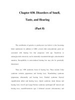
Fig. 4. Distribution of the patients with a postoperative air-bone gap (average of 0.5, 1,
2 and 3 kHz for air conduction minus the average for bone conduction) of 0–10 dB,
11–20 dB, 21–30 dB or Ͼ30 dB with a follow-up of at least 1 year postoperatively.
Jovanovic 262
Delayed Complications. Two additional patients underwent revision surgery
for conductive hearing loss 5–9 months postoperatively. One patient had a dis-
placed prosthesis, combined with total incus erosion due to a too short a prosthe-
sis. The new prosthesis could be fixed at the residual incus. One patient developed
a loose wire with prosthesis and incus fixation resulting from adhesions.
Lasering these adhesions and refixing the prosthesis at the incus improved the
conductive hearing loss.
Discussion
The aim of laser stapedotomy is to enable management of the stapes in such
a way as to ensure the greatest possible protection of the inner ear and to avoid
damage to residual middle ear structures. Advocates of the laser technique agree
that noncontact laser vaporization of the bone covering the vestibule is less trau-
matic for the inner ear than manual instrumental extraction or perforation of the
stapes footplate. It is also true, however, that the laser-related absorption of irra-
diation energy and generation of heat potentially endanger membranous inner
ear structures during perforation of the stapes footplate.
The energy setting should be such that a 0.5- to 0.7-mm perforation dia-
meter is achieved with a one-shot application. The laser perforation should
be circular with a clean-cut edge. This study demonstrated that an adequate
footplate perforation diameter of 0.5–0.7 mm could be achieved with a single
laser application by using a suitable scanner system.
Integrating the control of the scanner in the laser system (SurgiTouch
scanner) enabled synchronization of the spiral laser beam course with the trig-
gering of a laser impulse, so that the laser beam starts the spiral figure at the
same point and runs through the same figure each time. This results in higher
reproducibility of the laser-induced tissue effect. In addition, the laser beam is
moved at an increased speed, so that the spiral completes its course in only 0.04
or 0.05 s. With a maximal single-pulse energy of 1 J, the laser power of a sin-
gle scanner application can thus be increased to 20–22 W (power density of
80,000–88,000 W/cm
2
). In this way, the success rate of the one-shot technique,
i.e. creating an adequately large perforation with a single laser application,
could be increased to 68% of the cases. In 14%, the requisite perforation size
was achieved by a second application with the scanner at the same site, and in
18% the perforation was enlarged at the edge by slightly overlapping applica-
tions without using a scanner.
The results of previous studies support the use of both visible (argon and
KTP) and invisible, far-infrared (CO
2
and Er:YAG) laser systems for primary
otosclerosis surgery [1–7, 16–28].
Technical and Clinical Aspects of ‘One-Shot’ CO
2
Laser Stapedotomy 263
All studies use the multiple-application technique for footplate perfora-
tion. Since the beam of the argon or KTP laser has a diameter of about 0.15 mm,
most authors use the so-called rosette technique with a multiple circular appli-
cation pattern.
Argon and KTP lasers appear to be valuable tools in primary and revision
cases [1, 2, 5, 17, 29]. Here, the insertion of a fiber-optic microhandpiece
(Endo-Otoprobe) [2] is superior to laser application with micromanipulators
attached to the microscope, since the strong laser beam divergence at the exit of
the optical fiber rapidly decreases the power density in relation to the increase
in distance [21, 20]. This reduces the risk of inner ear damage associated with
the penetration depth and temperature problem in the perilymph. Moreover, the
use of the fiber-optic microhandpiece facilitates the vaporization, especially
also of the anterior crus, while reducing the amount of technical equipment
required [25].
The CO
2
laser is also widely applied in the clinical routine [6, 7, 16, 22, 30,
31, 34]. With a beam diameter of 0.18–0.2 mm, all authors use the multiple-
application technique for footplate perforation.
In the group of pulsed laser systems, the Er:YAG laser at first seemed to
possess the most suitable wavelength for middle ear surgery. The Er:YAG and
CO
2
lasers do not coincide in their tissue impact and effectiveness, since they
differ in their wavelength and irradiation time ratio. The continuous-wave CO
2
laser is suitable for use on soft tissue and, if well focussed, for vaporization of
thin bone structures [12], while the Er:YAG laser offers advantages mainly in
the treatment of bone structures [17, 23, 32]. However, as soon as bleeding
occurs, the oligothermic Er:YAG laser radiation is completely absorbed by
blood and no longer reaches the target area. It is then ineffective.
The introduction of new techniques in stapes surgery is always associated
with the question of possible risks to inner ear structures. The clinical applica-
tion must be preceded by experimental in vitro studies for risk assessment
[10–14]. In the final analysis, however, only the postoperative audiometric
results can provide information about the effects on inner ear structures. A com-
parison between post- and preoperative bone conduction auditory thresholds
showed that, on average, patients in the authors’ population had no postopera-
tive deterioration of inner ear function in the examined frequency range of
0.5–4 kHz. Thus, applying higher powers using the one-shot technique with the
scanner does not have a higher potential for damage than the multiple-application
technique [15].
Comparing published audiometric results after laser stapedotomy in relation
to mean differences in the bone conduction auditory thresholds in the main speech
region shows that postoperative improvements of 0.53–5.6dB in those thresholds
are achieved regardless of the laser system applied [6, 7, 22, 24, 28, 30, 31].
Jovanovic 264
The mean value for the frequency range of 0.5–4 kHz is 4.3 dB in our patient
population.
The higher sound level measured in Er:YAG laser therapy is associated
with the risk of inner ear trauma and tinnitus [13, 24, 32]. Moreover, it is sus-
pected that the pressure waves resulting from Er:YAG laser therapy may cause
transitory or even permanent inner ear damage such as high-frequency hearing
loss or tinnitus [24; our own experience]. Thus, the Er:YAG laser has a lower
application safety than the CO
2
laser and cannot be recommended for stapes
surgery at the present time.
The literature comparing hearing results after conventional and laser stape-
dotomy is not suitable for all series, since most authors dealt with more or less
selected groups. Older studies often averaged the air-bone gap for 0.5, 1 and
2 kHz, whereas the more recent ones include the frequencies of 3 or 4 kHz as
well.
In this study, 99% of the patients showed successful closure of the post-
operative air-bone gap to 20dB (average of 0.5, 1, 2 and 3 kHz). In the litera-
ture, closure of the air-bone gap to 10 dB was achieved by 67–99% and
closure to 20 dB by 85–99% of the patients who underwent laser stapedotomy
[1, 2, 4–7]. Assessing the results of conventional stapes surgery in the literature
showed that a mean residual air-bone gap of 10 dB was achieved by 40–96%
of the patients and a gap of 20 dB by 68–99% [25, 33–37]. These data are
comparable to those of laser stapes surgery.
Conclusion
Our findings as well as data in the literature suggest that CO
2
laser stapedo-
tomy is a safe procedure with a lower incidence and severity of intra- and post-
operative complications (e.g. floating footplate, accidental fracturing of a thin
footplate, vertigo) than conventional interventions [6, 7, 16]. Our results sup-
port these published data. No laser-induced sensorineural hearing loss could be
observed in our patients. The closure of the air-bone gap in our study is compa-
rable to conventional stapes surgery.
One-shot stapedotomy achieves an adequately large (0.5–0.7mm in
diameter) circular footplate perforation without appreciable thermal damage
to the surrounding area. It represents a considerable advance in CO
2
laser
stapedotomy.
The CO
2
laser combined with modern scanner systems is well suited for
application in stapes surgery, and, with strict adherence to the parameters, will
help to optimize this high-precision intervention and should reduce the inci-
dence of inner ear damage.
Technical and Clinical Aspects of ‘One-Shot’ CO
2
Laser Stapedotomy 265
References
1 McGee TM: The argon laser in surgery for chronic ear disease and otosclerosis. Laryngoscope
1983;93:1177–1182.
2 Horn KL, Gherini S, Griffin GM: Argon laser stapedectomy using an endo-otoprobe system.
Otolaryngol Head Neck Surg 1990;102:193–198.
3 Lesinski SG: Lasers for otosclerosis. Laryngoscope 1989;99(suppl 46):1–24.
4 Lesinski SG: Lasers for otosclerosis – Which one if any and why. Lasers Surg Med 1990;10:
448–457.
5 Vernick DM: A comparison of the results of KTP and CO
2
laser stapedotomy. Am J Otol
1996;17:221–224.
6 Shabana YK, Allam H, Pedersen CB: Laser stapedotomy. J Laryngol Otol 1999;113:413–416.
7 Buchman CA, Fucci MJ, Roberson JB Jr, De La Cruz A: Comparison of argon and CO
2
laser
stapedotomy in primary otosclerosis surgery. Am J Otolaryngol 2000;21:227–230.
8 Jovanovic S, Schönfeld U, Fischer R, Scherer H: CO
2
laser in stapes surgery. Proc SPIE
1993;1876:17–27.
9 Jovanovic S, Schönfeld U: Application of the CO
2
laser in stapedotomy. Adv Otorhinolaryngol
1995;49:95–100.
10 Jovanovic S, Schönfeld U, Prapavat V: Die Bearbeitung der Steigbügelfussplatte mit verschiede-
nen Lasersystemen. 1. Kontinuierlich strahlende Laser. HNO 1995;43:149–158.
11 Jovanovic S, Schönfeld U, Fischer R: Thermische Belastung des Innenohres bei der Laser-
Stapedotomie. 1. Kontinuierlich strahlende Laser. HNO 1995;43:702–709.
12 Jovanovic S, Schönfeld U, Prapavat V: Effects of continuous wave laser systems on stapes foot-
plate. Lasers Surg Med 1996;19:424–432.
13 Jovanovic S: Der Einsatz neuer Lasersysteme in der Stapeschirurgie; in Müller GJ, Berlien HP
(eds): Fortschritte der Lasermedizin 14. Landsberg, Ecomed, 1996.
14 Jovanovic S, Anft D, Schönfeld U: Influence of CO
2
laser application of the guinea-pig cochlea on
compound action potentials. Am J Otol 1999;20:166–173.
15 Jovanovic S: CO
2
laser in stapes surgery; in Oswal V, Remacle M, Jovanovic S, Krespi J (eds):
Principles and Practice of Lasers in Otolaryngology, Head and Neck Surgery. Den Haag, Kugler,
2002, pp 335–357.
16 Lesinski SG, Newrock R: Carbon dioxide lasers for otosclerosis. Otolaryngol Clin North Am
1993;26:417–441.
17 Perkins RC: Laser stapedotomy for otosclerosis. Laryngoscope 1980;90:228–241.
18 DiBartolomeo JR, Ellis M: The argon laser in otology. Laryngoscope 1980;90:1786–1796.
19 Palva T: Argon laser in otosclerosis surgery. Acta Otolaryngol (Stockh) 1987;104:153–157.
20 Causse JB, Gherini S, Horn KL: Surgical treatment of stapes fixation by fiberoptic argon laser
stapedotomy with reconstruction of the annular ligament. Otolaryngol Clin North Am 1993;26:
395–416.
21 Gherini S, Horn KL, Causse JB, McArthur GR: Fiberoptic argon laser stapedotomy: is it safe? Am
J Otol 1993;14:283–289.
22 Antonelli PJ, Gianoli GJ, Lundy LB: Early post-laser stapedotomy hearing thresholds. Am J Otol
1998;19:443–446.
23 Nagel D: The Er:YAG laser in ear surgery: first clinical results. Lasers Surg Med 1997;21:79–87.
24 Häusler R, Schar PJ, Pratisto H: Advantages and dangers of erbium laser application in stape-
dotomy. Acta Otolaryngol 1999;119:207–213.
25 Häusler R: Fortschritte in der Stapeschirurgie. Laryngorhinootologie 2000;79(suppl 2):95–139.
26 Huber A, Linder T, Fisch U: Is the Er:YAG laser damaging to inner ear function? Otol Neurotol
2001;22:311–315.
27 Lippert BM, Gottschlich S, Kulkens C: Experimental and clinical results of Er:YAG laser stape-
dotomy. Lasers Surg Med 2001;28:11–17.
28 Keck T, Wiebe M, Rettinger G, Riechelmann H: Safety of the erbium:yttrium-aluminium-garnet
laser in stapes surgery in otosclerosis. Otol Neurotol 2002;23:21–24.
29 Nissen RL: Argon laser in difficult stapedotomy cases. Laryngoscope 1989;108:1669–1673.
Jovanovic 266
30 Garin P, Van PK, Jamart J: Hearing outcome following laser-assisted stapes surgery. J Otolaryngol
2002;31:31–34.
31 Motta G, Moscillo L: Functional results in stapedotomy with and without CO
2
laser. ORL J
Otorhinolaryngol Relat Spec 2002;64:307–310.
32 Pratisto H, Frenz M, Ith M: Temperature and pressure effects during erbium laser stapedotomy.
Lasers Surg Med 1996;18:100–108.
33 Levy R, Shvero J, Hadar T: Stapedotomy technique and results: ten years’ experience and compar-
ative study with stapedectomy. Laryngoscope 1990;100:1097–1099.
34 Fisch U: Tympanoplasty, Mastoidectomy, and Stapes Surgery. Stuttgart, Thieme, 1994.
35 Somers T, Govaerts P, Marquet T, Offeciers E: Statistical analysis of otosclerosis surgery per-
formed by Jean Marquet. Ann Otol Laryngol 1994;103:945–951.
36 Persson P, Harder H, Magnuson B: Hearing results in otosclerosis surgery after partial stapedec-
tomy, total stapedectomy and stapedotomy. Acta Otolaryngol (Stock) 1997;117:94–99.
37 Ramsay H, Karkkainen J, Palva T: Success in surgery for otosclerosis: hearing improvement and
other indicators. Am J Otolaryngol 1997;18:23.
Prof. Dr. Sergije Jovanovic
Charité – Universitätsmedizin Berlin, Campus Benjamin Franklin
Hals-Nasen-Ohrenklinik mit Hochschulambulanz, Hindenburgdamm 30
DE–12200 Berlin (Germany)
Tel. 49 30 8445 2440, Fax 49 30 8445 4460, E-Mail
Arnold W, Häusler R (eds): Otosclerosis and Stapes Surgery.
Adv Otorhinolaryngol. Basel, Karger, 2007, vol 65, pp 267–272
Transient Depression of Inner Ear
Function after Stapedotomy: Skeeter
versus CO
2
Laser Technique
T. Somers, J.P. Vercruysse, A. Zarowski, M. Verstreken,
I. Schatteman, F.E. Offeciers
Univ ersity ENT Department, Sint-Augustinus Hospital, W ilrijk, Belgium
Abstract
Performing stapes surgery for otosclerosis is known to be potentially irreversibly harm-
ful to the inner ear function in about 1% of the cases. An early postoperative transient depres-
sion of the bone conduction thresholds is frequently detected after stapes surgery. The
purpose of this study was to compare the evolution of bone conduction thresholds after
primary stapedotomy with two different techniques: skeeter versus CO
2
laser stapedotomy.
Audiological data of 336 otosclerosis operations performed by 2 surgeons between 1997 and
2003 were subjected to analysis. The calibrated hole in the footplate was performed ran-
domly either with the skeeter drill or with the CO
2
laser. Preoperative bone conduction
thresholds were compared with the postoperative levels (day 2–3, week 2, week 6 and month 6)
in all patients. Evolution of the bone conduction was compared for the two studied subgroups
(laser versus skeeter).
Copyright © 2007 S. Karger AG, Basel
Performing stapes surgery for otosclerosis is known to be potentially irre-
versibly harmful to the inner ear function in about 1% of the cases. An early
postoperative transient depression of the bone conduction (BC) thresholds is
frequently detected after stapes surgery. The purpose of this study was to com-
pare the evolution of BC thresholds after primary stapedotomy with two differ-
ent techniques: skeeter versus CO
2
laser stapedotomy. Audiological data of 336
otosclerosis operations performed by 2 surgeons between 1997 and 2003 were
subjected to analysis. The calibrated hole in the footplate was performed
randomly either with the skeeter drill or with the CO
2
laser. Preoperative BC
thresholds were compared with the postoperative levels (day 2–3, week 2, week 6
Somers/Vercruysse/Zarowski/Verstreken/Schatteman/Offeciers 268
and month 6) in all patients. Evolution of the BC was compared for the two
studied subgroups (laser versus skeeter).
Three hundred and thirty-six patients were evaluated between 1997 and
2003. A CO
2
laser stapedotomy was performed in 205 patients (61%) and the
skeeter technique was used in 131 cases (39%).
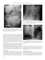
Figure 1 shows the mean preoperative and postoperative air (0.125–8kHz)
and BC thresholds (0.25–4 kHz). The average preoperative air conduction
thresholds revealed a Fletcher index (average threshold for 0.5, 1, and 2 kHz) of
55 dB and an air-bone gap of 29 dB in the Fletcher frequencies. The BC over-
closure for the Fletcher index was 4.2 dB. The average air conduction gain for
the Fletcher frequencies was 27.2 dB.
The evolution of the BC thresholds for the different frequencies is summa-
rized and magnified to a larger scale in figure 2 and shows a minimal but sig-
nificant downward shift (first arrow on the left) on days 2–3 in all frequencies
(p Ͻ 0.001). On days 2–3, an overall average loss of 1.8dB was measured in the
Fletcher frequencies. The temporary drop was minimal for frequencies 0.5, 1
and 2 kHz, but BC measured at 4 kHz dropped by 7dB.
The upward-directed arrows in figure 2 show the gradual BC recovery.
After 2 weeks, there was a partial BC recovery, but this was too slight to be sta-
tistically significant (p Ͼ 0.05). The most important recovery, with statistical
significance (p Ͻ 0.05), is visible between week 2 and week 6. Some further
slight improvement is noticed after 6 months.
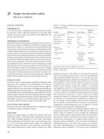
Figure 3 summarizes the evolution of the BC threshold shifts (postopera-
tive BC threshold minus preoperative BC) for the different frequencies. The
largest negative BC shifts were observed for the frequencies 250 and 4,000 Hz.
The residual BC loss at 6 months for 250 Hz averaged 1.6 dB, and for 4,000 Hz
0
10
20
30
40
50
60
70
80
90
0.125 0.25 0.5 1 2 4 8
Frequency (kHz)
Hearing level (dB)
Fig. 1. Mean preoperative thresholds for air conduction () and BC (᭡) and mean
postoperative thresholds for air conduction (ٗ) and BC (᭝) in 332 cases.
Stapes Surgery and Safety Issues 269
2.7 dB. On the other hand, the largest positive shift or overclosure is seen at fre-
quency 2,000Hz (the Carhart notch frequency).
Figure 4 shows how often and to which degree a negative BC shift was
observed on days 2–3 for the different frequencies. At 250 Hz, a BC drop, even
of the slightest degree, was seen in 63% of all cases. At 500 Hz, this was the
case in 50%, at 1,000 Hz in 41%, at 2,000Hz in 48% and at 4,000Hz in 64%.
If we only take the BC losses of Ն20 dB into consideration, the incidences are:
16% (250 Hz), 9% (500 Hz), 5% (1,000 Hz), 3.5% (2,000 Hz), and 13.5%
(4,000 Hz).
The evolution of the BC thresholds for the laser stapedotomy is seen in
figure 5 and for the skeeter stapedotomy in figure 6.
5
15
25
35
0.25 0.5 1 2 4
Frequency (kHz)
Hearing level (dB)
Fig. 2. Detailed view of the BC shifts: the first arrow pointing downward is the
BC shift from the preoperative BC line (ϫ) to early postoperative BC thresholds (2–3 days)
(᭜), the second arrow, which is now pointing upward, is the partial BC recovery at week
2(), and the third and fourth arrows are the further recoveries at 6 weeks (᭡) and
6 months (᭺).
Somers/Vercruysse/Zarowski/Verstreken/Schatteman/Offeciers 270
Ϫ8
Ϫ6
Ϫ4
Ϫ2
0
2
4
6
8
0.25 0.5
Hearing level (dB)
Frequency (kHz)
124
Days 2–3
Week 2
Week 6
Month 6
Fig. 3. This graph shows the evolution of the BC shift expressed as the difference
between the postoperative minus the preoperative BC thresholds and this for different fre-
quencies. A negative value is a BC loss (most obvious at 250 and 4,000 Hz) and a positive
value is a sign of BC overclosure (the most obvious being for 2,000Hz).
Frequency (kHz)
<10 dB
10 dB Ͻ x Ͻ 20 dB
20 dB Ͻ x Ͻ 30 dB
Ͼ30 dB
0
5
10
15
20
25
30
35
0.25 0.5
Incidence (%)
124
Fig. 4. Incidence of occurrence of BC for the different frequencies with different
grades of BC shift (Ͻ10 dB, between 10 and 20 dB, between 20 and 30db, Ͼ30 dB).
Stapes Surgery and Safety Issues 271
No statistically significant difference was detected between the skeeter and
laser technique groups in the downward shifting as well as in the recovery
(independent-sample t test: p Ͼ 0.05).
Conclusion
In conclusion, our study confirmed the frequent presence of a transient but
usually recoverable cochlear dysfunction after stapedotomy. Testing in a large
Frequency (kHz)
Ϫ8
Ϫ6
Ϫ4
Ϫ2
0
2
4
6
8
250
Hearing level (dB)
500 1 2 4
Days 2–3
Week 2
Week 6
Month 6
Fig. 5. Evolution of BC thresholds for the different frequencies after laser stapedotomy.
Frequency (kHz)
Ϫ8
Ϫ6
Ϫ4
Ϫ2
0
2
4
6
8
250
Hearing level (dB)
500 1 2 4
Days 2–3
Week 2
Week 6
Month 6
Fig. 6. Evolution of BC thresholds for the different frequencies after skeeter
stapedotomy.
Somers/Vercruysse/Zarowski/Verstreken/Schatteman/Offeciers 272
group showed significant bone conduction changes at all frequencies but
mainly at 0.25 and 4kHz. These usually recover after a few weeks and are with-
out clinical consequence. Early BC measurement can be used to monitor inner
ear function so as to detect those cases which may have been subjected to more
than usual inner ear trauma.
Dr. T. Somers, MD, PhD
University ENT Department, Sint-Augustinus Hospital
Oosterveldlaan 24
BE–2610 Wilrijk (Belgium)
Tel. ϩ32 34433712, Fax ϩ32 44 33 611, E-Mail
Arnold W, Häusler R (eds): Otosclerosis and Stapes Surgery.
Adv Otorhinolaryngol. Basel, Karger, 2007, vol 65, pp 273–277
Revision Stapes Surgery – Retrospective
Analysis of Surgical Findings in a Series
of 21 Otosclerosis Patients
Marcin Durko
a
, Dariusz Kaczmarczyk
b
, Tomasz Durko
a
Departments of
a
Otosurgery, and
b
Cytophysiology, Histology and Embryology,
Medical University of Lodz, Lodz, Poland
Abstract
Aim: Retrospective analysis of surgical findings in revision stapes surgery in a group of
21 otosclerosis patients qualified for the secondary procedure at the Otosurgery Department
of the Medical University of Lodz, Poland, from 1980 to 2002. Materials and Methods: 21
cases of revision stapes surgery out of a total of 350 surgically treated otosclerosis cases are
discussed. Group A consisted of 17 cases of revision surgery out of 274 patients who had
undergone total stapedectomy (1980–1995) and group B consisted of 4 cases out of 76
patients after stapedotomy (1996–2002). Results: In group A, 17 patients underwent revision
surgery, corresponding to 6.2% out of 274 total stapedectomy cases. Among the indications
for the secondary surgical procedure in this group of patients were: (a) platinum wire prosthe-
sis displacement with ossicular chain discontinuity (n ϭ 12); (b) perichondrium or adipose
tissue atrophy (n ϭ 3), and (c) incudostapedial joint luxation (n ϭ 2). Group B was composed
of 4 cases, i.e. 5.3% out of 76 stapedotomy patients (Teflon piston operation, 0.6 mm). For
both groups, the mean percentage of revision cases was 6% of all patients operated for oto-
sclerosis. Time from the initial surgical procedure to reoperation varied from 1 to 8 years.
Conclusions: (1) The most common indication for revision stapes surgery in patients after
total stapedectomy was prosthesis displacement and necrosis of the long crus of the incus. (2)
Obliteration of the stapes footplate after small fenestra operation was observed to be the most
frequent indication for the secondary stapes procedure in our patient groups.
Copyright © 2007 S. Karger AG, Basel
The growing number of stapes surgeries performed in an increasing num-
ber of otologic centers brings the inevitable risk of complications leading to the
decision to carry out a revision otosurgical procedure. Lack of hearing improve-
ment after the surgery or hearing deterioration, vertigo and tinnitus are the most
frequent signs and symptoms occurring in both the early and late postoperative
course in stapes surgery cases.
Stapes Revision Surgery and Complications
Durko/Kaczmarczyk/Durko 274
According to the literature, there is a significantly higher risk of perceptive
hearing loss, inner ear damage and vertigo of labyrinthine origin as a result of
revision stapes surgery compared to the primary operations [1–3]. Therefore, it
is extremely important to consider all pros and cons before making the decision
of performing a revision surgery [4].
The most common indications for performing revision stapes surgery
(despite the hearing gain) given by various authors are: fluctuation of hearing,
progressive hearing decrease, periodical or permanent vertigo, and increase in
air-bone gap [2, 4, 5].
However, it is very important to differentiate between the cochlear localiza-
tion of otosclerosis and cochlear hydrops coexisting with otosclerosis. The diagno-
sis of the above-mentioned pathologies may be a contraindication to performing a
secondary surgical procedure because of the very high risk of membranaceous
labyrinth injury leading to the deafness of the operated ear [6–8]. Therefore, radio-
logic studies are of growing importance in the management of otosclerosis.
The aim of the present study was a retrospective analysis of the surgical
findings in revision stapes surgery in a group of 21 otosclerosis patients quali-
fied for the secondary procedure at the Otosurgery Department of the Medical
University of Lodz from 1980 to 2002.
Materials and Methods
A series of 21 cases of revision stapes surgery out of a total of 350 patients surgically
treated for otosclerosis underwent a retrospective analysis (table 1). All studied patients were
divided into two groups according to the type of the primary stapes procedure. Group A con-
sisted of 17 cases of revision surgery out of 274 patients who had undergone total stapedec-
tomy (1980–1995) and group B consisted of 4 cases out of 76 patients after stapedotomy
(1996–2002). All the patients were operated by one surgeon. All subjects underwent a routine
audiologic examination performed by the same staff as before the primary surgery. The most
common indications for the revision surgery were conductive hearing loss with an air-bone
gap Ͼ20dB for the frequencies 0.5, 1, 2, 4 kHz and vertigo spells with progressive hearing
loss. Time from the initial surgical procedure to reoperation varied from 1 to 8 years.
Results
In group A, 17 patients underwent revision surgery corresponding to
6.2% out of 274 total stapedectomy cases. Among the indications for the sec-
ondary surgical procedure in this group of patients were: (a) platinum wire
prosthesis displacement with ossicular chain discontinuity (n ϭ 12); (b) peri-
chondrium or adipose tissue atrophy (n ϭ 3), and (c) incudostapedial joint lux-
ation (n ϭ 2).
Revision Stapes Surgery 275
Group B was composed of 4 cases, i.e. 5.3% out of 76 stapedotomy
patients (Teflon piston operation, 0.6 mm). Obliteration of the stapes footplate
was observed to be an indication for all the revision surgery cases in this group.
For both groups, the mean percentage of revision surgeries was 6% of all
treated patients.
Discussion
The percentage of revision cases found in the literature varies from 2 to 6%
[1, 2, 9, 10]. The main reasons to perform secondary surgeries are technical
problems with the stapes replacement prosthesis. In most cases, the connection
between the prosthesis and the long crus of the incus is too loose. Another very
common problem is the adequate length of the prosthesis.
In this case, the difficulty lies in the measurement of the distance between
the incudostapedial joint and the footplate of the stapes because this is not a
constant value due to the various materials used to seal the oval window.
In the presented series of patients, the perichondrium was used for oval
window sealing. Based on our own measurements, we can conclude that in the
studied material, the most frequently used length of the prosthesis varied from
3.75 to 4.00 mm due to the variable depth of the oval window niche [2]. We did
not encounter problems with the right length of the prosthesis, but only with its
displacement and fixation in the oval window niche.
Necrosis of the long process of the incus or vertigo and fluctuating hearing
loss (perilymphatic fistula) are other key issues considering indications for
Table 1. Summary of the retrospective analysis of intraoperative surgical findings in revision stapes
surgery in the series of 350 otosclerosis patients
Type of primary Total number of Total number of Intraoperative surgical
procedure operated patients revision cases findings
Total stapedectomy 274 17 (6.2%) (a) prosthesis displacement
(n ϭ 12; 70.5%)
(b) perichondrium flap replacement
(n ϭ 3; 17.7%)
(c) incudostapedial joint luxation
(n ϭ 2; 11.8%)
Stapedotomy 76 4 (5.3%) obliteration of the stapes footplate
(Teflon piston) (n ϭ 4; 100%)
Total number 350 21 21
Durko/Kaczmarczyk/Durko 276
revision surgery. During analysis of our series of patients, we observed 3 cases
of necrosis of the long process of the incus and 6 cases of perilymphatic fistula.
In the latter cases, the perilymph was not seen in the area of the oval window
niche. That is the reason we prefer not to remove the material sealing the oval
window niche but first to perform scarification of the mucosa and then to use a
fibrin glue as a sealing material. In our opinion, this significantly lowers the
risk of membranaceous labyrinth injury.
In 2 cases qualified for the secondary procedure due to unsatisfactory hearing
improvement, we decided to replace the prosthesis despite its adequate length and
position after the primary surgery. In both cases, the postoperative audiometric test
showed nonsignificant hearing improvement without any rational explanation con-
cerning the surgical technique or audiometric preoperative evaluation.
Analyzing revision surgeries in patients after stapedotomy (Teflon piston
operation in all 4 secondary cases), we observed obliteration of the small fenes-
tra in the footplate of the stapes. The most probable explanation for this was
prosthesis displacement in the upward direction. In such a case, the procedure
of choice is prosthesis replacement as well as restoration of the small fenestra
hole, resulting in significant postoperative hearing improvement [8].
Based on our clinical experience, we think that it is advisable that revision
surgery must be performed by the same surgeon as the primary procedures.
Removing the tissue sealing the vestibule window niche requires extreme preci-
sion and accuracy from the surgeon in order to prevent profound hearing
impairment as a complication. Performing the stapedotomy procedures with
Teflon piston prostheses considerably reduces the possibility of such a postop-
erative complication [9].
Conclusions
(1) The most common indication for revision stapes surgery in patients
after total stapedectomy was prosthesis displacement and necrosis of the long
crus of the incus.
(2) Obliteration of the stapes footplate after small fenestra operation was
observed to be the most frequent indication for the secondary stapes procedure
in our patient groups.
Acknowledgement
This study was supported by grants from the Medical University of Lodz (No. 502-11-
454).
Revision Stapes Surgery 277
References
1 Glasscock ME 3rd, Storper IS, Haynes DS, Bohrer PS: Twenty-five years of experience with
stapedectomy. Laryngoscope 1995;105:899.
2 Latkowski B, Durko T, Pajor A, Morawiec-Bajda A, Kornatowski T, Modrzewska H:
Niepowodzenia i powik5ania po stapedektomiach. Otolaryngol Pol 1992;(suppl 14):230.
3 Shea JJ: Stapedectomy – a long term report. Ann Otol Rhinol Laryngol 1982;91:516.
4 Sheehy J, Nelson R: Revision stapedectomy: a review of 258 cases. Laryngoscope 1981;91:43.
5 Sommers T, Govartes P, DeVarebeke SJ, Offeciers E: Revision stapes surgery. J Laryng Otol
1997;111:233.
6 Fisch U, Acar GO, Huber AM: Malleostapedotomy in revision surgery for otosclerosis. Otol
Neurotol 2001;22:776–785.
7 Wiet RJ, Kubek DC, Lemberg P, Byskosh AT: A meta-analysis review of revision stapes surgery
with argon laser: effectiveness and safety. Am J Otol 1997;18:166–171.
8 Magliulo G, Cristofari P, Terranova G: Functional hearing results in revision stapes surgery. Am J
Otol 1997;18:408–412.
9 Kos MI, Montandon PB, Guyot JP: Short- and long-term results of stapedotomy and stapedectomy
with a teflon-wire piston prosthesis. Ann Otol Rhinol Laryngol 2001;110:907–911.
10 Persson P, Harder H, Magnuson B: Hearing results in otosclerosis surgery after partial stapedec-
tomy, total stapedectomy and stapedotomy. Acta Otolaryngol 1997;117: 94–99.
Marcin Durko, MD
Otosurgery Department, Medical University of Lodz
ul. Kopcinskiego 22
PL–90-153 Lodz (Poland)
Tel./Fax ϩ48 42 678 57 85, E-Mail
Arnold W, Häusler R (eds): Otosclerosis and Stapes Surgery.
Adv Otorhinolaryngol. Basel, Karger, 2007, vol 65, pp 278–284
How to Prevent a Stapes Gusher
C.W.R.J. Cremers
Department of Otolaryngology, University Medical Center St. Radboud,
Nijmegen, The Netherlands
Abstract
A stapes gusher is the result of a congenital inner ear anomaly showing at tone audio-
metry a conductive or mixed hearing loss. The conductive part of the hearing loss could lead
to the thought to explore the middle ear. The congenital origin should lead to a high resolu-
tion. CT-scanning to evaluate a widening of the internal acoustic canal. Repeated audiometry
could show especially a large conductive impairment in the lowest frequencies with a closure
of the airbone gap at 2 khz and a high sensorineural high frequency loss at 4 and 8 khz.
Contralateral stapedial reflexes may be present. Since the x-recessive mixed deafness syn-
drome (DFN3) frequently involves males with an early childhood hearing impairment, clini-
cal suspicion should be high. When stapes surgery is considered a precise medical history is
essential regarding on the start of the hearing impairment. A continuous suspicion will guide
to the audiological, radiological and molecular genetic clues to trace the correct diagnosis
before embarking on stapes surgery.
Copyright © 2007 S. Karger AG, Basel
A stapes gusher is a gusher of perilymph, i.e. cerebrospinal fluid filling
the external ear canal after opening the stapedial footplate [1–3]. It has been
shown to be the result of a too wide communication between the intracranial
space and the vestibule along the internal acoustic canal [1]. A bony widening
of the lateral part of the internal acoustic canal has been shown especially in the
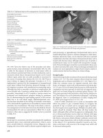
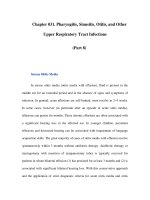
X-recessive stapes gusher syndrome (fig. 1) [3–7].
Prevention of a stapes gusher may be possible in the preoperative diagnos-
tic evaluation focussing on features in the medical history, audiometry results
and CT scanning. Genetic testing of the X chromosome may confirm the clini-
cal diagnosis of the X-linked stapes gusher syndrome.
How to Prevent a Stapes Gusher 279
Medical History
A mainly conductive or mixed progressive hearing impairment already
present in childhood without any indication for an acquired etiology in a male
subject should evoke a first suspicion to diagnose a stapes gusher syndrome.
In the X-recessive stapes gusher syndrome, the hearing impairment is more
severe in the males compared to the affected females as a result of the mode of
inheritance [1–3, 8–11]. Over decades, the hearing impairment is progressive
leading to profound deafness. Head trauma may evoke a deterioration of the
hearing level (fig. 2). In the medical history, affected males may be found in the
family of the mother who is an obligate carrier as a result of the mode of inher-
itance. Her father is affected in case he has transmitted an affected X chromo-
some to her.
A history of a stapes gusher during stapes surgery may be present in that
family.
13.22
01.02
13
01.04
13
01.01
13
22.01
13.22
13.03
13.08
13.08.03.02
13.08.03.03
13.08.03.05
13.08.01
13
08.03
13
08.05
13
08.0502
13
08.0505
13
08.07
13.10
13.24
13.16
Male, deaf or hard of hearing
Male, female, normal by history
Female, carrierFemale, deaf due to meningitis or to
autosomal recessive inheritance
Female, abnormal audiogram
Proband
Dead
Male, female, normal audiogram
Fig. 1. Pedigree of a Dutch family with the X-recessive mixed deafness syndrome
(taken from Cremers et al. [3]).
Cremers 280
Audiometric Evaluation
Preoperative audiograms of adolescents exhibiting a perilymphatic gusher
immediately after opening the stapes footplate have shown remarkably large
air-bone gaps for the low frequencies (0.5, 1.0 kHz) and an almost nonpresent
air-bone gap at 2 kHz (fig. 2). At the higher frequencies, there is again a larger
hearing impairment. Another remarkable finding may be the presence of con-
tralaterally evoked stapedial reflexes [12].
The stapedial footplate may be fixed, which contributes to a large air-bone
gap (figs. 2–4). In case the air-bone gap is smaller, i.e. about 20 dB, this may be
the result of the congenital widened bony vestibule that leads to the presence of
a third window (fig. 2–4). As a result, acoustic energy is lost on its way to the
tectorial membrane. In the pure-tone audiogram, this is reflected in a conduc-
tive component, while brainstem audiometry indicates an inner ear hearing loss.
High-Resolution CT Scanning
A widening of the vestibule, and especially a widening of the internal
acoustic canal, is seen on high-resolution CT scans (figs. 5–7). This has been
0.25
Frequency (kHz)
0.51248
Threshold (dB)
120
100
80
60
40
20
0
Ϫ10
AC
0
10
20
50
60
70
30
40
2
0.25
Frequency (kHz)
0.5 1 2 4 8
Threshold (dB)
120
100
80
60
40
20
0
Ϫ10
BC
0
10
20
50–70
30
40
3
Fig. 2. Air conduction (AC) hearing levels related to age in a Dutch family with the
X-recessive mixed deafness syndrome (DFN3) (taken from Cremers et al. [17]).
Fig. 3. Bone conduction (BC) hearing levels related to age from a Dutch family and an
isolated case with the X-recessive mixed deafness syndrome (DFN3) (taken from Cremers
et al. [17]).
How to Prevent a Stapes Gusher 281
shown to be the case in males affected by the X-recessive stapes gusher syn-
drome. The typical CT image of this anomaly has been used by several authors,
even as a diagnostic case of the month in ORL journals, to help raise suspicion
of this anomaly (figs. 3–7). Before embarking on an exploratory tympanotomy
0.25
Frequency (kHz)
0.51248
Threshold (dB)
120
100
80
60
40
20
0
Ϫ10
ABG
0Ð70
4
Fig. 4. Air-bone gap (ABG) related
to frequency from a Dutch family and an
isolated case with the X-recessive mixed
deafness syndrome (DFN3) (taken from
Cremers et al. [17]).
5
Fig. 5. Three-dimensional picture of part of the normal temporal bone (left side) and a
temporal bone observed in a typical DFN3 patient (right side). In the DFN3 patient, the inner ear
canal as well as its connection to the inner ear are substantially widened (taken from Tang and
Parnes [7] and Cremers et al. [17] with permission).
6
Fig. 6. High-resolution CT scanning of the temporal bone, slice thickness 1 mm. Axial
slice from the right ear at the level of the internal acoustic canal. At the level of the lateral end
(asterisk), the canal is widened and lacks a bony border with the cochlea (small arrows). The
cochlea itself is dysplastic as well because at the side of the modiolus, no bone is visible (long
arrow) (taken from Cremers et al. [17]).
Cremers 282
in patients with a congenital ossicular chain anomaly, it is wise to have per-
formed high-resolution CT scanning of the temporal bone, in case there is a
remarkable inner ear component in the hearing loss. There is a severe argument
to suspect an inner ear anomaly.
Molecular Diagnosis
The DFN3 gene was mapped to the Xq21 region by genetic linkage analy-
ses and the identification of deletions in syndromic and nonsyndromic DFN3
patients [13–16]. The underlying gene POU3F4 was identified. POU3F4 muta-
tions or deletions are the cause of 60% of all stapes gusher cases investigated.
Deletions located far upstream of the POU3F4 gene likely disrupt a transcrip-
tional regulator element. The Nijmegen otogenetic lab has the facilities to per-
form a search for POU3F4 mutations and deletions.
Surgical Aspects
A long-standing perilymphatic gusher may cause a deterioration of the sen-
sorineural component in the hearing loss. As a result of the stapedectomy, the
conductive part may increase. A stapes gusher may continue for about a week dur-
ing hospitalization. A lumbal CSF tap could shorten that period. So, it is desirable
to stop that stapes gusher immediately after its presentation. Nowadays, openings
of the stapes footplate are limited in size by use of a small microdiamond drill or
laser. By an endaural incision, fibrous tissue can be gained and placed directly in
the oval window niche helped to stay in place by the long process of the incus.
Fig. 7. High-resolution CT scanning
of the temporal bone, slice thickness 1 mm.
Axial slice from the left ear of the same
patient as in figure 6. The internal acoustic
canal is clearly depicted and shows enlarge-
ment at the lateral end (asterisk). The first
part of the facial nerve canal (open arrow)
as well as the vestibular nerve canal (long
arrow) are dilated. The vestibulum is
enlarged as well (curved arrow) (taken from
Cremers et al. [17]).
How to Prevent a Stapes Gusher 283
Additional placement of Oxycel
®
proved to be of help according to our experi-
ence to completely stop the gusher surgically. In case the stapes is not fixed as
may occur in the stapes gusher syndrome, perilymphatic fluid may be seen during
testing of the mobility of the stapes footplate. This sign could help the surgeon to
stop the scheduled surgical intervention of a stapedotomy.
Discussion
A good preoperative evaluation may be of help to diagnose the X-linked
stapes gusher syndrome and so to prevent an unneeded stapedotomy procedure.
In case a stapes gusher occurs, it will be helpful for the surgeon to understand
the origin of this complication of stapes surgery. Packing of the oval window
with fibrous tissue has shown to be successful to stop this stapes gusher. Doing
so, it will be helpful to prevent additional inner ear damage.
References
1 Glasscock ME: The stapes gusher. Arch Otolaryngol 1973;98:82–91.
2 Nance WE, Setleff R, McLeod A, Sweeney A, Cooper C, McConnell F: X-linked mixed deafness
with congenital fixture of the stapedial footplate and perilymphatic gusher. Birth Defects Orig
Artic Ser 1971;7:64–69.
3 Cremers CWRJ, Hombergen GCJH, Scaff JJ, Huygen PLM, Volkers WS, Pinckers AJLG: X-
linked progressive mixed deafness with perilymphatic gusher during stapes surgery. Arch
Otolaryngol 1985;111:249–254.
4 Phelps PD, Reardon W, Pembrey M, Bellman S, Luxon L: X-linked deafness, stapes gushers and a
distinctive defect of the inner ear. Neuroradiology 1991;33:326–330.
5 Michel O, Breunsbach J, Matthias R: Das angeborene Liquordrucklabyrinth. HNO 1991;39:
486–490.
6 Talbot JM, Wilson DF: Computed tomographic diagnosis of X-linked congenital mixed deafness,
fixation of the stapedial footplate, and perilymphatic gusher. Am J Otol 1994;15:177–182.
7 Tang A, Parnes LS: X-linked progressive mixed hearing loss: computed tomography findings. Ann
Otol Rhinol Laryngol 1994;103:655–657.
8 Cremers CWRJ: Audiological features of the X-linked progressive mixed deafness syndrome with
perilymphatic gusher during stapes surgery. Am J Otol 1985;6:243–246.
9 Snik AFM, Hombergen GCJH, Mylanus EAM, Cremers CWRJ: Air-bone gap in patients with the
X-linked stapes gusher syndrome. Am J Otol 1995;16:241–246.
10 Cremers CWRJ, Huygen PLM: Clinical features of female heterozygotes in the X-linked mixed
deafness syndrome (with perilymphatic gusher during stapes surgery). Int J Pediatr Otolaryngol
1983;6:179–185.
11 Snik A, Mylanus E, Cremers CWRJ: Audiological characteristics of patients with X-linked stapes
gusher syndrome; in Ernst A, Marchbanks R, Samii M (eds): Intracranial and Intralabyrinthine
Fluids. Basic Aspects and Clinical Applications. Berlin, Springer, 1996, pp 239–243.
12 Cremers CWRJ, Hombergen GCJH, Wentges RTLR: Perilymphatic gusher and stapes surgery.
A predictable complication? Clin Otolaryngol 1983;8:235–240.
13 De Kok YJM, van der Maarel SM, Bitner-Glindzicz M, Huber I, Monaco AP, Malcolm S, Pembrey
ME, Ropers HH, Cremers FPM: Association between X-linked mixed deafness and mutations in
the POU domain gene POU3F4. Science 1995;267:685–688.
Cremers 284
14 De Kok YJM, Vossenaar ER, Cremers CWRJ, Dahl N, Laporte J, Hu LJ, Lacombe D, Fischel-
Ghodsian N, Friedman RA, Parnes LS, Thorpe P, Bitner-Glindzicz M, Pander HJ, Heilbronner H,
Gravelin J, den Dunnen JT, Brunner HG, Ropers HH, Cremers FPM: Identification of a hot spot
for microdeletions in patients with X-linked deafness (DFN3) 900 kb proximal to the DFN3 gene
POU3F4. Hum Mol Genet 1996;5:1229–1235.
15 De Kok YJM, Cremers CWRJ, Ropers HH, Cremers FPM: The molecular basis of X-linked deaf-
ness type 3 (DFN3) in two sporadic cases: identification of a somatic mosaicism for a POU3F4
missense mutation. Hum Mutat 1997;10:207–211.
16 Cremers FPM, Cremers CWRJ, Ropers HH: The ins and outs of X-linked deafness type 3;
in Kitamura K, Steel KP (eds): Genetics in Oto-Rhino-Laryngology. Basel, Karger, 2000, vol 56,
pp 184–195.
17 Cremers CWRJ, Snik AFM, Huygen PLM, Joosten FBM, Cremers FPM: X-linked mixed deafness
syndrome with congenital footplate fixation of the stapedial footplate and perilymphatic gusher
(DFN3) Adv Otorhinolaryngol 2002;61:161–167.
Prof. Dr. C.W.R.J. Cremers
Department of Otolaryngology, University Medical Center St. Radboud
PO Box 9101
NL–6500 HB Nijmegen (The Netherlands)
Tel. 31 24 361 44 50, Fax 31 24 354 02 51, E-Mail
Arnold W, Häusler R (eds): Otosclerosis and Stapes Surgery.
Adv Otorhinolaryngol. Basel, Karger, 2007, vol 65, pp 285–295
Postoperative Granuloma after
Stapedectomy: Is It Destiny or
Avoidable?
C. Batman
a
, Ö. Öztürk
a
, S.S. Ramadan
b
a
Department of Otorhinolaryngology Head and Neck Surgery,
b
Department of
Pathology, Marmara University Hospital, Istanbul, Turkey
Abstract
Objective: The aims of this study were (1) to investigate the pathophysiological charac-
teristics of the middle ear mucoperiosteum against the caustic nature of the gastric content
(GC), which consists largely of acid and pepsin components, and (2) to investigate the possible
role of gastroesophageal reflux and postoperative vomiting (POV) in the etiology of post-
stapedectomy granuloma. Methods: 40 Spraque-Dawley rats of either sex and with a body
weight of 200–300 g were used, and divided into different study groups: group 1: GC adminis-
tration to the middle ear (n ϭ 8); group 2: phosphate-buffered saline administration to the mid-
dle ear (n ϭ 8); group 3: GC (pH: 2) administration in the presence of a Teflon piston (TP)
(n ϭ 6); group 4: phosphate-buffered saline administration in the presence of a TP (n ϭ 6);
group 5: GC administration in the presence of a wired piston (WP) (n ϭ 6); group 6: phos-
phate-buffered saline administration in the presence of a WP (n ϭ 6). GC was administrated to
the middle ear cavities by way of the eustachian tube (ET). In order to overcome the pressure of
the ET, a pump mechanism was used. The increased nasopharyngeal pressure caused a passive
opening of the ET, and transferred a bolus to the middle ear. The animals were decapitated after
1 week, and the bullae were isolated. The tympanic bullae were serially cut and examined with
light microscopy. Results: In the saline controls, there was only a mild amount of polymor-
phonuclear cell (PMN) infiltration in the mildly thickened subepithelial space, indicating a less
pronounced inflammation as compared to the gastric acid group. In the GC group, in addition
to focal hemorrhage and severe subepithelial infiltration of PMNs, the middle ear mucosa was
dramatically thickened with subepithelial edema and dilated capillaries. In the subepithelial tis-
sue, retention cysts and granulation tissue were present. In the piston groups (TP and WP),
there was extensive subepithelial inflammation and edema after GC and saline administrations.
Granulation tissue filling the entire bulla around the piston segments was detected.
Conclusion: The relationship between the administration of GC and middle ear inflammation,
and the possible role of POV in the etiology of poststapedectomy granuloma are emphasized
with our experimental study. The length of the TP may be considerably important to prevent
POV which may lead to gastric reflux to the middle ear.
Copyright © 2007 S. Karger AG, Basel