Stem Cells in Endocrinology - part 3 doc
Bạn đang xem bản rút gọn của tài liệu. Xem và tải ngay bản đầy đủ của tài liệu tại đây (567.58 KB, 29 trang )
44 Nagano
25. Shim H, Gutierrez-Adan A, Chen LR, BonDurant RH, Behboodi E, Anderson GB. Isolation
of pluripotent stem cells from cultured porcine primordial germ cells. Biol Reprod
1997;57:1089–1095.
26. Shamblott MJ, Axelman J, Wang S, et al. Derivation of pluripotent stem cells from cultured
human primordial germ cells. Proc Natl Acad Sci USA 1998;95:13726–13731.
27. Stewart CL, Gadi I, Bhatt H. Stem cells from primordial germ cells can reenter the germ line.
Dev Biol 1994;161:626–628.
28. Labosky PA, Barlow DP, Hogan BL. Mouse embryonic germ (EG) cell lines: transmission
through the germline and differences in the methylation imprint of insulin-like growth factor
2 receptor (Igf2r) gene compared with embryonic stem (ES) cell lines. Development
1994;120:3197–3204.
29. Smith A 2001 Embryoinc stem cells. In: Marshak DR, Garnder RL, Gottlieb D, eds. Stem Cell
Biology. Cold Spring Harbor, Cold Spring Harbor Laboratory Press, 2001, pp. 205–230.
30. Brook FA, Gardner RL. The origin and efficient derivation of embryonic stem cells in the
mouse. Proc Natl Acad Sci USA 1997;94:5709–5712.
31. Tsunoda Y, Tokunaga T, Imai H, Uchida T. Nuclear transplantation of male primordial germ
cells in the mouse. Development 1989;107:407–411.
32. Yamazaki, Y, Mann RW, Lee SS, et al. 2003 Reprogramming of primordial germ cells begins
before migration into the gential ridge, making these cells inadequate donors for reproductive
cloning. Proc Natl Acad Sci USA 2003;100:12207–12212.
33. Wakayama T, Yanagimachi R1 Mouse cloning with nucleus donor cells of different age and
type. Mol Reprod Dev 2001;58:376–383.
34. Constancia M, Pickard B, Kelsey G, Reik W Imprinting mechanisms. Genome Res
1998;8:881–900.
35. Monk M. Epigenetic programming of differential gene expression in development and evo-
lution. Dev Genet 1995;17:188–197.
36. Gage FH. Mammalian neural stem cells. Science 2000;287:1433–1438.
37. Brinster RL, Zimmermann JW. Spermatogenesis following male germ-cell transplantation.
Proc Natl Acad Sci USA 1994;91:11298–11302.
38. Brinster RL, Avarbock MR. Germline transmission of donor haplotype following spermatogo-
nial transplantation. Proc Natl Acad Sci USA 1994;9124:11303–11307.
39. Ogawa T, Arechaga JM, Avarbock MR, Brinster RL. Transplantation of testis germinal cells
into mouse seminiferous tubules. Int J Dev Biol 1997;41:111–122.
40. Nagano MC. Spermatogonial transplantation. In: Gardner DK, Lane M, Watson A, eds. A
Laboratory Guide to the Mammalian Embryo. Oxford, UK, Oxford University Press, 2004,
pp. 334–351.
41. Ryu BY, Orwig KE, Avarbock MR, Brinster RL. Stem cell and niche development in the
postnatal rat testis. Dev Biol 2003;263:253–263.
42. Johnston DS, Russell LD, Friel PJ, Griswold MD. Murine germ cells do not require functional
androgen receptors to complete spermatogenesis following spermatogonial stem cell trans-
plantation. Endocrinology 2001;142:2405–2408.
43. Mahato D, Goulding EH, Korach KS, Eddy EM. Spermatogenic cells do not require estrogen
receptor-α for development or function. Endocrinology 2000;141:1273–1276.
44. Zhang X, Ebata KT, Nagano MC. Genetic analysis of the clonal origin of regenerating mouse
spermatogenesis following transplantation. Biol Reprod 2003;69:1872–1878.
45. Dobrinski I, Ogawa T, Avarbock MR, Brinster RL. Computer assisted image analysis to assess
colonization of recipient seminiferous tubules by spermatogonial stem cell from transgenic
donor mice. Mol Reprod Dev 1999;53:142–148.
Chapter 2 / Germ Line Stem Cells 45
46. Nagano MC. Homing efficiency and proliferation kinetics of male germ line stem cells fol-
lowing transplantation in mice. Biol Reprod 2003;69:701–707.
47. Shinohara T, Orwig KE, Avarbock MR, Brinster RL. Remodeling of the postnatal mouse testis
is accompanied by dramatic changes in stem cell number and niche accessibility. Proc Natl
Acad Sci USA 2001;98:6186–6191.
48. Orwig KE, Shinohara T, Avarbock MR, Brinster RL. Functional analysis of stem cells in the
adult rat testis. Biol Reprod 2002;66:944–949.
49. Ogawa T, Ohmura M, Yumura Y, Sawada H, Kubota Y. Expansion of murine spermatogonial
stem cells through serial transplantation. Biol Reprod 2003;68:316–322.
50. Franca LR, Ogawa T, Avarbock MR, Brinster RL, Russell LD. Germ cell genotype controls
cell cycle during spermatogenesis in the rat. Biol Reprod 1998;59:1371–1377.
51. Ogawa T, Dobrinski I, Avarbock MR, Brinster RL. Transplantation of male germ line stem
cells restores fertility in infertile mice. Nat Med 2000;6:29–34.
52. de Rooij DG, Okabe M, Nishimune Y. Arrest of spermatogonial differentiation in jsd/jsd,
Sl17H/Sl17H, and cryptorchid mice. Biol Reprod 1999;61:842–847.
53. Boettger-Tong HL, Johnston DS, Russell LD, Griswold MD, Bishop CE. Juvenile spermatogo-
nial depletion (jsd) mutant seminiferous tubules are capable of supporting transplanted sper-
matogenesis. Biol Reprod 2000;63:1185–1191.
54. Meng X, Lindahl M, Hyvonen ME, et al. Regulation of cell fate decision of undifferentiated
spermatogonia by GDNF. Science 2000;287:1489–1493.
55. Furuchi T, Masuko K, Nishimune Y, Obinata M, Matsui Y Inhibition of testicular germ cell
apoptosis and differentiation in mice misexpressing Bcl-2 in spermatogonia. Development
1996;122:1703–1709.
56. Matzuk MM, Lamb DJ. Genetic dissection of mammalian fertility pathways. Nat Cell Biol
2002;4(Suppl.):s41–s49.
57. Nagano M, Shinohara T, Avarbock MR, Brinster RL. Retrovirus-mediated gene delivery into
male germ line stem cells. FEBS Lett 2000;475:7–10.
58. Nagano M, Watson DJ, Ryu BY, Wolfe JH, Brinster RL. Lentiviral vector transduction of
male germ line stem cells in mice. FEBS Lett 2002;524:111–115.
59. Nagano M, Ryu BY, Brinster CJ, Avarbock MR, Brinster RL. Maintenance of mouse male
germ line stem cells in vitro. Biol Reprod 2003;68:2207–2214.
60. Mather JP, Attie KM, Woodruff TK, Rice GC, Phillips DM. Activin stimulates spermatogo-
nial proliferation in germ-Sertoli cell cocultures from immature rat testis. Endocrinology
1990;127:3206–3214.
61. Yomogida K, Yagura Y, Tadokoro Y, Nishimune Y. Dramatic expansion of germinal stem
cells by ectopically expressed human glial cell line-derived neurotrophic factor in mouse
Sertoli cells. Biol Reprod 2003;69:1303–1307.
62. Zhang J, Niu C, Ye L, et al. Identification of the haematopoietic stem cell niche and control
of the niche size. Nature 2003;425:836–841.
63. Kanatsu-Shinohara M, Ogonuki N, Inoue K, et al. Long-term proliferation in culture and
germline transmission of mouse male germline stem cells. Biol Reprod 2003;69:612–616.
64. Pawliuk R, Eaves C, Humphries RK. Evidence of both ontogeny and transplant dose-regulated
expansion of hematopoietic stem cells in vivo. Blood 1996;88:2852–2858.
65. Iscove NN, Nawa K. Hematopoietic stem cells expand during serial transplantation in vivo
without apparent exhaustion. Curr Biol 1997;7:805–808.
66. Watt FM, Hogan BLM. Out of Eden: stem cells and their niches. Science 2000;287:1427–1430.
67. Calvi LM, Adams GB, Weibrecht KW, et al. Osteoblastic cells regulate the haematopoietic
stem cell niche. Nature 2003;425:841–846.
46 Nagano
68. Ohta H, Yomogida K, Dohmae K, Nishimune Y. Regulation of proliferation and differentia-
tion in spermatogonial stem cells: the role of c-kit and its ligand SCF. Development
2000;127:2125–2131.
69. Vidal F, Lopez P, Lopez-Fernandez LA, et al. Gene trap analysis of germ cell signaling to
Sertoli cells: NGF-TrkA mediated induction of Fra1 and Fos by post-meiotic germ cells. J Cell
Sci 2001;114:435–443.
70. Giuili G, Tomljenovic A, Labrecque N, Oulad-Abdelghani M, Rassoulzadegan M, Cuzin F.
Murine spermatogonial stem cells: targeted transgene expression and purification in an active
state. EMBO Rep 2002;3:753–759.
71. Shinohara T, Avarbock MR, Brinster RL. β1- and α6-integrin are surface markers on mouse
spermatogonial stem cells. Proc Natl Acad Sci USA 1999;96:5504–5509.
72. Yoshinaga K, Nishikawa S, Ogawa M, et al. Role of c-kit in mouse spermatogenesis: identi-
fication of spermatogonia as a specific site of c-kit expression and function. Development
1991;113:689–699.
73. Shinohara T, Orwig KE, Avarbock MR, Brinster RL. Spermatogonial stem cell enrichment by
multiparameter selection of mouse testis cells. Proc Natl Acad Sci USA 2000;97:8346–8351.
74. Shinohara T, Avarbock MR, Brinster RL. Functional analysis of spermatogonial stem cells in
Steel and cryptorchid infertile mouse models. Dev Biol 2000;220:401–411.
75. Kubota H, Avarbock MR, Brinster RL. Spermatogonial stem cells share some, but not all,
phenotypic and functional characteristics with other stem cells. Proc Natl Acad Sci USA
2003;100:6487–6492.
76. Kanatsu-Shinohara M, Toyokuni S, Shinohara T. CD9 is a surface marker on mouse and rat
male germline stem cells. Biol Reprod 2004;70:70–75.
77. Orwig KE, Ryu BY, Avarbock MR, Brinster RL. Male germ-line stem cell potential is pre-
dicted by morphology of cells in neonatal rat testes. Proc Natl Acad Sci USA 2002;99:11706–
11711.
78. Ramalho-Santos M, Yoon S, Matsuzaki Y, Mulligan RC, Melton DA. “Stemness:” transcrip-
tional profiling of embryonic and adult stem cells. Science 20002;298: 97–600.
79. Ivanova NB, Dimos JT, Schaniel C, Hackney JA, Moore KA, Lemischka IR. A stem cell
molecular signature. Science 2002;298:601–604.
80. Fortunel NO, Otu HH, Ng HH, et al. Comment on “ ‘Stemness’: transcriptional profiling of
embryonic and adult stem cells” and “a stem cell molecular signature.” Science 2003;302:393.
81. Evsikov AV, Solter D. Comment on “ ‘Stemness’: transcriptional profiling of embryonic and
adult stem cells” and “a stem cell molecular signature.” Science 2003;302:393.
82. Vogel G. ‘Stemness’ genes still elusive. Science 2003;302:371.
83. Ivanova NB, Dimos JT, Schaniel C, et al. Response to comments on “ ‘stemness’: transcrip-
tional profiling of embryonic and adult stem cells” and “a stem cell molecular signature”
Science 2003;302:393.
84. Crow JF. The origins, patterns and implications of human spontaneous mutation. Nat Rev
Genet 2000;1:40–47.
85. Crow JF. There’s something curious about paternal-age effects. Science 2003;301:606–607.
86. Goriely A, McVean GA, Rojmyr M, Ingemarsson B, Wilkie AO. Evidence for selective
advantage of pathogenic FGFR2 mutations in the male germ line. Science 2003;301:643–646.
87. Tiemann-Boege I, Navidi W, Grewal R, et al. The observed human sperm mutation frequency
cannot explain the achondroplasia paternal age effect. Proc Natl Acad Sci USA 2002;99:
14952–14957.
88. Oldridge M, Lunt PW, Zackai EH, et al. Genotype-phenotype correlation for nucleotide
substitutions in the IgII-IgIII linker of FGFR2. Hum Mol Genet 1997;6:137–143.
Chapter 2 / Germ Line Stem Cells 47
89. Vajo Z, Francomano CA, Wilkin DJ. The molecular and genetic basis of fibroblast growth
factor receptor 3 disorders: the achondroplasia family of skeletal dysplasias, Muenke cranio-
synostosis, and Crouzon syndrome with acanthosis nigricans. Endocr Rev 2000;21:23–39.
90. Santoro M, Carlomagno F, Romano A, et al. Activation of RET as a dominant transforming
gene by germline mutations of MEN2A and MEN2B. Science 1995;267:381–383.
91. Santoro M, Melillo RM, Carlomagno F, Fusco A, Vecchio G. Molecular mechanisms of RET
activation in human cancer. Ann N Y Acad Sci 2002;963:116–121.
92. Takahashi M. The GDNF/RET signaling pathway and human diseases. Cytokine Growth
Factor Rev 2001;12:361–373.
93. Nagano M, Brinster CJ, Orwig KE, Ryu BY, Avarbock MR, Brinster RL. Transgenic mice
produced by retroviral transduction of male germ-line stem cells. Proc Natl Acad Sci USA
2001;98:13090–13095.
94. Hamra FK, Gatlin J, Chapman KM, Grellhesl DM, Garcia JV, Hammer RE, Garbers DL.
Production of transgenic rats by lentiviral transduction of male germ-line stem cells. Proc Natl
Acad Sci USA 2002;99:14931–14936.
95. Donovan PJ. Growth factor regulation of mouse primordial germ cell development. Curr Top
Dev Biol 1994;19:189–225.
96. Nagano M, Avarbock MR, Brinster RL. Pattern and kinetics of mouse donor spermatogonial
stem cell colonization in recipient testes. Biol Reprod 1999;60:1429–1436.
Chapter 3 / Umbilical Cord Stem Cells 49
49
From: Contemporary Endocrinology: Stem Cells in Endocrinology
Edited by: L. B. Lester © Humana Press Inc., Totowa, NJ
3
Umbilical Cord Stem Cells
Kathy E. Mitchell
CONTENTS
INTRODUCTION
STRUCTURE AND DEVELOPMENT OF THE UMBILICAL CORD
STEM CELLS DERIVED FROM EXTRAEMBRYONIC TISSUES
RELATIONSHIP TO ES, EG, AND ADULT STEM CELLS
UMBILICAL CORD STEM CELLS AND THE IMMUNE SYSTEM
POTENTIAL FOR CELL-BASED THERAPIES
SUMMARY
REFERENCES
1. INTRODUCTION
The two most basic properties of stem cells are the capacities to self-renew and
to differentiate into multiple cell or tissue types (1–3). Generally, stem cells are
categorized as one of three types: embryonic stem cells (ES), embryonic germ
cells (EG), or adult stem cells. ES cells are derived from the inner cell mass of
the blastula (Fig. 1). They proliferate indefinitely and can differentiate sponta-
neously into all three tissue layers of the embryo (4) and into germ cells as well
(5–7). EG cells are derived from primordial germ cells (see Fig. 1), a small set
of stem cells that reside in the protected environment of the yolk stalk, so that they
remain undifferentiated during embryogenesis. As with ES cells, EG cells have the
capacity to differentiate into all three tissue layers (8). Adult stem cells are found
in most tissues and in the circulation. They may have less replicative capacity
than ES or EG cells and, until recently, were thought to have restricted develop-
mental fates (9). This classification system omits a significant source of stem
cells derived from the extraembryonic tissues (umbilical cord, placenta and
amniotic tissues/fluids), which are derived from neither the adult organism nor
the embryo proper. This review will describe studies of stem cells derived from
50 Mitchell
Fig. 1. Stem cells and origins from inner cell mass (ICM) and extraembryonic mesoderm.
ES cells arise from cells derived from the ICM. EG cells, umbilical cord matrix cells, cells
from amniotic tissues, and early hematopoietic stem cells (HSC) arise from extraembry-
onic mesoderm.
Chapter 3 / Umbilical Cord Stem Cells 51
extraembryonic tissues with an emphasis on cells derived from umbilical cord,
their developmental origins, and relationships to other types of stem cells and
potential in regenerative medicine.
2. STRUCTURE AND DEVELOPMENT
OF THE UMBILICAL CORD
The fully developed umbilical cord has one vein and two arteries surrounded
by mucous or gelatinous connective tissue also known as Wharton’s jelly and is
covered with amnion (Fig. 2). There are three distinct zones of stromal cells and
matrix that can be identified: subamniotic layer, Wharton’s jelly, and media and
adventitia surrounding the vessels but no differences along the longitudinal axis
(10). The Wharton’s jelly region, the most abundant, has cleft-like spaces of
stroma matrix molecules of collagens type I, III, and VI, with collagen type VI,
laminin, and heparin sulphate proteoglycan around the clefts. The jelly-filled,
cleft-like spaces are surrounded by stromal cells that are slender and spindle-
shaped myofibroblasts that express vimentin and smooth muscle actin as well as
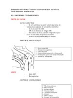
Fig. 2. Human umbilical cord matrix cells. (A) Umbilical cords have two arteries and one
vein surrounded by Wharton’s jelly. (B) Pockets of cobblestone-appearing cells between
the adventitia and Wharton’s jelly. (C) Umbilical cord matrix cells in culture. (D) Human
umbilical cord cells treated by neural induction method of Woodbury et al. (33).
52 Mitchell
desmin (11). Earlier cords have only vimentin and desmin. The structure and
composition of the umbilical cord, rich in highly resilient matrix and myofibro-
blasts, protects the vessels from compression and may also facilitate an exchange
between cord blood and amniotic fluid.
The umbilical cord is derived from extraembryonic mesoderm (see Fig. 1).
After the blastula develops, cells from the inner cell mass (from which ES cells
are derived) form the epiblast (12). Cells destined to become the extraembryonic
mesoderm arise from the proximal epiblast and are the earliest mesoderm to
migrate through the primitive streak (13). Extraembryonic mesoderm increases
over the next few stages of embryogenesis to line the trophectoderm shell, the
amniotic ectoderm, and the yolk sac endoderm and form the connecting stalk as
well. Thus extraembryonic mesoderm contributes to the chorion, amnion, yolk
sac, and, eventually, the umbilical cord (14).
Primordial germ cells (from which EG stem cells are derived) and early
hematopoietic stem cells arise from extraembryonic mesoderm (see Fig. 1).
Hematopoiesis occurs in the yolk sac blood islands 8–8.5 days postconception
in the mouse (15,16). These yolk sac hematopoietic stem cells provide early,
local hematopoiesis during development and circulate through the embryo to
provide oxygen and nutrients. Primordial germ cells arise from the extraembry-
onic mesoderm and appear in the yolk sac as distinguishable entities at about 7
days postconception in the mouse (17). They migrate to the genital ridges of the
developing fetus by about 11.5–12.5 days postconception. Primordial germ cells
retrieved from the genital ridges and cultured in vitro are multipotential (8). The
migration of primordial germ cells is controlled by a number of factors, including
c-Kit and members of the nanos family (18). Primordial germ cells, which do not
home correctly to the genital ridges, undergo apoptosis. If apoptosis does not
occur, these cells can form pediatric germ cell tumors (19).
Recent work has shown that the umbilical cord is a rich source of stem cells.
Ende coined the term Berashis cells, meaning “beginning cells,” to describe the
primitive multipotential cells found in human umbilical cord blood and sug-
gested that they may be related to fetal stem cells (20,21). Three types of stem
cells have been identified in umbilical cord: myofibroblast-like cells from the
umbilical cord matrix, and hematopoietic and mesenchymal stem cells from cord
blood. Stem cells obtained from umbilical cord and placental blood express low
levels of human leukocyte antigens (HLA) and have a universal donor potential
(22). This is an important source of stem cells for bone marrow replacement
when HLA-matched donors cannot be found. The properties of umbilical cord
stem cells, their relationship to other types of stem cells, and their immunogenic
properties are areas of much interest in the emerging fields of stem cell biology
and regenerative medicine.
Chapter 3 / Umbilical Cord Stem Cells 53
3. STEM CELLS DERIVED FROM EXTRAEMBRYONIC TISSUES
3.1. Umbilical Cord Matrix Cells
Umbilical cord matrix may be the remnants of the yolk stalk, the protected
environment where early hematopoietic stem cells and primordial germ cells
arise. As such, it may be a reservoir of cells with stem cell-like characteristics that
can migrate into the developing fetus at appropriate times during development.
Umbilical cord matrix cells express markers for stem cells, including many that
are expressed in ES, EG, and neural precursor or stem cells (Table 1). In addition,
umbilical cord matrix cells can be easily expanded and maintained in culture for
more than 80 population doublings. They express low levels of telomerase. They
also form structures reminiscent of embryoid bodies when cultured past
confluence. They express smooth muscle actin and vimentin, markers for
myofibroblasts; nestin, neuron-specific enolase (NSE), and glial fibrillary acidic
protein (GFAP), markers for neural stem cells; and c-Kit, Oct-4, Tra-1-60, markers
expressed in ES and EG cells. Importantly, umbilical cord matrix cells do not form
teratomas in nude mice (23) or when injected into rat brain or muscle (24).
Pluripotency of ES cells has been linked to expression of Oct-4, a Pit-Oct-Unc
transcription factor (25). Until recently, it was believed that Oct-4 expression in
mature animals was confined exclusively to germ cells (26). Initially expressed
in all cells in the morula, Oct-4 becomes restricted to the inner cell mass at the
blastula stage. Oct-4 is expressed by nearly 100% of isolated umbilical cord
matrix cells after 10 passages and is localized to the nucleus. The full-length
transcript was cloned from umbilical cord matrix cells and has 100% homology
to the reported human embryonic form of Oct-4 (23). The role of Oct-4 in umbilical
cord matrix cells is not known. In ES cells, the precise level of Oct-4 expression
seems to determine cell fate with high levels of Oct-4 expression pushing ES cells
toward extraembryonic mesoderm or endodermal lineages and low Oct-4
expression resulting in cells that become trophectoderm (27). Only ES cells
expressing normal Oct-4 levels remained pluripotent. Recently, a population of
bone marrow stromal cells was isolated after serum deprivation that expressed
Oct-4 (28). Oct-4 expression was also found in amniotic fluid cells (29). Taken
together, these findings suggest that Oct-4 may play a role in nonembryonic stem
cells. This is being investigated for umbilical cord matrix cells in our laboratory.
Umbilical cord matrix cell express many of the markers Shamblott et al. (30)
identified in derivatives of cultured EG cells including NSE, vimentin, and
nestin—markers for neural precursors—and glial markers, 2',3'-cyclic nucle-
otide 3'-phosphodiesterase, and GFAP, also expressed in early neural precursors
(see Table 1). In addition, umbilical cord matrix cells express c-Kit, which is
important for proper migration of primordial germ cells. Expression of these
54 Mitchell
proteins, including Oct-4, by both umbilical cord matrix cells and EG cells
suggests a possible relationship between the two cell types, particularly in light
of their residing in the same region of the developing fetus and common origin
from extraembryonic mesoderm.
Umbilical cord matrix cells can be differentiated to form neuron-like cells
based on morphology, expression of neuron-specific proteins, and development
of voltage-gated potassium channels found in early neurons that are important
for development of electrical excitability (31,32). Some cells differentiate spon-
taneously to express neuronal markers. Induction by the method of Woodbury et
al. (33) greatly enhances the number of cells that differentiate into a neuron-like
cell (approximately 80%) (31). Umbilical cord matrix cells induced by this
method form primitive networks between the cells with long axon-like pro-
cesses, refractile cell bodies and dendrite-like processes, highly reminiscent of
primary neurons in culture (Fig. 2D). The induced umbilical cord matrix cells
express neurofilament M, Tuj1, growth cone-associated protein (GAP43), and
tyrosine hydroxylase, which are markers for more mature neurons. Thus, as with
many stem cells, umbilical cord matrix stem cells appear to differentiate along
a neuronal fate readily, with some differentiation occurring spontaneously.
Umbilical cord matrix cells have also been used in in vivo xenotransplantation.
Studies by Weiss et al. (24) suggest that porcine umbilical cord matrix cells
survive, migrate, and begin to express markers for mature neurons when trans-
planted into rat brain. Umbilical cord matrix cells loaded with the fluorescent
dye, PKH26, were transplanted into rat brains and detectable at periods from 2
to 6 weeks after transplantation. After 4 weeks, the umbilical cord matrix cells
were detected primarily along the injection tract and were small and spherical,
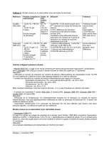
Table 1
Comparison of Markers for Stem Cells Expressed in ES, EG, UCM, Amniotic,
and NS Cells
ES cells EG cells UCM cells Amniotic NS cells
Oct-4 + + + + NA
Telomerase + + + + –
Vimentin NA + + + +
Nestin NA + + + +
NSE NA + + + +
GFAP NA + + + +
ES, embronic stem; EG, embryonic germ; UCM, umbilical cord matrix; NS, neural stem;
NSE, neuron-specific enolase; GFAP, glial fibrillary acidic protein; NA, not applicable.
Chapter 3 / Umbilical Cord Stem Cells 55
with very few processes. However, the transplanted umbilical cord cells did
express neuronal filament 70 (NF70) based on detection with an antibody spe-
cific for porcine but not rodent NF70. In contrast, 6 weeks after injection, about
10% of the detectable umbilical cord matrix cells had migrated away from the
injection site and into the region just ventral to the corpus callosum. These
umbilical cord matrix cells also expressed NF70. Taken together, these studies
suggest that umbilical cord matrix cells may have the capacity to differentiate
into neurons in vitro and in vivo. More work needs to be done to establish that
the umbilical cord matrix cells can generate action potentials in vitro and form
new neuronal connections in vivo. Studies are under way to address these issues
and to establish whether umbilical cord matrix cells can ameliorate neural defi-
cits after oxygen deprivation of the brain or in a Parkinson’s disease model in rat.
3.2. Umbilical Cord Blood Cells
Umbilical cord blood is a rich source of hematopoietic stem/progenitor cells
and has been used successfully as an important source of cells for hematopoietic
stem cell (HSC) transplantation (34). Although somewhat controversial, umbili-
cal cord blood is also thought to be a source of mesenchymal stem cells (MSC).
MSC can be differentiated into cells other than blood, but may also be important
for long-term engraftment in bone marrow transplants with umbilical cord blood
(35). There is much interest in the potential of umbilical cord blood as a source
of multipotential stem cells; umbilical cord blood is often banked and cryogeni-
cally stored for use by the individual from whom the cord blood was taken or as
a source for donation to other individuals in need of bone marrow transplants or
other cell-based therapies.
Umbilical cord blood is an important source of HSC for bone marrow trans-
plants for which HLA-matched donors cannot be found. Umbilical cord blood
stem cell progenitors are used now routinely as an alternative to bone marrow
transplant (36). There are many potential advantages in using the HSC from cord
blood as compared with HSC derived from bone marrow. First, HSC in umbilical
cord blood occur at higher frequency than in peripheral blood (37) and at com-
parable levels to their occurrence in bone marrow, making up about 2% of the
total mononuclear cell population (38). Importantly, umbilical cord HSC have a
greater ability to replicate than bone marrow-derived HSC and can be manipu-
lated genetically as well (39). They can be collected noninvasively with no risk
to mother or child. Because of their increased proliferative rate, HSC can be
expanded ex vivo, unlike adult hematopoietic stem cells (40,41). This potential
for expansion can be augmented by treatment with a cocktail of growth factors
(thrombo poetin, stem cell factor, interleukin-3, flt3-ligand, and basic fibroblas-
tic growth factor) allowing for a 500-fold expansion of CD34
+
HSC from umbili-
56 Mitchell
cal cord blood (42). CD34
+
umbilical cord cells may also have potentials beyond
the hematopoietic lineages. Pesce et al. (43) showed that CD34
+
umbilical cord
cells can differentiate into muscle fibers in immune-suppressed mice and can
also form myotubes when cocultured with muscle cells in vivo. The abilities to
expand ex vivo, genetically manipulate, and cryogenically store umbilical cord
blood HSC in addition to their potential to contribute to repair of other tissues
holds great promise for future stem cell-based therapies.
Although umbilical cord blood is known to be a rich source of HSC (44,45),
the existence of MSC in umbilical cord blood has been somewhat controversial
(46). However, in recent studies, MSC have been isolated from cord blood
through methods used for isolation of MSC from bone marrow (47). The umbili-
cal cord-derived MSC displayed a fibroblast-like morphology and were smooth
muscle actin and fibronectin positive. This suggests that they may be related to
the cells isolated from umbilical cord matrix, which may migrate into the cord
blood circulation. Other groups have isolated MSC from umbilical cord blood
that could be expanded in culture and induced to differentiate into osteocytes,
chondrocytes, and adipocytes as well as hepatocytes of mesenchymal origin
(48). They were also able to induce the cells to express markers for neurons and
glia. Hou et al. isolated MSC from umbilical cord blood by negative selection.
These cells do not express CD34, CD11a, or CD11b, but do express CD29 and
CD71, which is identical to markers of MSC derived from bone marrow (49).
Hou et al. also isolated clonal populations of MSC that could differentiate into
adipocytes, chondrocytes, osteocytes, hepatocytes, neuronal, and glial cells based
on expression of specific markers.
Cells that resemble neural stem cells have been isolated from umbilical cord
blood (50). Nestin, an intermediate filament expressed in neural precursors, is
expressed by a large percentage of human cord blood monocytes that also
coexpress CD133. However, nestin expression was not detected in adult mono-
cytes (50). Buzanska et al. (51) showed that nestin-expressing cells from umbili-
cal cord blood could be directed to differentiate into early neurons that expressed
TUJ1 (a neuron-specific class III β-tubulin), astrocytes expressing GFAP, and
galactocerebrosidase expressing oligodendrocytes by treatment with brain-
derived neurotrophic factor and retinoic acid. Similarly, other studies have
shown that CD45-negative cells from umbilical cord blood could be expanded
in culture and then be induced to form cells that express neuronal and glial
markers TUJ1 and GFAP (52). Many other studies have shown the potential for
cells from cord blood to differentiate into cells that express neuronal or glial
proteins using a number of different induction protocols (53). Interestingly,
many of the proteins are expressed in umbilical cord cells without any treatment
to induce them. For example, GFAP was expressed in about one-third of the
isolated cells. This was increased by treatment with retinoic acid. Similar results
Chapter 3 / Umbilical Cord Stem Cells 57
were found for expression of NeuN. These studies show that a population of cells
within umbilical cord blood express markers and have properties very similar to
those of umbilical cord matrix cells and neural precursor cells (see Table 1).
3.3. Other Extraembryonic Stem Cells
Other cells with stem cell-like properties have been identified in the extraem-
bryonic tissues. Oct-4-expressing cells have been identified in human amniotic
fluid (29). Amniotic fluid cells express stem cell factor, smooth muscle actin, and
vimentin and are rapidly proliferating compared with adult cells (54). They may
also express telomerase as telomerase activity has been detected in amniotic fluid
(55). Amniotic cells also express a number of glial and neuronal proteins, includ-
ing neurofilament proteins, microtubule-associated protein 2, GFAP, 2',3'-cyclic
nucleotide 3'-phosphodiesterase, myelin basic protein, and galactocerebroside
(56,57). These properties are similar to those of cells isolated from umbilical cord
matrix, suggesting that they may have a common origin.
An interesting observation made by several investigators is that many neu-
ronal and glial proteins are expressed in extraembryonic tissues. Initially,
expression of some neuronal and glial proteins, NSE and S100, in cord blood
and amniotic fluid was thought to be indicative of neonatal neuronal damage
(58–61). But recent studies have shown that high levels of NSE and S-100 are
expressed in umbilical cord blood after normal delivery. They are expressed at
higher levels in the artery than venous blood, suggesting fetal origin (62).
Wijnberger et al. did a more extensive analysis of neuronal and glial protein
expression in the placenta and umbilical cord, looking for expression of S-100,
NSE, GFAP, and GAP43 (63). They found that many cell types, including
myofibroblasts of Wharton’s jelly, are positive for NSE and S-100, as are cells
of the vascular wall, amnion epithelium, and macrophages and monocytes in
umbilical cord blood. GFAP and GAP43 were not detected, however. S-100 is
also expressed in placental tissues (64). These results suggest that extraembry-
onic tissues are possibly a rich source of stem cells with neural precursor type
properties.
4. RELATIONSHIP TO ES, EG, AND ADULT STEM CELLS
ES cells are derived from the inner cell mass of the blastula. EG cells are
derived proximal to the epiblast, residing temporarily in a protected environment
of the yolk stalk so that they remain undifferentiated. Adult stem cells are found
in most tissues, as well as in circulation. Adult stem cells are usually quiescent
but become activated under conditions of stress or injury. What are the origins
of adult stem cells and how do they keep from differentiating? These are some
of the most critical questions in stem cell biology. It has been suggested that stem
58 Mitchell
cells may not be the first cells to show up in a tissue, but rather may appear later
in development when they can populate adult niches. Adult stem cells may be
differentiated appropriately for their tissue, but also have other potentials if in a
different microenvironment.
Are multipotential adult stem cells related to the primitive stem cells of the
umbilical cord? Most multipotential adult stem cells share common characteris-
tics with the myofibroblast-like cells isolated from umbilical cord matrix (31).
Postnatal stem cells in the adult, from a wide variety of sources, appear to be
capable of differentiation into multiple tissue types. Cells derived from bone
marrow (65), skin (66), astrocytes (67), synoviocytes (68), adipose (69), and
dental pulp (70) have recently been shown to be multipotential. Many of these
multipotential stem cells may have a common precursor in that they are tissue-
specific myofibroblasts. Myofibroblasts are found throughout the body and
include bone marrow stromal cells, astrocytes, synoviocytes, and pericytes (71).
Myofibroblasts in the adult take part in growth, development, and repair of
normal tissue. They can also be the cause of organ fibrosis, scar formation, and
tumors. Myofibroblasts have some tissue-specific functions but are similar in
morphology, function, and biochemistry regardless of their location (71). Per-
haps myofibroblasts or their precursors exist as a pool of pluripotent stem cells
that exist in equilibrium between stem cells that are buried in the diverse organs
and those that circulate from the bone marrow, similar to monocytes and mac-
rophages as suggested by Labat for adult stem cells (72). There are intrinsic
differences in fetal versus adult myofibroblasts that regulate their responses to
cytokines, which in turn may account for the ability for scarless repair by fetal
myofibroblasts (73). This may be a critical characteristic that favors younger
myofibroblasts, such as those isolated from umbilical cord matrix for therapeutic
applications.
5. UMBILICAL CORD STEM CELLS AND THE IMMUNE
SYSTEM
Although much of the enthusiasm about the potentials for therapeutic appli-
cations of ES cells is based on the hope that they will evade the immune system,
very little work has been done to investigate this potential. Immunological rejec-
tion may be an important barrier for ES cell-based therapies if MHC molecules
responsible for immune-mediated graft rejection are expressed by ES cells after
they differentiate. Human ES cells express HLA class I but not class II molecules.
Expression of both classes of molecules increases with differentiation in vitro or
in vivo (74). As with ES cells, mesenchymal stem cells express low levels of
HLA class I molecules but not class II (75). Importantly, they were able to
Chapter 3 / Umbilical Cord Stem Cells 59
suppress mixed lymphocytic cultures and retained this capability even after dif-
ferentiation.
Tumor formation (teratomas) by ES cells is a major hurdle that needs to be
overcome before this source of cells can be used in therapeutic applications.
Preliminary findings show that, unlike ES cells, human umbilical cord matrix
cells do not form tumors in immune compromised mice (23). Porcine umbilical
cord matrix cells do not illicit an immune response when injected into rat brain
or muscle, nor are they rejected at 4 weeks (24). The mechanism of this immune
evasion is not known but may involve the low expression of HLA class I mol-
ecules and expression of HLA-G (23), a nonclassical HLA that suppresses
immune response at the maternal–fetal interface (76) and in muscle (77).
Umbilical cord blood HSC have low immunogenicity with a lower incidence
of graft-vs-host disease when used for transplantation in cancer patients, even
when the number of HLA markers that are matched are lower (78). The mecha-
nism of this potential to evade the immune system is not understood. However,
β2-microglobulin is expressed constitutively in cord blood cells (79) and is
known to be an integral part of MHC expression in killer T cells and, thus, may
play a role in immune evasion of umbilical cord blood HSC (80). Stem cells from
umbilical cord appear to have the unique ability to evade the immune system,
which makes their use therapeutically particularly exciting. More research on the
mechanisms by which umbilical cord stem cells suppress immune response and
how long after differentiation this is maintained is essential.
6. POTENTIAL FOR CELL-BASED THERAPIES
Umbilical cord blood is commonly used in cell-based therapies today for
reconstitution of the bone marrow after bone marrow ablation for cancers of the
blood (36). There are some new experimental therapies using bone marrow
transplant with cord blood cells being developed for other diseases. Umbilical
cord blood transplantation in Wiskott Aldrich syndrome, which results in severe
immune deficiency and early death if not treated, was found to result in rapid and
reliable recovery of immune function, with low risk of graft-vs-host disease (81).
Using umbilical cord blood stem cells taken from unrelated donors, Staba et al.
(82) treated children with Hurler’s syndrome, who lack of a functional enzyme,
alpha-
L-iduronidase. These researchers were able to treat these patients without
bone marrow ablation and to have improvement in survival and less neuronal
degeneration than Hurler’s patients who received bone marrow transplants. The
researchers speculate that stem cells from cord blood may transport α-
L-
iduronidase across the blood–brain barrier more effectively. In addition, they are
younger cells and do not have to be matched as closely. Research is under way
to expand the use of umbilical cord blood cells to treat other disorders such as β-
thalassemia (83).
60 Mitchell
Animal models suggest that umbilical cord blood cells may be useful in treat-
ment of amyotrophic lateral sclerosis by slowing motor neuron degeneration
when injected intravenously (84). Ende and coworkers found that intravenous
injection of umbilical cord blood cells could extend the survival of several mouse
knockout models of human disease, including amyotrophic lateral sclerosis (85),
Alzheimer’s (85), Huntington’s (86), Parkinson’s (87), and type 1 diabetes (88).
Human umbilical cord blood cells also improve the mobility of rats with spinal
cord injuries when injected intravenously. Cord blood cells were observed in the
areas of injury of spinal cord but not others and never seen in the control, unin-
jured animals (89). Similarly, umbilical cord blood cells were able to improve
function in a stroke model in the rat when injected intravenously. The human
umbilical cord blood cells differentiated into cells that expressed glial or neu-
ronal markers (90). This suggests that umbilical cord blood cells have the ability
to target to and heal neurologic defects.
Cells from umbilical cord matrix may also be a source of cells for treatment
of neurodegenerative disease. Medicetty et al. (91) treated rats with a unilateral
6-hydroxydopamine (6-OHDA) lesion that caused parkinsonian-like symptoms.
Four weeks after the 6-OHDA lesion, rats were injected with umbilical cord
matrix cells or sham transplants. Four weeks after transplantation, there was a
significant decrease in apomorphine-induced rotatory behavior in the parkinso-
nian rats that received umbilical cord matrix cell transplants as compared
with parkinsonian rats that received a sham transplant. Normal rats, without
6-OHDA lesions, were transplanted with umbilical cord matrix cells but
showed no changes in behavior. This work suggests that umbilical cord matrix
cells can target areas of neurodegeneration and play a role in healing of neural
tissue. Amniotic cells may have a similar potential (92). Labeled amniotic epi-
thelial cells were injected into monkeys with spinal cord injuries. Some labeled
neurons were subsequently found in the spinal cord. Glial scar formation was
decreased compared with animals that did not receive amniotic epithelial cells.
More importantly, the function of the animals improved suggesting that amniotic
epithelial cells help in axon regrowth. These studies suggest that cells from
umbilical cord blood and other cells from extraembryonic tissues may be an
important source of stem cells for a variety of therapeutic applications.
7. SUMMARY
There is much hope today for the many potential benefits that can be achieved
through stem cell research, including a better understanding of the basic biology
of stem cells that may provide insights into cancer when proper control of pro-
liferation and differentiation have gone awry, for developmental processes, and
for drug discovery. There is significant potential to discover new drugs through
Chapter 3 / Umbilical Cord Stem Cells 61
stem cell research that will increase the proliferative capacity of specific popu-
lations of cells in the brain to ameliorate Parkinson’s disease or in the islets to
produce new insulin-producing cells or discover new chemotherapeutic agents
that target the cancer stem cell and thus improve long-term survival of cancer
patients. What is clear is that there is much yet to be learned; stem cell biology
and regenerative medicine are in their infancy. We need to study cells from many
sources to be able to harness these potentials. The cells from the umbilical cord
and other extraembryonic tissues are a particularly exciting and promising source
of primitive stem cells based on their ready availability, low immunogenicity,
and lack of tumorigenicity. The study of extraembryonic stem cells may also
reveal the origins of the adult stem cell. Extraembryonic stem cells may also be
a particularly useful tool in drug development because of their ready availability,
making it possible to harvest cells that represent a genetically diverse population
or stem cells that carry specific genetic defects.
ACKNOWLEDGMENTS
Supported by P20 RR 15563-02 COBRE-NIH and RO1-NS/HL36124.
Jeremy Traas is also acknowledged for his research contributions.
REFERENCES
1. McKay R. Stem cells in the central nervous system. Science 1997;276:66–71.
2. Gordon MY, Blackett NM. Reconstruction of the hematopoietic system after stem cell trans-
plantation. Cell Transplant 1998;7:339–344.
3. Scheffler B, Horn M, Blumcke I, et al. Marrow-mindedness: a perspective on neuropoiesis.
Trends Neurosci 1999;22:348–357.
4. Smith A. Embryo-derived stem cells: of mice and men. Annu Rev Cell Dev Biol 2001;17:435–462.
5. Hubner K, Fuhrmann G, Christenson LK, et al. Derivation of oocytes from mouse embryonic
stem cells. Science 2003;300:1251–1256.
6. Toyooka Y, Tsunekawa N, Akasu R, Noce T. Embryonic stem cells can form germ cells in
vitro. PNAS 2003;100:11457–11462.
7. Clark AT, Bodnar MS, Fox M, et al. Spontaneous differentiation of germ cells from human
embryonic stem cells in vitro. Hum Mol Genet 2004;13:727–739.
8. Shamblott MJ, Axelman J, Wang S, et al. Derivation of pluripotent stem cells from cultured
human primordial germ cells. PNAS 1998;95:13726–13731.
9. Paul G, Li JY, Brundin P. Stem cells: hype or hope? Drug Discov Today 2002;7:295–302.
10. Nanaev AK, Kohnen G, Milovanov AP, Domogatsky SP, Kaufmann P. Stromal differentia-
tion and architecture of the human umbilical cord. Placenta 1997;18:53–64.
11. Takechi K, Kuwabara Y, Mizuno M. Ultrastructural and immunohistochemical studies of
Wharton’s jelly umbilical cord cells. Placenta 1993;14:235–245.
12. Gardner RL. Cell lineage and cell commitment in the early mammalian embryo. Mead Johnson
Symp Perinat Dev Med 1979;16:18–24.
13. Lawson KA, Meneses JJ, Pedersen RA. Clonal analysis of epiblast fate during germ layer
formation in the mouse embryo. Development 1991;113:891–911.
62 Mitchell
14. Vogler H. Human Blastogenesis. Formation of the Extraembryonic Cavities. Bibliotheca
Anatomica 30. Karger, Basel, 1987.
15. Moore MA, Metcalf D. Ontogeny of the haemopoietic system: yolk sac origin of in vivo and in
vitro colony forming cells in the developing mouse embryo. Br J Haematol 1970;18:279–296.
16. Weissman IL, Warnke R, Butcher EC, Rouse R, Levy R. The lymphoid system. Its normal
architecture and the potential for understanding the system through the study of lymphopro-
liferative diseases. Hum Pathol 1978;9:25–45.
17. Mintz B, Russell ES. Gene-induced embryological modifications of primordial germ cells in
the mouse. J Exp Zool 1957;134:207–237.
18. Tres, LL, Rosselot C, Kierszenbaum AL Primordial germ cells: what does it take to be alive?
Mol Reprod Dev 2004;68:1–4.
19. Schneider DT, Schuster AE, Fritsch MK, et al. Multipoint imprinting analysis indicates a
common precursor cell for gonadal and nongonadal pediatric germ cell tumors. Cancer Res
2001;61:7268–7276.
20. Ende, N. History of umbilical cord blood transplantation. Lancet 1995;346:1161.
21. Ende N. Berashis cells in human umbilical cord blood vs. embryonic stem cells. J Med
2002;33:167–171.
22. von Drygalski A, Adamson J. Placental/umbilical cord blood (PCB) stem cells for transplan-
tation: early clinical outcomes and the status of ex vivo expansion strategies. Keio J Med
2000;49:141–151.
23. Traas J, Kaptur R, Schermerhorn T, Chun R, Mitchell KE Stem cell gene array analysis of Oct-
4 positive human umbilical cord matrix cells. Mol Cell Biol 2003;14:115a.
24. Weiss ML, Mitchell KE, Hix JE, et al. Transplantation of porcine umbilical cord matrix cells
into the rat brain. Exp Neurol 2003;182:288–299.
25. Suzuki N, Rohdewohld H, Neuman T, Gruss P, Scholer HR. Oct-6: a POU transcription factor
expressed in embryonal stem cells and in the developing brain. EMBO J 1990;9:3723–3732.
26. Pesce M, Scholer HR. Oct-4: control of totipotency and germline determination. Mol Reprod
Dev 2000;55:452–457.
27. Niwa H, Miyazaki J, Smith AG. Quantitative expression of Oct-3/4 defines differentiation,
dedifferentiation or self-renewal of ES cells. Nat Genet 2000;24:372–376.
28. Pochampally RR, Smith JR, Ylostalo J, Prockop DJ. Serum deprivation of human marrow
stromal cells (hMSCs) selects for a subpopulation of early progenitor cells with enhanced
expression of OCT-4 and other embryonic genes. Blood 2004;103:1647–1652.
29. Prusa AR, Marton E, Rosner M, Bernaschek G, Hengstschlager M. Oct-4-expressing cells in
human amniotic fluid: a new source for stem cell research? Hum Reprod 2003;18:1489–1493.
30. Shamblott MJ, Axelman J, Littlefield JW, et al. Human embryonic germ cell derivatives
express a broad range of developmentally distinct markers and proliferate extensively in vitro.
Proc Natl Acad Sci USA 2001;98:113–118.
31. Mitchell KE, Weiss ML, Mitchell BM, et al. Matrix cells from Wharton’s jelly form neurons
and glia. Stem Cells 2003;21:50–60.
32. Helwig B, Van Wye T, Hoynowski S, Mitchell KE. Defining key proteins in stem cell based
neuronal development using proteomics. Mol Cell Biol 2003;14:115a.
33. Woodbury D, Schwarz EJ, Prockop DJ, Black IB. Adult rat and human bone marrow stromal
cells differentiate into neurons. J Neurosci Res 2000;61:364–370.
34. Broxmeyer HE, Douglas GW, Hangoc G, et al. Human umbilical cord blood as a potential
source of transplantable hematopoietic stem/progenitor cells. Proc Natl Acad Sci USA
1989;86:3828–3832.
35. Sirchia G, Rebulla P. Placental/umbilical cord blood transplantation. Haematologica
1999;84:738–747.
Chapter 3 / Umbilical Cord Stem Cells 63
36. Gluckman E, Rocha V, Boyer-Chammard A, et al. Outcome of cord-blood transplantation
from related and unrelated donors. Eurocord Transplant Group and the European Blood and
Marrow Transplantation Group. N Engl J Med 1997;337:373–381.
37. Murohara T, Ikeda H, Duan J, et al. Transplanted cord blood-derived endothelial precursor
cells augment postnatal neovascularization. J Clin Invest 2000;105:1527–1536.
38. Wu AG, Michejda M, Mazumder A, et al. Analysis and characterization of hematopoietic
progenitor cells from fetal bone marrow, adult bone marrow, peripheral blood, and cord blood.
Pediatr Res 1999;46:163–169.
39. Mayani H, Lansdorp PM. Biology of human umbilical cord blood-derived hematopoietic
stem/progenitor cells. Stem Cells 1998;16:153–165.
40. Lansdorp PM, Poon S, Chavez E, et al. Telomeres in the haemopoietic system. Ciba Found
Symp 1997;211:209–218; discussion 219–222.
41. Globerson A. Hematopoietic stem cells and aging. Exp Gerontol 1999;34:137–146.
42. Kashiwakura I, Takahashi TA. Basic fibroblast growth factor-stimulated ex vivo expansion
of haematopoietic progenitor cells from human placental and umbilical cord blood. Br J
Haematol 2003;122:479–488.
43. Pesce M, Orlandi A, Iachininoto MG, et al. Myoendothelial differentiation of human umbilical
cord blood-derived stem cells in ischemic limb tissues. Circ Res 2003;93:e51–e62.
44. Hows JM. Status of umbilical cord blood transplantation in the year 2001. J Clin Pathol
2001;54:428–434.
45. Benito AI, Diaz MA, Gonzalez-Vicent M, Sevilla J, Madero L. Hematopoietic stem cell
transplantation using umbilical cord blood progenitors: review of current clinical results.
Bone Marrow Transplant 2004;33:675–690.
46. Mareschi K, Biasin E, Piacibello W, Aglietta M, Madon E, Fagioli F. Isolation of human
mesenchymal stem cells: bone marrow versus umbilical cord blood. Haematologica
2001;86:1099–1100.
47. Romanov YA, Svintsitskaya VA, Smirnov VN. Searching for alternative sources of postnatal
human mesenchymal stem cells: candidate MSC-like cells from umbilical cord. 2003;Stem
Cells 21:105–110.
48. Lee OK, Kuo TK, Chen WM, Lee KD, Hsieh SL, Chen TH. Isolation of multipotent mesen-
chymal stem cells from umbilical cord blood. Blood 2004;103:1669–1675.
49. Hou L, Cao H, Wang D, et al. Induction of umbilical cord blood mesenchymal stem cells into
neuron-like cells in vitro. Int J Hematol 2003;78:256–261.
50. Ha Y, Lee JE, Kim KN, Cho YE, Yoon DH. Intermediate filament nestin expressions in human
cord blood monocytes (HCMNCs). Acta Neurochir (Wien) 2003;145:483–487.
51. Buzanska L, Machaj EK, Zablocka B, Pojda Z, Domanska-Janik K. Human cord blood-
derived cells attain neuronal and glial features in vitro. J Cell Sci 2002;115:2131–2138.
52. Bicknese AR, Goodwin HS, Quinn CO, Henderson VC, Chien SN, Wall DA. Human umbili-
cal cord blood cells can be induced to express markers for neurons and glia. Cell Transplant
2002;11:261–264.
53. Sanchez-Ramos JR, Song S, Kamath SG, et al. Expression of neural markers in human um-
bilical cord blood. Exp Neurol 2001;171:109–115.
54. Kaviani A, Perry TE, Dzakovic A, Jennings RW, Ziegler MM, Fauza DO. The amniotic fluid
as a source of cells for fetal tissue engineering. J Pediatr Surg 2001;36:1662–1665.
55. Mosquera A, Fernandez JL, Campos A, Goyanes VJ, Ramiro-Diaz J, Gosalvez J. Simulta-
neous decrease of telomere length and telomerase activity with ageing of human amniotic fluid
cells. J Med Genet 1999;36:494–496.
56. Sakuragawa N, Thangavel R, Mizuguchi M, Hirasawa M, Kamo I. Expression of markers for
both neuronal and glial cells in human amniotic epithelial cells. Neurosci Lett 1996;209:9–12.
64 Mitchell
57. Uchida S, Inanaga Y, Kobayashi M, Hurukawa S, Araie M, Sakuragawa N. Neurotrophic
function of conditioned medium from human amniotic epithelial cells. J Neurosci Res
2000;62:585–590.
58. Zinsmeyer J, Marangos PJ, Issel EP, Gross J. Neuron specific enolase in amniotic fluid—a
possible indicator for fetal distress and brain implication. J Perinat Med 1987;15:199–202.
59. Elimian A, Figueroa R, Verma U, Visintainer P, Sehgal PB, Tejani N. Amniotic fluid neuron-
specific enolase: a role in predicting neonatal neurologic injury? Obstet Gynecol 1998;92:546–
550.
60. Kintzel K, Sonntag J, Strauss E, Obladen M. Neuron-specific enolase: reference values in cord
blood. Clin Chem Lab Med 1998;36:245–247.
61. Gazzolo D, Vinesi P, Marinoni E, et al. S100B protein concentrations in cord blood: correla-
tions with gestational age in term and preterm deliveries. Clin Chem 2000;46:998–1000.
62. Amer-Wahlin I, Herbst A, Lindoff C, Thorngren-Jerneck K, Marsal K, Alling C. Brain-
specific NSE and S-100 proteins in umbilical blood after normal delivery. Clin Chim Acta
2001;304:57–63.
63. Wijnberger LD, Nikkels PG, van Dongen AJ, et al. Expression in the placenta of neuronal
markers for perinatal brain damage. Pediatr Res 2002;51:492–496.
64. Marinoni E, Di Iorio R, Gazzolo D, et al. Ontogenetic localization and distribution of S-
100beta protein in human placental tissues. Obstet Gynecol 2002;99:1093–1099.
65. Jiang Y, Jahagirdar BN, Reinhardt RL, et al. Pluripotency of mesenchymal stem cells derived
from adult marrow. Nature 2002;418:41–49.
66. Toma JG, Akhavan M, Fernandes KJ, et al. Isolation of multipotent adult stem cells from the
dermis of mammalian skin. Nat Cell Biol 2001;3:778–784.
67. Laywell ED, Rakic P, Kukekov VG, Holland EC, Steindler DA. Identification of a multipotent
astrocytic stem cell in the immature and adult mouse brain. Proc Natl Acad Sci USA
2000;97:13883–13888.
68. De Bari C, Dell’Accio F, Tylzanowski P, Luyten FP. Multipotent mesenchymal stem cells
from adult human synovial membrane. Arthritis Rheum 2001;44:1928–1942.
69. Zuk PA, Zhu M, Ashjian P, et al. Human adipose tissue is a source of multipotent stem cells.
Mol Biol Cell 2002;13:4279–4295.
70. Miura M, Gronthos S, Zhao M, et al. SHED: stem cells from human exfoliated deciduous teeth.
Proc Natl Acad Sci USA 2003;100:5807–5812.
71. Powell, DW, Mifflin RC, Valentich JD, Crowe SE, Saada JI, West AB. Myofibroblasts. I.
Paracrine cells important in health and disease. Am J Physiol 1999;277:C1–C9.
72. Labat ML. Stem cells and the promise of eternal youth: embryonic versus adult stem cells.
Biomed Pharmacother 2001;55:179–185.
73. Moulin V, Tam BY, Castilloux G, et al. Fetal and adult human skin fibroblasts display intrinsic
differences in contractile capacity. J Cell Physiol 2001;188:211–222.
74. Bradley JA, Bolton EM, Pedersen RA. Stem cell medicine encounters the immune system. Nat
Rev Immunol 2002;2:859–871.
75. Le Blanc K, Tammik C, Rosendahl K, Zetterberg E, Ringden O. HLA expression and immu-
nologic properties of differentiated and undifferentiated mesenchymal stem cells. Exp Hematol
2003;31:890–896.
76. Rouas-Freiss N, Kirszenbaum M, Dausset J, Carosella ED. Fetomaternal tolerance: role of
HLA-G molecule in the protection of the fetus against maternal natural killer activity). C R
Acad Sci III 1997;320:385–392.
77. Wiendl H, Mitsdoerffer M, Hofmeister V, et al. The non-classical MHC molecule HLA-G
protects human muscle cells from immune-mediated lysis: implications for myoblast trans-
plantation and gene therapy. Brain 2003;126:176–185.
Chapter 3 / Umbilical Cord Stem Cells 65
78. Barker JN, Wagner JE. Umbilical cord blood transplantation: current practice and future
innovations. Crit Rev Oncol Hematol 2003;48:35–43.
79. Beerheide W, von Mach MA, Ringel M, et al. Downregulation of beta2-microglobulin in
human cord blood somatic stem cells after transplantation into livers of SCID-mice: an escape
mechanism of stem cells? Biochem Biophys Res Commun 2002;294:1052–1063.
80. Hoglund P, Glas R, Menard C, et al. Beta2-microglobulin-deficient NK cells show increased
sensitivity to MHC class I-mediated inhibition, but self tolerance does not depend upon target
cell expression of H-2Kb and Db heavy chains. Eur J Immunol 1998;28:370–378.
81. Knutsen AP, Steffen M, Wassmer K, Wall DA. Umbilical cord blood transplantation in
Wiskott Aldrich syndrome. J Pediatr 2003;142:519–523.
82. Staba SL, Escolar ML, Poe M, et al. Cord-blood transplants from unrelated donors in patients
with Hurler’s syndrome. N Engl J Med 2004;350:1960–1969.
83. Hall JG, Martin PL, Wood S, Kurtzberg J. Unrelated umbilical cord blood transplantation for
an infant with beta-thalassemia major. J Pediatr Hematol Oncol 2004;26:382–385.
84. Garbuzova-Davis S, Willing AE, Zigova T, et al. Intravenous administration of human um-
bilical cord blood cells in a mouse model of amyotrophic lateral sclerosis: distribution, migra-
tion, and differentiation. J Hematother Stem Cell Res 2003;12:255–270.
85. Ende N, Weinstein F, Chen R, Ende M. Human umbilical cord blood effect on sod mice
(amyotrophic lateral sclerosis). Life Sci 2000;67:53–59.
86. Ende N, Chen R. Human umbilical cord blood cells ameliorate Huntington’s disease in
transgenic mice. J Med 2001;32:231–240.
87. Ende N, Chen R. Parkinson’s disease mice and human umbilical cord blood. J Med
2002;33:173–180.
88. Ende N, Chen R, Mack R. NOD/LtJ type I diabetes in mice and the effect of stem cells
(Berashis) derived from human umbilical cord blood. J Med 2002;33:181–187.
89. Saporta S, Kim JJ, Willing AE, Fu ES, Davis CD, Sanberg PR. Human umbilical cord blood
stem cells infusion in spinal cord injury: engraftment and beneficial influence on behavior. J
Hematother Stem Cell Res 2003;12:271–278.
90. Chen J, Sanberg PR, Li Y, et al. Intravenous administration of human umbilical cord blood
reduces behavioral deficits after stroke in rats. Stroke 2001;32:2682–2688.
91. Medicetty S, Bledsoe AR, Mitchell KE, Troyer D, Weiss ML. Transplantation of human
umbilical cord matrix stem cells alleviates apomorphine-induced rotations in Parkinsonian
rats. Neuroscience Meeting Abstract, 2003.
92. Sankar V, Muthusamy R. Role of human amniotic epithelial cell transplantation in spinal cord
injury repair research. Neuroscience 2003;118:11–17.
Chapter 4 / Differentiation Potential of Adult Stem Cells 67
67
From: Contemporary Endocrinology: Stem Cells in Endocrinology
Edited by: L. B. Lester © Humana Press Inc., Totowa, NJ
4
Differentiation Potential
of Adult Stem Cells
Henry E. Young and Asa C. Black, Jr.
CONTENTS
ADULT PRECURSOR CELLS
USE OF ADULT PRECURSOR CELLS FOR THERAPEUTIC MODALITIES
CONCLUSION
REFERENCES
1. ADULT PRECURSOR CELLS
Stem cells are a subcategory of cells designated as “precursor” cells. Precursor
cells provide the cellular building blocks to maintain the tissues and organs of
the body throughout the life-span of an individual. Precursor cells also provide
the cellular building blocks for tissue replacement and repair following injury.
There are three basic categories of precursor cells: lineage-uncommitted pluri-
potent stem cells; germ layer lineage-committed ectodermal, mesodermal, and
endodermal stem cells; and lineage-committed progenitor cells. These three
categories of precursor cells are based on their life-span, the nature of their
lineage commitment, their ability to form various differentiated cell types, and
their programmed developmental lineage pattern (Fig. 1).
1.1. Life Span
Differentiated cells and lineage-committed cells have a finite life span. These
tissue-specific cells have a “mitotic clock” of 50–70 population doublings before
programmed replicative cell senescence and cell death occurs. The mitotic clock
for these tissue-specific cells begins at birth. From birth to approximately 20
years of age, about the time an individual attains full stature, there is an exponen-
tial increase in the mitotic clock of these cells to about 30 population doublings.
From this point, there is an inverse relationship between the increasing age of
68 Young and Black