VASCULAR COMPLICATIONS OF DIABETES - PART 7 pptx
Bạn đang xem bản rút gọn của tài liệu. Xem và tải ngay bản đầy đủ của tài liệu tại đây (476.7 KB, 23 trang )
SECTION III • DIABETIC RETINOPATHY AND ASSOCIATED OPHTHALMIC DISORDERS
140
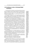
Microaneurysms
These are the very earliest clinically detectable lesions of diabetic retinopathy
(Fig. 16.1). They appear as small, round, red dots and may be found in any
part of the retina although they predominate in the posterior pole of the eye.
They are not associated with any visible blood vessels and represent localized
dilatations of retinal capillaries. The number of microaneurysms increases
with increasing severity of retinopathy. A microaneurysm indicates a local-
ized area in the microvascular circulation where the blood-retinal barrier is
deficient and may therefore be associated with abnormal vascular leakage.
The pathogenesis of microaneurysms is unclear but they may represent
outpouchings of capillaries at areas of relative weakness where there is peri-
cyte loss. Pericytes are cells which partly enclose retinal capillaries and may be
considered the smooth muscle equivalent of the microvasculature; pericyte
numbers diminish early in the development of diabetic retinopathy.
Microaneurysms may also represent a localized response to surrounding
hypoxia, i.e a limited proliferative process, as they tend to predominate in
areas where there is closure of surrounding capillary beds.
Haemorrhages
Haemorrhages co-exist with microaneurysms but are more variable in their
appearance (Fig. 16.1). At their smallest, they may be difficult to differentiate
from microaneurysms. A haemorrhage, unlike a microaneurysm, is not nec-
Fig. 16.1 Moderate background diabetic retinopathy with microaneurysms,
haemorrhages and exudates.
CHAPTER 16 • CLASSIFICATION AND DIAGNOSIS
141
essarily round and may take on a variety of outlines; the phrase ‘dot and blot’
is an apt description. Haemorrhages can occur within the retina, where they
remain confined by the retina, or they can occur on the retinal surface (flame-
shaped haemorrhage) where they spread out over the superficial nerve fibre
layer taking on a characteristic flame appearance. This latter form of haem-
orrhage is less obviously a feature of diabetic retinopathy and may suggest the
co-existence of hypertensive vessel damage.
Haemorrhages probably occur from rupture of microaneurysms or other
weak-walled vascular abnormalities. Small intraretinal haemorrhages occur
early in diabetic retinopathy and their numbers increase with increasing
severity. In more advanced disease, large dark blot intraretinal haemorrhages
suggest severe retinal ischaemia with arteriolar occlusion, a feature of pre-
proliferative disease.
Exudates
These are usually small collections of lipoprotein which have accumulated
within the retina from abnormal vascular leakage, and are therefore found in
the vicinity of microaneurysms (Fig. 16.1). They are usually reflective and may
appear to have a rigid, multifaceted contour, ranging in colour from white to
yellow. They were previously referred to as ‘hard’ exudates to differentiate
them from soft exudates (now called cotton wool spots); however, this sepa-
ration is now redundant since it is well established that cotton-wool spots are
not the products of exudation.
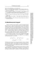
Like microaneurysms, exudates are most frequently detected in the pos-
terior pole and may be distributed in the form of a whole or partial ring
appearance (Fig. 16.2). Such ‘circinate’ ring arrangements usually have
microaneurysms in the centre, which are responsible for the vascular leak-
age that gives rise to the exudates at the margins. The number of exudates
may paradoxically increase as the degree of extravascular fluid diminishes
due to precipitation of lipids and proteins, analogous to a saline solution
depositing salt upon drying. There may, therefore, be a transient increase
in the number of exudates following laser treatment as the macula
becomes drier.
What is diabetic maculopathy?
The term macula refers to the important centre of the retina. It measures
approximately 5 mm in diameter and is the area centred upon the fovea with
a radius that extends to the temporal margin of the optic disc.The fovea itself
is about the same size as the optic disc (1.5–1.7 mm in diameter), with its cen-
tre (foveola) recognizable in normal eyes by the foveolar reflex. More practi-
cally, the macula can be considered as the area within the major temporal vas-
cular arcades.
SECTION III • DIABETIC RETINOPATHY AND ASSOCIATED OPHTHALMIC DISORDERS
142
Diabetic maculopathy can be defined as any retinopathy lesion located
within the macula. However, the term maculopathy is usually reserved for
sight-threatening lesions close to the centre of the macula. The Early
Treatment Diabetic Retinopathy Study (ETDRS) group produced the follow-
ing list of criteria, any one of which is sufficient to diagnose clinically signifi-
cant macular oedema (CSMO) requiring laser treatment:
• Thickening of the retina located less than 0.5 mm from the centre of the
macula.
• Exudates (with thickening of adjacent retina) located less than 0.5 mm
from the centre of the macula.
• An area of retinal thickening 1 disc diameter in size located less than 1 disc
diameter from the centre of the macula.
For practical purposes, sight-threatening maculopathy is any retinopathy lesion
within
1
⁄2 a disc diameter from the centre of the macula; this simplified defini-
tion will assist the non-ophthalmologist in identifying what may be sight-
threatening but this would not necessarily be an indication for laser treatment.
PREPROLIFERATIVE DIABETIC RETINOPATHY
Although classified as a sub-category of background retinopathy, preprolifer-
ative retinopathy is a sight-threatening condition that is usually considered
separately from background disease. It also differs from background
Fig. 16.2 Diabetic maculopathy with a circinate exudate ring.
retinopathy in having four new features, i.e. cotton wool spots, retinal venous
abnormalities, large blot intraretinal haemorrhages and intraretinal
microvascular abnormalities.
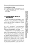
Cotton wool spots
Cotton wool spots appear as pale cream patches of variable sizes (Fig. 16.3).
They do not have clearly defined outlines and are most frequently seen in the
posterior pole. A cotton wool spot is an area of infarction in the nerve fibre
layer, and the appearance is due to swollen nerve axons with impaired axo-
plasmic flow. It therefore represents an area of localized retinal ischaemia and
suggests the presence of arteriolar occlusion. Cotton wool spots persist for a
long time, ranging from 8–17 months. Five or more cotton wool spots are
generally required to suggest preproliferative disease.
Intraretinal microvascular abnormalities (IRMA)
These usually appear as irregular loops of vessels within the retina which may
straddle normal vessels (Fig. 16.3). IRMA occur adjacent to areas of capillary
bed closure and their origin is unclear. Unlike ‘new vessels’, IRMA do not
always leak fluorescein, although some leakage may occur at their growing
tips. At least two different theories exist as to what abnormal vasculature are
presently classified as IRMA: shunt vessels and intraretinal new vessels.
Fig. 16.3 Early preproliferative retinopathy with intraretinal retinal microvascular
abnormalities associated with cotton wool spots.
CHAPTER 16 • CLASSIFICATION AND DIAGNOSIS
143
SECTION III • DIABETIC RETINOPATHY AND ASSOCIATED OPHTHALMIC DISORDERS
144
Venous abnormalities
Various abnormalities occur in the retinal veins in response to the hypoxic
environment (Fig. 16.4). These take the form of:
• beading, e.g. the ‘string-of-sausages’ appearance;
• reduplication of veins, whereby the vein appears to divide into two paral-
lel channels over a short segment; and
• venous loops, where the vein makes a sudden deviation in the form of a loop.
Venous abnormalities, particularly those of beading and reduplication, are
strong indicators of hypoxia and suggest that new vessel development is
imminent.
Deep retinal haemorrhages
These are large dark haemorrhages within the retina representing haemor-
rhagic infarction secondary to retinal arteriolar occlusion.
PROLIFERATIVE DIABETIC RETINOPATHY
Ischaemia within the retina due to widespread closure of capillary beds leads
to newly formed blood vessels appearing on the retinal surface, or overlying
the optic disc. These vessels extend in the plane between the retina and the vit-
reous and are accompanied by a supporting network of fibroglial proliferation.
Fig. 16.4 Severe preproliferative retinopathy venous abnormalitits.
CHAPTER 16 • CLASSIFICATION AND DIAGNOSIS
New vessels developing from the vasculature of the optic disc are called
‘disc new vessels’ (NVD) (Fig. 16.5) whilst those developing on the surface of
the retina are called ‘new vessels elsewhere’ (NVE) (Fig. 16.6). It is thought that
NVD represents severe generalized ischaemia of the retina, whereas NVE are a
response to local ischaemia in the quadrant of the retina where they occur.
New vessels usually arise from a vein and have a haphazard growth pattern.
As they grow, the combination of new vessels and supporting fibroglial tissue
becomes adherent to both the retinal and posterior vitreous surfaces, inducing
the vitreous to detach from the retina. The subsequent traction may cause
haemorrhage either because the fragile new vessels break or because they are
avulsed from their point of origin on the main retinal vessel. If bleeding is con-
fined to the space between the retina and the vitreous, a preretinal or retro-
hyaloid haemorrhage is clearly visible on ophthalmoscopy (the so-called ‘boat
shaped’ haemorrhage with a fluid level appearance). Depending on whether
the haemorrhage obscures the macula, vision may be severely affected or min-
imally compromised. If the haemorrhage is to break through into the main
body of the vitreous, the view of the retina may be variably obscured, likewise
the patient’s vision. At worse, no view may be possible of the retina.
The second outcome of neovascular traction is a retinal detachment. A
tractional retinal detachment usually occurs slowly, and may remain stable for
years assuming laser treatment has been applied to control the neovascular
process. A tractional retinal detachment affects vision in two ways. Firstly, if it
directly affects the fovea, vision will be reduced; if extrafoveal traction exists,
145
Fig. 16.5 Proliferative retinopathy with disc new vessels (NVD).
SECTION III • DIABETIC RETINOPATHY AND ASSOCIATED OPHTHALMIC DISORDERS
146
tension induced retinal folds may secondarily affect the fovea, producing visu-
al distortion. Secondly, a stable tractional detachment may suddenly become
unstable if a full thickness hole occurs in the retina, leading to a rhegmatoge-
nous retinal detachment which may spread to involve the fovea.
The proliferative process may not be confined to the posterior segment of
the eye. Iris neovascularization is a feared complication because of the risk of
neovascular or thrombotic glaucoma; a form of glaucoma which is difficult to
manage once established. As with retinal neovascularization, fibrous tissue
eventually develops which can occlude the trabecular meshwork and the
anterior chamber angle leading to uncontrolled neovascular glaucoma or sec-
ondary angle closure glaucoma, resulting in a painful, red blind eye.
INTERNATIONAL CLINICAL DIABETIC RETINOPATHY DISEASE
SEVERITY SCALE
The International Council of Ophthalmology has produced a new classifica-
tion for diabetic retinopathy, in an attempt to standardize terminology. The
classification comprises five stages from no retinopathy to proliferative
retinopathy (Table 16.2). The Council has also proposed a classification for
diabetic maculopathy based on whether it is absent or present; the latter is
then subclassified into three grades of severity (Table 16.3).
Fig. 16.6 Proliferative retinopathy with retinal new vessels (NVE).
CHAPTER 16 • CLASSIFICATION AND DIAGNOSIS
Diagnosis of diabetic retinopathy
It is essential that patients with diabetes are regularly examined for the pres-
ence of symptomless retinopathy. Retinopathy screening is presently per-
formed by health care professionals from a variety of disciplines, including
optometrists, nurses, medical photographers, general practitioners and dia-
betologists using a variety of techniques.
147
Table 16.3 International clinical classification of the severity of diabetic
macular oedema.
Table 16.2 International clinical diabetic retinopathy disease classification scale.
International clinical diabetic retinopathy disease classification scale
Disease severity level Findings observable upon dilated ophthalmoscopy
No apparent retinopathy No abnormalities
Mild non-proliferative Microaneurysms only
diabetic retinopathy
Moderate non-proliferative More than just microaneurysms but less than severe
diabetic retinopathy NPDR
Severe non-proliferative Any of the following:
diabetic retinopathy • More than 20 intraretinal haemorrhages in each of 4
quadrants
• Definite venous beading in 2+ quadrants
• Prominent IRMA in 1+ quadrant
And no signs of proliferative retinopathy
Proliferative diabetic One or more of the following:
retinopathy • Neovascularization
• Vitreous/preretinal haemorrhage
International clinical classification of the severity of
diabetic macular oedema
Classification Findings observable upon dilated ophthalmoscopy*
Diabetic macular • Mild diabetic macular oedema
oedema present Some retinal thickening or hard exudates in posterior pole but
distant from the macula
• Moderate diabetic macular oedema
Retinal thickening or hard exudates approaching the centre of
the macula but not involving the centre
• Severe diabetic macular oedema
Retinal thickening or hard exudates involving the centre of the
macula
Screening for diabetic retinopathy
Diabetic retinopathy is a disease which fulfills all the necessary criteria for a
screening programme: Those at risk form an identifiable population; it has a
recognized disease pattern; and laser treatment, if performed early, is effective
in preventing loss of vision, particularly in proliferative disease. Advanced
disease, when diagnosed late, is less amenable to treatment and much more
costly, both economically and in terms of the patient’s quality of life.
In the UK and many other countries, however, there is no national strate-
gy for the screening of diabetic retinopathy. In any one year in the UK, the
proportion of diabetic patients who receive retinopathy screening varies from
38%–85%, and from 14%–97% between different primary care practices.
A variety of methods are in use in the UK at present, e.g. selected
optometrists accredited to perform the screening using slit-lamp biomi-
croscopy, and schemes that are based on retinal photography, either fixed-site
or via a mobile unit.
Techniques for screening
Direct ophthalmoscopy using a hand-held ophthalmoscope is used to a vary-
ing extent, and with varying degrees of success, by general practitioners,
optometrists and diabetologists. It is technically difficult, allowing only a two-
dimensional view of the retina. Therefore retinal oedema cannot be accurate-
ly diagnosed using this technique. The peripheral parts of the retina are diffi-
cult to examine using this technique. It is a form of examination that has
largely been abandoned by ophthalmologists, who now mainly examine the
retina by slit-lamp biomicroscopy.
Slit-lamp biomicroscopy provides a much wider three-dimensional view
of the retina using a 78 or 90 dioptre lens. Although an effective technique, it
is very skill-dependent and the operator requires extensive training. Both
methods of ophthalmoscopy have to be performed with mydriasis (dilated
pupils) and both have the disadvantage of not providing a hard record for
qualitative assessment and for monitoring signs of progression of disease.
Retinal photography is a technique that is more easily acquired, and the
image can be interpreted later by another health professional, e.g. diabetol-
ogist, ophthalmologist or a specially trained grader. The number of
ungradeable photographs ranges from 3.7% to 20%, the failure rate being
lower with mydriasis. Increasingly, digital photography is supplanting the
analogue techniques of slides and Polaroid photography. It has major
advantages in its ease of image acquisition, data storage and there is also the
option of electronic data transfer. The computerized interpretation of
images is a real possibility and the screening process may eventually become
entirely electronic.
SECTION III • DIABETIC RETINOPATHY AND ASSOCIATED OPHTHALMIC DISORDERS
148
CHAPTER 16 • CLASSIFICATION AND DIAGNOSIS
The British Diabetic Association (BDA) has proposed that a screening test for
diabetic retinopathy should have at least 80% sensitivity and 95% specificity. In
a wide-ranging review of multiple studies examining the effectiveness of direct
ophthalmoscopy, indirect ophthalmoscopy and retinal photography, it has been
reported that retinal photography with a dilated pupil is the most effective. Most
of the studies which employed retinal photography had sensitivity levels of over
80%, and mydriatic photographs gave an even higher level of sensitivity.
Proposed UK national screening scheme
Screening for retinopathy in the UK is likely to change as part of the
“Preservation of sight in diabetes: a risk reduction programme” initiative. A
nationwide screening programme will be established by 2005/2006 with clear
aims: (1) to reduce the rate of avoidable visual loss by early detection of sight-
threatening retinopathy so that it can be treated promptly; and (2) detection
of any retinopathy, so that the diabetic patient can be made aware that
changes have begun to occur in their eyes, and attempts can be made to
improve glycaemic and blood pressure control. It is anticipated that screen-
ing will be offered to all diabetic patients by 2007.
The main features of this screening program will include:
• Target population: all diabetic patients, types 1 and 2, over 12 years of age,
or post-puberty.
• Frequency: annually, initially, but after a few screening rounds those
deemed to be at low risk can probably be screened less frequently whilst
those with more severe disease may be screened more frequently.
• Technique: the Steering Committee has recommended mydriatic digital
photography (two fields, macular and nasal) with visual acuity measure-
ment (visual acuity alone will not lead to referral to an assessment clinic
unless accompanied by retinopathy, although the general practitioner will
be informed if visual acuity falls below 6/12 in either eye).
• Examination: grading of photographs by specially trained graders using a
standardized grading scheme employing reference images, with opinions
from ophthalmologists and/or diabetologists if required.
• Positive patients: referral to special assessment clinics at convenient oph-
thalmology departments.
149
FURTHER READING
Bachmann MO, Nelson SJ. Impact of diabetic retinopathy screening on a British district
population: case detection and blindness prevention in an evidence-based model. J
Epidemiol Community Health 1998; 52: 45–52.
Bagga P, Verma D, Walton C, Masson EA, Hepburn DA. Survey of diabetic retinopathy
screening services in England and Wales. Diabetic Medicine 1998; 15: 780–782.
Garvican L, Clowes J, Gillow T. Preservation of sight in diabetes: developing a national risk
reduction programme. Diabetic Medicine 2000; 17: 627–634.
Hart PM, Harding S. Is it time for a national screening programme for sight threatening
retinopathy? Eye 1999; 13: 129–130.
Hutchinson A, McIntosh A, Peters J et al. Effectiveness of screening and monitoring tests
for diabetic retinopathy—a systematic review. Diabetic Medicine 2000; 17: 495–506.
CURRENT ISSUES
• The classification of diabetic retinopathy into background and
proliferative stages is well established and assists in the management of
sight-threatening retinopathy.
• There has been a recent attempt by the International Council of
Ophthalmology to standardize the terminology employed in the
classification of diabetic retinopathy.
• Screening for diabetic retinopathy in many countries, including the UK, is
not sufficiently well established or co-ordinated to achieve the standards
required by the St Vincent declaration of 1989.
• The most sensitive screening technique for diabetic retinopathy is
mydriatic fundal photography and the least effective is direct
ophthalmoscopy.
• Successful retinopathy screening is principally technique-dependent and
less personnel-dependent.
150
SECTION III • DIABETIC RETINOPATHY AND ASSOCIATED OPHTHALMIC DISORDERS
CHAPTER 17
DIABETIC MACULOPATHY
Hean-Choon Chen FRCS, FRCOphth
151
INTRODUCTION
Diabetic maculopathy is the commonest cause of visual loss in patients with
diabetes and it can be defined as the presence of sight-threatening lesions
within the macula. These lesions commonly consist of microaneurysms,
haemorrhages and exudates. The cause of visual loss is a consequence of
either or both of the following:
• leaking blood vessels leading to exudate formation and accumulation of
extracellular fluid causing macular oedema (exudative diabetic macu-
lopathy or diabetic macular oedema); and
• capillary closure giving rise to macular ischaemia (ischaemic diabetic
maculopathy).
Loss of vision from exudation and oedema is the commoner of the two mech-
anisms and is fortunately responsive to laser treatment, at least in part. There
is no treatment for macular ischaemia.
EPIDEMIOLOGY
The Wisconsin Epidemiologic Study of Diabetic Retinopathy (WESDR)
reported an overall prevalence rate of 10% for macular oedema; predictably,
the prevalence increases with increasing severity of overall retinopathy status,
ranging from approximately 2% in those with mild background retinopathy
to 20–37% in those with moderate to severe background disease, and in those
with proliferative retinopathy the prevalence of macular oedema was approx-
imately 70% (Table 17.1). The prevalence of maculopathy also increases with
duration of diabetes, and is higher in type 2 compared with type 1 diabetes
(Figs 17.1 & 17.2).
DEFINITIONS
Exudative maculopathy
The Early Treatment Diabetic Retinopathy Study (ETDRS) group produced
a list of criteria to denote clinically significant macular oedema, i.e. macu-
lopathy for which laser treatment is indicated (see chapter 16).
Ischaemic maculopathy
The normal fovea possesses a central avascular area known as the foveolar
avascular zone (FAZ); this exists so as to provide the centre of the fovea with
the least possible impedance to incident light. In the normal eye, the FAZ
varies significantly in size with an average of approximately 0.5–0.6 mm. In
Vascular Complications of Diabetes: Current Issues in Pathogenesis and Treatment, Second Edition
Edited by Richard Donnelly, Edward Horton
Copyright © 2005 by Blackwell Publishing Ltd
SECTION III • DIABETIC RETINOPATHY AND ASSOCIATED OPHTHALMIC DISORDERS
152
Fig. 17.1 Frequency of macular oedema by duration of diabetes in years for insulin-
taking early onset persons. Ophthalmology 1984; 91: 1464–1474.
Per cent with macula oedema
40
30
20
10
0
05 1510
Duration (years)
2520 30 35
Table 17.1 Relationship of diabetic retinopathy with macular oedema* and
duration of diabetes (Wisconsin, HAS-1, 1980–82). The Wisconsin Epidemiologic
Study of Diabetic Retinopathy. IV. Diabetic macular oedema. Ophthalmology 1984;
91: 1464–1474.
Relationship of Diabetic Retinopathy with Macular Oedema* and
Duration of Diabetes (Wisconsin, HAS-1, 1980-82)
Younger onset Older onset
Duration 10+ years Duration 0–14 years Duration 15+ years
Retinopathy % Macular % Macular % Macular
status No. oedema P No. oedema P No. oedema P
Non-proliferative
Mild 172 1.7 – 152 2.6 – 126 6.3 –
Moderate
to severe 128 20.3 <0.001 60 36.7 <0.001 87 63.2 <0.001
Proliferative 85 69.7 – 26 73.1 – 35 74.3
*Percentage with macular oedema = (number of persons with macular oedema status 2
or 3 in either or both eyes/number of persons with macular oedema status 0 in both eyes +
number of persons with macular oedema status 2 or 3 in either or both eyes) × 100.
Fig. 17.2 Frequency of macular oedema by duration of diabetes for insulin- and
non-insulin-taking older onset persons. The Wisconsin Epidemiologic Study of Diabetic
Retinopathy. IV. Diabetic macular oedema. Ophthalmology 1984; 91: 1464–1474.
Per cent with macula oedema
40
30
20
10
0
0510
Duration (years)
Insulin taking
Non-insulin taking
15 302520
CHAPTER 17 • DIABETIC MACULOPATHY
ischaemic maculopathy, there is a gradual increase in the size of the FAZ.
Although there is no defining measurement of FAZ for a diagnosis of
ischaemic maculopathy, when the diameter of FAZ exceeds 1 mm, vision is
usually compromised (Fig 17.3).
DIAGNOSIS
The diagnosis of diabetic maculopathy is made clinically and, when neces-
sary, with the aid of fluorescein angiography. More recently, a new imaging
technique called optical coherence tomography (OCT) has provided a means
of objectively assessing macular thickening non-invasively. Changes in visual
function may raise the suspicion of macular disease:
• reduced visual acuity;
• reduced contrast sensitivity; and
• colour vision defects, usually along the blue-yellow (tritan-like) axis, may
occur from an early stage in the disease and may even predate clinically
visible lesions.
Clinically, the presence of microaneurysms and exudates indicates the presence
of pathological vascular leakage, although these changes alone may not reduce
visual acuity. The retinal pigment epithelial ‘pump’ and surrounding competent
153
SECTION III • DIABETIC RETINOPATHY AND ASSOCIATED OPHTHALMIC DISORDERS
154
capillaries are able to remove extravasated fluid and extracellular fluid accumu-
lates when the rate of leakage exceeds the capacity for fluid removal, giving rise
to retinal thickening. Vision is affected when this occurs at the centre of the mac-
ula. However, exudates in large numbers, and especially at the centre of the mac-
ula, can also affect vision in the absence of retinal thickening (probably due to
direct photoreceptor damage). When large numbers of exudates are present,
macular oedema is usually also present. It is not unusual to observe a paradoxi-
cal increase in the number of exudates as oedema dissipates, since lipoproteins
are precipitated within the retina as the water component of oedema is removed.
The diagnosis of macular thickening requires stereoscopic views of the
macula. This is possible with slit-lamp biomicroscopy using either a non-
contact 78 or 90 dioptre lens, or with a fundus contact lens. The contact lens-
es provide better stereopsis and may be better at detecting subtle degrees of
retinal thickening. Fluorescein angiography is usually not necessary in the
diagnosis of macular oedema. However, when there is an unexplained loss of
vision, i.e. when it cannot be attributed to vascular leakage, fluorescein
angiography is helpful. The diagnosis of macular ischaemia can only be defin-
itively made with fluorescein angiography.
Fluorescein angiography
Fluorescein angiography outlines the retinal circulation, illuminating the
otherwise invisible microvasculature. It is a photographic investigation tech-
nique that uses an adapted fundus camera. Fluorescein, a vegetable dye
extract, is able to absorb light with a wavelength of approximately 490 nm
(blue light) and in response emits light at a longer wavelength of 530 nm
(green light). Fluorescein is injected into an antecubital vein (usually 5 ml of
Fig. 17.3 This fluorescein
angiogram of the
posterior pole of the eye
demonstrates the
presence of ischaemic
maculopathy with an
enlarged foveal avascular
zone.
CHAPTER 17 • DIABETIC MACULOPATHY
a 10% solution), where it becomes 70–85% plasma protein bound, and pho-
tographs are taken of the fundus as it traverses the retinal circulation. The
adapted fundus camera possesses two filters to ensure that blue light enters
the eye and yellow-green light enters the camera, where it is captured on film.
Photographs are usually taken from about 10 seconds after injection and
thereafter at one to two second intervals. Late pictures may be taken after a
few minutes to determine the possible presence of late leakage.
Fluorescein angiography provides assistance in the diagnosis of:
• Capillary closure or non-perfusion, especially in the macula, where capil-
laries exist as a monolayer with an increased melanin background, provid-
ing greater contrast. This is particularly helpful in the diagnosis of
ischaemic maculopathy, where, in good quality fluorescein angiographic
pictures, FAZ is clearly delineated.
• Vascular leakage, although fluorescein angiography is seldom required to
make this diagnosis (Fig. 17.4). Cystoid macular oedema takes on a char-
acteristic petalloid appearance (Fig. 17.5).
• Subtle neovascularization, which may not be immediately obvious on clin-
ical examination. New vessels do not possess a normal blood-retinal bar-
rier and are hyperpermeable, therefore giving rise to extensive fluorescein
leakage. Several other retinopathy lesions can also be highlighted, e.g.
microaneurysms and intraretinal neovascular abnormalities (IRMA).
Optical coherence tomography
This is a relatively novel non-invasive, non-contact imaging technique based
on interferometry whereby high-resolution, cross-sectional images of the
retina are obtained. It is similar to ultrasound except that light is used in place
155
Fig. 17.4 Fluorescein
angiogram demonstrating
diffuse macular leakage.
SECTION III • DIABETIC RETINOPATHY AND ASSOCIATED OPHTHALMIC DISORDERS
156
of sound; ‘echoes’ of light are produced at junctions of tissue layers with dif-
ferent density. It allows for objective measurements of retinal, in particular
macular, thickness. The technology is expensive but is becoming more wide-
ly available. It is in certain circumstances a substitute for fluorescein angiog-
raphy, which is a more invasive technique and non-quantitative.
PATHOGENESIS
Exudative maculopathy or macular oedema is the consequence of a break-
down of the blood-retinal barrier (BRB), more specifically the inner BRB, i.e.
the retinal capillary endothelium. This is usually at sites of microaneurysms
although other retinopathy lesions also signify breaches in the BRB such as
dilated capillaries, IRMA and retinal neovascularization.
There is however, also some evidence suggesting a breakdown in the outer
BRB, i.e. the retinal pigment epithelium (RPE), as well. The RPE forms a bar-
rier between the neural retina and the choriocapillaris, the capillary network
of the choroid, which is highly permeable to macromolecules (unlike retinal
capillaries). The normally tight junctions of the RPE, therefore, maintain a
diffusion gradient for water from retina to choroid because of the higher
oncotic pressure within the choroid.
It has also recently been demonstrated that a taut, thickened posterior
hyaloid (vitreous), which is still attached to the retina, can give rise to trac-
tion upon the macula producing intraretinal cystic spaces, and possibly a
tractional detachment. These changes in the posterior hyaloid usually occur
in response to ischaemic disease and therefore this form of macular oedema
more frequently occurs in severe forms of retinopathy.
Fig. 17.5 Fluorescein
angiogram demonstrating
cystoid macular oedema
with its characteristic
petal-like leakage pattern.
CHAPTER 17 • DIABETIC MACULOPATHY
TREATMENT
Ischaemic maculopathy is not amenable to treatment. Exudative maculopa-
thy may, for treatment purposes, be subclassified into two varieties:
• Focal oedema. This is a localised area of leakage, usually from a cluster of
microaneurysms which exist in the centre of an area of oedema. The area
affected is usually circular and its periphery often delineated by a ring of
exudates, the so-called ‘circinate of exudates’.
• Diffuse oedema. Leakage appears to occur from a large area of dilated cap-
illaries, sometimes with little evidence of exudate formation. This is possi-
bly due to capillary beds dilating to compensate for surrounding areas of
capillary occlusion, or there may be an autoregulatory increase in local
retinal blood flow. Prolonged diffuse oedema can lead to the formation of
cystic spaces in the centre of the macula, known as cystoid macular oede-
ma (CMO), and is more likely to be associated with systemic problems
such as renal failure and uncontrolled hypertension.
Both forms of oedema can coexist. There will usually be areas of focal leakage
in patients with diffuse oedema but an area of focal oedema frequently exists
in isolation. The visual deficit is usually more severe in cases of diffuse oede-
ma, which is unfortunately also more refractory to treatment, particularly in
the presence of cystoid oedema.
Laser therapy
Laser therapy is effective in both focal and diffuse macular oedema. In focal
oedema, the treatment is applied directly at the area(s) of leakage, for example
the microaneurysms at the centre of a ring of exudates. An attempt may be
made to coagulate the microaneurysm. The ETDRS protocol included direct
treatment of microaneurysms greater than 40μm in diameter. The technique
employed to do this is to initially ‘whiten’ the underlying retinal pigment
epithelium using a 100μm spot size. The aim of this step is to ensure that the
whitened underlying tissue will no longer absorb the energy from subsequent
laser shots. The spot size should then be reduced to 50μm and aimed at the
microaneurysm, with the aim of turning it white or a darker shade of red.
In diffuse oedema the method used is known as ‘grid macular treatment’.
It involves applying laser burns with a spot size of between 100μm and
200μm, aiming for a blanching effect of the retinal pigment epithelium, i.e. a
grey-white effect. The spots are applied to cover the part of the macula that is
thickened, sparing the fovea; treatment should commence approximately
500μm from the centre of the macula and be spaced about one burn-width
apart. If oedema persists, retreatment with laser burns applied closer to the
centre of the macula can be applied; the ETDRS protocol allowed for treat-
ment from 300μm from the centre of the macula, unless there was perifoveal
157
capillary dropout. Treatment over the papillo-macular bundle is possible
since the nerve fibre layer is spared, as the level of laser energy used affects
only the pigment epithelium and its immediate neighbours. The ETDRS pro-
tocol advocated treating not just the areas that were oedematous but also any
adjacent areas of capillary non-perfusion.
Treatment should be applied using an appropriate contact lens to provide
good visibility of the macula, e.g. the wide-angle Volk Transequator. The type
of thermal laser wavelength used should be either green (e.g. argon and dou-
ble-frequency YAG lasers) or yellow (dye laser); blue light laser should be
avoided in treatment of the macula because the absorption of blue light by
abundant macular xanthophyll pigment can cause greater degrees of collater-
al damage to the retina. The red light of the krypton laser will not be absorbed
by haemoglobin and therefore will not be useful in closing microaneurysms.
At the commencement of treatment, it is prudent to assess the energy
requirement away from areas close to the fovea, perhaps with a trial shot a
short distance away. An initial energy setting of approximately 80–100 mW
should be considered with a pulse duration of 0.1 seconds; these settings may
then be modified according to the response.
Following treatment, an appropriate review interval will be between two
and four months. It often takes this period of time for any effect on oedema
resolution to become obvious.
Surgical Procedures
If a taut posterior hyaloid is visible (possibly with the aid of OCT), pars plana
vitrectomy has been found to be effective in resolving macular oedema.
Features supporting this particular treatment option may include oedema
resistant to grid laser treatment and the presence of cystoid oedema.
Vitrectomy for macular oedema even in the absence of overt posterior
hyaloid traction has been reported to produce a beneficial effect although this
is not widely carried out in the absence of significant clinical evidence. The
removal of large subfoveal exudates has also been reported with some success.
THE EFFECTIVENESS OF LASER TREATMENT
In treating focal leakage, successful closure of the leaking points, i.e. the
microaneurysms, implies that leakage stops. However, part of the success is
likely to be due to those factors which lead to the resolution of oedema fol-
lowing treatment for diffuse leakage.
Why laser treatment works in the treatment of diffuse leakage is unclear.
Because the laser is targeted at the retinal pigment epithelium, the actual leak-
ing points, i.e. the retinal capillaries, are not directly treated. Various
hypotheses have been formulated to explain the therapeutic benefits of laser:
SECTION III • DIABETIC RETINOPATHY AND ASSOCIATED OPHTHALMIC DISORDERS
158
CHAPTER 17 • DIABETIC MACULOPATHY
• Laser-damaged pigment epithelial cells regenerate and the new cells that
fill the gaps created by thermal necrosis may create a more effective outer
BRB.
• The laser also damages and causes loss of retinal photoreceptor cells,
which are the major consumers of oxygen within the neural retina. This
may relieve hypoxia and reduce the autoregulatory increment in blood
flow through dilated capillary beds, thereby reducing leakage.
• The effect of the laser on the pigment epithelium may release a diffusible
factor that induces retinal capillary repair, leading to a restoration of the
inner BRB.
Laser treatment of diabetic maculopathy is not always successful; it is fre-
quently only able to maintain current levels of vision, or slow the rate of pro-
gression, rather than lead to an improvement in vision.
If macular oedema coexists with a degree of ischaemia, it is reasonable to
apply laser treatment although the prognosis is poorer compared with eyes
without ischaemia. The diagnosis of ischaemia may be difficult in the pres-
ence of significant leakage since this obscures the view of the retinal micro-
circulation on fluorescein angiograms.
Complications of macular laser treatment
• Accidental damage to the foveola.
• Rupture of Bruch’s membrane with use of high laser energy, which may
lead to the formation of a choroidal neovascular membrane.
• Development of paracentral scotomata; these may be subtle and detectable
only with more specialized perimetric techniques, such as blue-on-yellow
perimetry.
• Submacular fibrosis from laser scars which appear to spread and merge.
WHEN SHOULD LASER TREATMENT BE APPLIED?
If clinically significant macular oedema (CSMO) exists, laser treatment
should be applied soon, i.e. within weeks, and this is especially so if the cen-
tre of the macula is involved or directly threatened. When macular oedema
coexists with proliferative retinopathy, the ETDRS group has clearly demon-
strated that scatter photocoagulation leads to a worsening of the macular sit-
uation. It is recommended that whenever possible the macula should be
treated first, or simultaneously, with the scatter treatment initially confined
to the nasal quadrants.
Intraocular surgery, for example cataract extraction, has been shown to
worsen maculopathy and, if the view allows it, macular laser treatment should
be applied before the patient undergoes cataract surgery. If maculopathy is
present but has not yet reached the severity of CSMO, the patient should be
159
SECTION III • DIABETIC RETINOPATHY AND ASSOCIATED OPHTHALMIC DISORDERS
160
closely observed in the postoperative period as the level of maculopathy is
very likely to increase in severity. A significant proportion of patients who
develop macular oedema after cataract surgery will experience a spontaneous
regression in their oedema, usually over several months after surgery, partic-
ularly if the underlying retinopathy status is mild. However, if oedema was
present preoperatively, any worsening is likely to be persistent.
MICROPULSED DIODE LASER
This is a relatively novel approach using a diode laser with a wavelength of 810
nm (invisible, infrared wavelength) and the laser applied in a ‘micropulse’
fashion, i.e. frequent, short (microsecond) pulses delivering subthreshold
laser energy (an energy level that does not produce an immediate, clinically
evident effect). The energy is delivered in ‘envelopes’, each consisting of mul-
tiple short pulses. The advantage over conventional macular laser treatment
is the specific targeting of the laser on the retinal pigment epithelium, there-
fore inducing less damage to surrounding tissues with fewer complications.
TREATMENTS UNDER CONSIDERATION
The intravitreal injection of triamcinolone (a long-acting steroid) has been
demonstrated to have some effect on reducing macular oedema. However, not
all patients respond and the effect may be relatively short-lived. The efficacy of
a protein kinase C inhibitor in reducing macular oedema is being examined.
CURRENT DEVELOPMENT
• The ETDRS group has recommended laser treatment for those patients
with features fulfilling the ‘clinically significant macular oedema’ criteria.
• Laser treatment is most likely to maintain current levels of vision, or
retard the rate of progression of visual loss, rather than produce an
improvement in vision.
• Laser treatment should be applied using a green or yellow wavelength,
and may be applied in a focal and/or grid fashion for localized areas of
leakage or diffuse leakage, respectively.
• Pars plana vitrectomy is effective in reducing diffuse macular oedema if a
taut thickened posterior hyaloid is present. There is also evidence that
persistent diabetic cystoid macular oedema may respond to pars plana
vitrectomy, even if there is no clinical evidence of vitreous traction upon
the macula.
• There is some evidence that intravitreal triamcinolone can reduce macular
oedema with an improvement in vision, at least in the shorter term.
CHAPTER 17 • DIABETIC MACULOPATHY
FURTHER READING
Early Treatment Diabetic Retinopathy Study Research Group: Photocoagulation for dia-
betic macular oedema: Early Treatment Diabetic Retinopathy Study report number 1.
Arch Ophthalmol 1985; 103: 1796–1806.
Ikeda T, Sato K, Katano T, Hayashi Y. Vitrectomy for cystoid macular oedema with
attached posterior hyaloid membrane in patients with diabetes. Br J Ophthalmol 1999;
83: 12–14.
Klein R, Klein BEK, Moss SE, Davis MD, DeMets DL. The Wisconsin Epidemiologic Study of
Diabetic Retinopathy. IV. Diabetic macular oedema. Ophthalmology 1984; 91: 1464–1474.
Lewis H, Abrams GW, Blumenkranz MS, Campo R. Vitrectomy for diabetic macular trac-
tion and oedema associated with posterior hyaloidal traction. Ophthalmology 1992; 99:
753–759.
Moorman CM, Hamilton AM. Clinical applications of the MicroPulse diode laser. Eye 1999;
13: 145–150.
161
CHAPTER 18
PROLIFERATIVE DIABETIC RETINOPATHY
Hean-Choon Chen FRCS, FRCOphth
163
INTRODUCTION
The development of newly formed blood vessels either on the retina or on the
optic disc, with or without fibrous tissue, signifies the presence of prolifera-
tive retinopathy. New vessels develop in response to retinal ischaemia, and
the presence of neovasularization carries a high risk of significant loss of
vision due to various complications:
• Vitreous haemorrhage.
• Traction upon the fovea.
• Retinal detachment.
• Neovascular glaucoma.
EPIDEMIOLOGY
The Wisconsin Epidemiological Study of Diabetic Retinopathy (WESDR)
showed that the prevalence of proliferative retinopathy in type 1 diabetic
patients is 0% in the first five years of diagnosis, increasing to 67% in those
with diabetes of >35 years duration. In type 2 diabetes, the corresponding
prevalence rates are 2% rising to 16% among those with diabetes for >15 years.
Because type 2 diabetes is much more common, the absolute numbers of
patients with proliferative eye disease is similar for the two types of diabetes.
In the WESDR, approximately 43% of those examined with proliferative dis-
ease had type 1 diabetes whilst 42% had insulin-treated type 2 diabetes. The
severity of proliferative disease is generally greater in type 1 diabetes, and the
WESDR also showed that among patients with severe background retinopa-
thy the 4-year incidence of proliferative retinopathy is around 40–50%.
In the Diabetic Retinopathy Study (DRS), the risk of severe visual loss
(worse than 5/200) in eyes with proliferative disease was 16% over two years.
The risk was highest in patients with new vessels on the optic disc and those
with vitreous or pre-retinal haemorrhages. The appearance of new vessels on
the retina did not further increase the risk of severe visual loss in eyes with
established new vessels on the disc.
DEFINITIONS
Pathological newly formed blood vessels in the eye can be classified into three
groups:
• New vessels on the retina, commonly called new vessels elsewhere (NVE),
usually defined as being greater than one disc diameter from the optic disc
(Fig. 18.1).
Vascular Complications of Diabetes: Current Issues in Pathogenesis and Treatment, Second Edition
Edited by Richard Donnelly, Edward Horton
Copyright © 2005 by Blackwell Publishing Ltd