Báo cáo y học: "Musculoskeletal ultrasound imaging of the plantar forefoot in patients with rheumatoid arthritis: inter-observer agreement between a podiatrist and a radiologist" pptx
Bạn đang xem bản rút gọn của tài liệu. Xem và tải ngay bản đầy đủ của tài liệu tại đây (317.87 KB, 7 trang )
BioMed Central
Page 1 of 7
(page number not for citation purposes)
Journal of Foot and Ankle Research
Open Access
Research
Musculoskeletal ultrasound imaging of the plantar forefoot in
patients with rheumatoid arthritis: inter-observer agreement
between a podiatrist and a radiologist
Catherine J Bowen*
1
, Keith Dewbury
2
, Madeline Sampson
2
, Sally Sawyer
3
,
Jane Burridge
1
, Christopher J Edwards
3,4
and Nigel K Arden
3,4
Address:
1
School of Health Professions and Rehabilitation Sciences, University of Southampton, Southampton, UK,
2
Ultrasound Imaging,
Department of Radiology, Southampton University Hospitals NHS Trust, Southampton, UK,
3
Department of Rheumatology, Southampton
University Hospitals NHS Trust, Southampton, UK and
4
MRC Epidemiology Resource Centre, University of Southampton, Southampton, UK
Email: Catherine J Bowen* - ; Keith Dewbury - ;
Madeline Sampson - ; Sally Sawyer - ; Jane Burridge - ;
Christopher J Edwards - ; Nigel K Arden -
* Corresponding author
Abstract
Background: The use of musculoskeletal ultrasound (MSUS) in the diagnosis and management of
foot and ankle musculoskeletal pathology is increasing. Due to the wide use of MSUS and the depth
and breadth of training required new proposals advocate tailored learning of the technique to
discrete fields of practice. The aims of the study were to evaluate the inter-observer agreement
between a MSUS radiologist and a podiatrist, who had completed basic skills training in MSUS, in
the MSUS assessment of the forefoot of patients with Rheumatoid Arthritis.
Methods: A consecutive sample of thirty-two patients with rheumatoid arthritis was assessed for
presence of synovitis, erosions and bursitis within the forefoot using MSUS. All MSUS assessments
were performed independently on the same day by a podiatrist and one of two Consultant
Radiologists experienced in MSUS.
Results: Moderate agreement on image acquisition and interpretation was achieved for bursitis
(kappa 0.522; p < 0.01) and erosions (kappa 0.636; p < 0.01) and fair agreement for synovitis (kappa
0.216; p < 0.05) during the primary assessments. Following a further training session, substantial
agreement (kappa 0.702) between the two investigators was recorded. The sensitivity of the
podiatrist using MSUS was 82.4% for detection of bursitis, 83.0% for detection of erosion and 84.0%
for detection of synovitis. Specificity of the podiatrist using MSUS was 88.9% for detection of
bursitis, 80.7% for detection of erosion and 35.9% for detection of synovitis.
Conclusion: This study demonstrated good inter-observer agreement between a podiatrist and
radiologist on MSUS assessment of the forefoot, particularly for bursitis and erosions, in patients
with rheumatoid arthritis. There is scope to further evaluate and consider the role of podiatrists
in the MSUS imaging of the foot following appropriate training and also in the development of
reliable protocols for MSUS assessment of the foot.
Published: 28 July 2008
Journal of Foot and Ankle Research 2008, 1:5 doi:10.1186/1757-1146-1-5
Received: 16 May 2008
Accepted: 28 July 2008
This article is available from: />© 2008 Bowen et al; licensee BioMed Central Ltd.
This is an Open Access article distributed under the terms of the Creative Commons Attribution License ( />),
which permits unrestricted use, distribution, and reproduction in any medium, provided the original work is properly cited.
Journal of Foot and Ankle Research 2008, 1:5 />Page 2 of 7
(page number not for citation purposes)
Introduction
As technology has improved, clinical expertise in perform-
ing musculoskeletal ultrasound (MSUS) has also
advanced dramatically [1,2]. The advantages of ultra-
sound imaging over conventional radiography, computed
tomography, radioisotope scan and MRI (Magnetic Reso-
nance Imaging) are reported as being that it is: painless,
does not use ionising radiation, less expensive, can be per-
formed real time, needs no special environment and is
clinically readily accessible [1].
There have been a number of studies that have utilised
MSUS to diagnose specific soft tissue foot pain in healthy
volunteers [3-5] and soft tissue problems in Rheumatoid
Arthritis (RA) [6,7]. More specifically for the forefoot
ultrasonographic findings of metatarsophalangeal joint
(MTPJ) synovitis [8,9], plantar flexor tenosynovitis [10]
and plantar bursitis have been described [11].
MSUS is, however, affirmed as being highly operator
dependent [12]. The procedure itself has no known spe-
cific side effects, although harm that may result from
incorrect acquisition and interpretation of images has
been highlighted [2]. The reported challenges in training
for this skill are primarily due to the quality and interpre-
tation of the ultrasound images that are acknowledged as
being greatly dependent on the expertise and experience
of the operator [13]. Knowledge about the basic principles
relevant to sound waves and a detailed anatomical knowl-
edge of the structures under investigation are therefore
mandatory [14].
Models for the use of MSUS by clinicians other than radi-
ologists, have been demonstrated [15,16] and specific
competencies for MSUS performed by non radiologists
have been rigorously developed [17-19]. Within the most
recent proposed framework for development of compe-
tencies in MSUS scanning techniques, due to the training
challenges, tailored learning to areas directly relevant to a
clinician's discrete field of practice is proposed [19].
This study provided an opportunity to evaluate the inter-
observer agreement between a radiologist and podiatrist
who had followed recommended training guidelines for
MSUS, in the MSUS assessment of the forefoot of patients
with RA. This work forms part of an ultrasound based
research programme investigating the clinical relevance of
soft tissue inflammation within the forefoot of patients
with RA.
Materials and methods
Patient selection
The initial approach for recruitment was based upon
potential participants with rheumatoid arthritis diag-
nosed according to the American College of Rheumatol-
ogy (ACR) criteria, [20] and starting anti-TNF-α therapy
(infliximab, etanercept, adalimumab) all of whom were
attending the Southampton University Hospitals NHS
Trust Rheumatology Department. Patients were excluded
if they had previous surgery to the forefoot, had corticos-
teroid injection therapy to the forefoot within the previ-
ous 3 months prior to commencement of the study; had
concomitant musculoskeletal disease (eg, primary oste-
oarthritis, gout, Pagets disease, systemic lupus erythema-
tosus), had a serious medical or psychological disorder
that would affect the study protocol or were unable to give
informed consent. Local research ethics committee
approval was secured and all patients gave informed writ-
ten consent prior to their participation.
Confirmation of RA diagnosis and disease activity
Prior to data collection, the diagnosis of RA was con-
firmed by the supervising rheumatology consultant using
the ACR criteria. Following acceptance into the study all
participants were assessed by a trained specialist rheuma-
tology nurse and Disease Activity Scores (DAS28) were
calculated [21].
Data collection
Data collection took place between March 2004 and
August 2007. On each occasion, the same treatment bays
and ultrasound facilities were utilised in an attempt to
standardize environmental factors, i.e. room temperature
and scanning positions. All investigators performing
MSUS were blinded to the other's results in order to min-
imize the risk of bias.
Assessment of demographic and clinical characteristics
General demographic data of age, gender, disease dura-
tion, presence of rheumatoid factor, weight and limb
dominance were obtained from the clinical notes and
rheumatology department database. Clinical characteris-
tics included visual analog scale (VAS 100 mm) assess-
ment for the patient's global impression of health and
disease activity and the number of painful, tender and
swollen joints calculated as DAS28.
Imaging Data
The presence and location of any bursitis (see Figure 1)
across the plantar forefoot region and any synovial thick-
ening/synovitis and erosion (see Figure 2) within the sec-
ond and fifth MTPJs identified by MSUS was recorded.
Thirty minutes was allowed for each assessment and thus
was limited to two joints within each foot. The second
MTPJ was selected for investigation because it is in line
with the centre of load through the forefoot during gait
[22], it is easily accessible for the MSUS probes, and was
considered representative of the MTPJs. The first MTPJ was
excluded due to the sesamoid bones underlying its plantar
aspect. The fifth MTPJ was also selected for its ease of
Journal of Foot and Ankle Research 2008, 1:5 />Page 3 of 7
(page number not for citation purposes)
accessibility with the MSUS probes and as it has been
reported as being the most common site of radiographic
and sonographic erosion in RA [23].
Imaging Equipment
A Diasus diagnostic ultrasound scanner (Dynamic Imag-
ing, Livingston, Scotland UK), was used by the podiatrist.
The Diasus scanner operates as a system with dual probe,
but for the study, a L5–10 MHz 26 mm ultra wideband
linear array probe was used.
A Philips HDI 5000 System was used by the radiologists
(Royal Philips Electronics, Netherlands). The Philips sys-
tem also operates with broadband linear probes and in
order for direct comparisons to be made within the study
a L5 – 12 MHz probe was used.
MSUS Scanning Training and Protocol Development
The Podiatrist attended the BSR (British Society for Rheu-
matology) core MSUS course that is organised by experts
in MSUS providing hands on experience at different levels
of basic, intermediate and advanced specifically for non-
radiologists within the UK [24]. MSUS practice and men-
torship in scanning technique from two experienced
MSUS Radiologists took place over a further period of six
months following the BSR course.
On the same day the MSUS foot scans were performed
independently and without access to the clinical findings
by the podiatrist and one of two Consultant Radiologists
experienced in MSUS (Table 1). Scanning was in B-Mode
and recorded according to the EULAR (European League
Against Rheumatism) standard approaches (i.e. from the
sole of the foot) [14]. Each MTPJ and inter-metatarsal
space was scanned longitudinally and transversely from
the plantar view.
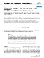
A MSUS image of the left foot plantar metatarsal area of a study participant with RAFigure 1
A MSUS image of the left foot plantar metatarsal
area of a study participant with RA. The MSUS image
demonstrates bursitis as a demarcated complex mass pro-
truding beyond the 3rd and 4th metatarsal heads with hyper-
trophied synovium and anechoic spaces containing synovial
fluid (arrow). The image is seen from the plantar aspect and
in the transverse plane. M4: 4
th
metatarsal head; M3: 3
rd
met-
atarsal head; P: plantar fat pad.
A MSUS image of the plantar aspect of the right forefootFigure 2
A MSUS image of the plantar aspect of the right forefoot. The MSUS image demonstrates synovial thickening, joint
effusion and bone changes within the right fifth plantar MTPJ of a study participant with RA. The image is seen from the plantar
aspect and in the longitudinal plane *:proximal phalanx; **:metatarsal head, S:synovial thickening; E:joint effusion; B:bone
changes.
Journal of Foot and Ankle Research 2008, 1:5 />Page 4 of 7
(page number not for citation purposes)
At the end of the data collection period, a consensus meet-
ing took place between one of the radiologists and the
podiatrist. During this meeting, all archived study images
recorded by the Diasus (Dynamic Imaging Ltd, Living-
ston, Scotland, UK) ultrasound system were reviewed and
discussed and confirmed by the radiologist as being bur-
sitis, synovitis, erosion, or normal.
Following the consensus meeting, thirty six images were
randomly selected from the Diasus (Dynamic Imaging
Ltd, Livingston, Scotland, UK) ultrasound system
archived data by an independent research assistant. All
thirty six images were numbered, logged and the sequence
for image viewing was randomized in an unrestricted ran-
dom method of allocation by the research assistant. Both
the primary investigator and consultant radiologist were
blinded to the image selection procedure.
The two investigators independently scored all 36 images
for the presence of bursitis, synovitis, erosion or normal
and both were blind to each others findings.
Statistical analysis
Data evaluation and statistical analysis were performed
using SPSS version 14.0 software (SPSS, Chicago IL). The
data was initially examined using histograms and scatter
plots to identify 'outliers' that may have occurred due to
data entry bias or normal biological outliers.
Inter-observer agreements were calculated by overall
agreement (percentage of observed exact agreement) and
kappa statistics (unweighted for dichotomous scoring, ie.
presence or absence of bursitis, synovitis and erosion).
Sensitivity and specificity of the podiatrist's results were
calculated from cross-tabulation against the radiologist's
results as the gold standard.
Results
Thirty two patients with RA were recruited. One patient
withdrew due to time issues during the visit. Thirty one
patients completed the study. There were 24 female and 7
male patients, 12 seronegative and 19 seropositive. The
mean age was 59.58 (SD 10.1) years and all patients had
active disease with mean DAS scores of 5.8 (SD 0.9)
(Table 2). Bursitis was present in 51/62 feet, synovitis in
81/124 MTP joints and erosions in 53/124 MTP joints
scanned by the Radiologist (Table 3).
Overall agreement was 83.3% for presence or absence of
bursitis, 81.8% for presence or absence of erosion and
68.3% presence or absence of synovitis. Kappa scores for
from the primary data revealed moderate agreement for
bursitis (kappa 0.522; p < 0.01) and erosions (kappa
0.636; p < 0.01) and fair agreement for synovitis (kappa
0.216; p < 0.05).
The sensitivity of the podiatrist using MSUS was 82.4% for
detection of bursitis, 83.0% for detection of erosion and
84.0% for detection of synovitis. Specificity of the podia-
trist using MSUS was 88.9% for detection of bursitis,
80.7% for detection of erosion and 35.9% for detection of
synovitis.
Following the consensus meeting and test on a ran-
domised selection of thirty six images from the DIASUS
machine, good levels of agreement were achieved between
the podiatrist and radiologist for 8/9 bursitis, 4/10 syno-
vitis, 6/9 erosion, 8/8 normal images (kappa 0.702; p <
0.01).
Discussion
The use of MSUS assessment of the foot and ankle in clin-
ical practice would be beneficial to patients with RA in
facilitating more effective timely referral and management
of foot problems. This is the first study to investigate tai-
lored learning of MSUS to the discrete field of foot and
ankle practice in evaluating the inter-observer agreement
in the use of MSUS between an allied health professional
(podiatrist) and an expert radiologist on imaging of the
foot. We have demonstrated good agreement for bursitis
and erosions, but only fair agreement for the presence of
synovitis.
Competency assessment in MSUS is an important issue
[17]. Usually, reliability in technique is reported by rheu-
matologists who have trained or are being trained in
Table 1: Overview of MSUS Scanning Protocol
1. The nature of the test was explained
2. The forefeet of all participants were scanned by the investigator using a broadband linear probe of 5 – 10 MHz frequency.
3. The participant was asked to sit in a supine position on the bed.
4. The participant's hosiery was removed and the ultrasound probe placed on the plantar aspect of each foot.
5. Scans were taken according to the EULAR standard approach from the plantar aspect of the foot [14].
6. Each MTP joint and inter-metatarsal space was scanned longitudinally and transversely from the plantar view.
7. Images of the plantar aspects of the forefoot in both feet were recorded in transverse and longitudinal aspects and saved on the ultrasound
machine hard drive.
8. Observations of synovitis and erosions of the second and fifth metatarsophalangeal joints and any evidence of bursitis were noted on a
separate data collection sheet.
Journal of Foot and Ankle Research 2008, 1:5 />Page 5 of 7
(page number not for citation purposes)
MSUS that are tested against experienced MSUS sonogra-
phers or radiologists [25-28]. The podiatrist in this study
had followed recommended BSR training in MSUS tech-
niques followed by further training and mentorship from
expert radiologists. Podiatrists are regularly involved in
the assessment and management of musculoskeletal foot
and ankle pathology. With extended scope practice in the
use of MSUS by podiatrists there are the potentially valu-
able benefits to patients mentioned above, as well as
lower costs in service provision.
In our study overall exact agreement between the radiolo-
gist and podiatrist was recorded as 83.3% for bursitis,
68.3% for synovitis and 81.8% for erosions. Acceptable
sonographic images were obtained by the podiatrist with
moderate agreements for bursitis (kappa 0.522, p <0.01)
and erosions (kappa 0.636, p <0.01). Low agreements for
synovitis (kappa 0.216, p <0.05) were obtained initially,
however following further training, levels of agreement
for all three variables increased to a good standard (kappa
0.702, p <0.01).
Within the MSUS literature the foot is under-investigated
and those who have reported on assessments of the foot
joints have observed similar low agreement scores for syn-
ovitis [25-28]. Inter-observer reliability among 14 experts
in MSUS produced overall good agreements for all exam-
ined joints (kappa 0.76) although low agreement for
ankle/toe joints (0.28 kappa) was reported [27]. In evalu-
ating scanning technique and diagnostic criteria, a group
of 23 experts in MSUS who scanned shoulder, wrist/hand,
ankle/foot and knee joints, reported exact overall agree-
ment of 91% for synovitis, 87% for cortical abnormalities
and 83.5% for bursitis but only 'fair' agreements for the
ankle and foot region (kappa 0.54) [28]. In MSUS exami-
nation of synovitis of the metacarpophalangeal joints and
MTPJs the learning curve of three rheumatologists in
MSUS techniques was investigated [26]. The agreements
at the final evaluations were good for two of the fellows,
(kappa 0.63 and 0.62), consistent with our findings, how-
ever for the third fellow was poor (kappa 0.18). This high-
lights the variability of learning requirements for the
technique [26].
The use of MSUS by the podiatrist within this study did
reveal good sensitivity and specificity for detecting fore-
foot bursitis (82.4% and 88.9%) and erosion (83.0% and
80.7%), although in detection of MTP synovitis sensitivity
was good, specificity was low (84.0% and 35.9%) with
over-reporting of false positives. As reported by others
[27] we did encounter difficulties in using MSUS to detect
synovitis in the MTPJs from the plantar aspect of the foot,
especially if deformities and/or subluxation of those
joints were present. Interestingly others have scanned
each joint in the dorsal aspect of the hands and feet,
reporting that this technique was preferred to a palmer or
plantar scan because of its reliability for detecting synovi-
tis [26] although they did not mention technique where
deformity and subluxation existed in the MTPJs. From our
experiences within this study we recommend that further
Table 2: Demographic characteristics of the study participants
Variable No Mean (SD) Range
Age (years) 31 59.58 (10.14) 37–76
Time since RA diagnosis (years) 31 11.32 (10.57) 1–39
Weight (Kg) 31 70.66 (15.35) 47.70–107.50
Overall well being (100 mm VAS) 28 60.29 (21.12) 20–100
Disease activity Score-28 joints 29 5.76 (0.93) 3.91–7.52
* SD = standard deviation; RA = Rheumatoid arthritis; VAS = visual analog score.
Table 3: Relation between the podiatrist's and radiologist's results of MSUS scans for the detection of plantar forefoot
Pod presence (N) Pod Absence (N) Total (N)
Rad Bursitis Presence (N) 42 9 51
Rad Bursitis Absence (N) 189
Rad Erosion Presence (N) 44 9 53
Rad Erosion Absence (N) 11 46 57
Rad Synovitis Presence (N) 68 13 81
Rad Synovitis Absence (N) 25 14 39
bursitis (N = 62 feet), erosion (N = 124 joints) and synovitis (N = 124 joints) in the study participants.
*Pod:Podiatrist; Rad = Radiologist
Journal of Foot and Ankle Research 2008, 1:5 />Page 6 of 7
(page number not for citation purposes)
work on establishing reliability of protocols for MSUS
assessment of the foot and ankle be conducted.
A number of potential limitations within this current
study should be acknowledged. The prevalence of synovi-
tis, erosion and bursitis within the forefoot was not vali-
dated by any other 'gold standard' imaging technique
such as MRI. Other authors have not attempted to address
this issue either although results from the OMERACT
MSUS special interest group highlighted the need to con-
sider comparison of MSUS data with histology and MRI
[29]. MRI however remains more costly and less readily
accessible than MSUS which made it less feasible to be
included in this study. Limitations relating to the sample
should also be highlighted. The sample was relatively
small and comprised patients with severe disease there-
fore generalizibility to the whole population of patients
with RA needs to be confirmed. Finally, the images
selected for analysis after the consensus meeting were
taken from part of the cohort that the podiatrist and radi-
ologist used for the initial reliability analyses. Whilst the
consensus meeting did take place once data collection was
complete, an element of recall bias may have been intro-
duced and thus should be acknowledged.
Conclusion
This study was the first to attempt to investigate inter-
observer agreement in the use of diagnostic ultrasound
between an allied health professional (podiatrist) and an
expert radiologist. Performance of MSUS in image acqui-
sition and interpretation by the podiatrist was of an
acceptable standard during the primary investigation and
following further training levels of agreement increased to
a good standard. The foot is a complex structure with
many small joints and podiatrists are well placed to be
involved in its MSUS assessment, however we have high-
lighted the importance of appropriate training and further
mentorship from experts in MSUS imaging techniques.
Finally, we have recommended that further work is under-
taken in establishing reliable protocols for MSUS assess-
ment of the foot.
List of Abbreviations
MSUS: Musculskeletal ultrasound; MRI: Magnetic Reso-
nance Imaging; MTPJ: Metatarso-phalangeal joint; RA:
Rheumatoid Arthritis; ACR: American College of Rheuma-
tology; TNF: Tumour necrosis factor; DAS28: 28 joint Dis-
ease Activity Score; VAS: Visual Analog Scale; BSR: British
Society for Rheumatology; RCR: Royal College of Radiol-
ogists; EULAR: European League Against Rheumatism;
SD: Standard Deviation; OMERACT: Outcome Measures
in Rheumatology Clinical Trials.
Competing interests
No benefits in any form have been received or will be
received from a commercial party related directly or indi-
rectly to the subject of this article.
The authors declare that they have no competing interests.
Authors' contributions
CB conceived of the study and participated in its design,
coordination and collection of clinical variables, MSUS
assessments and writing of the manuscript. KD partici-
pated in the design of the study, MSUS assessments, con-
sensus meeting and helped to draft the manuscript. MS
participated in the MSUS assessments, and helped to draft
the manuscript. SS participated in study design and coor-
dination. JB participated in the design of the study and
helped to draft the manuscript. CE participated in the
design of the study, coordination, confirmation of RA
diagnosis and helped to draft the manuscript. NA partici-
pated in the design of the study, coordination and helped
to draft the manuscript. All authors read and approved the
final manuscript
Acknowledgements
This study was supported by the Southampton Rheumatology Trust and
Arthritis Research Campaign.
References
1. Gibbon WW: Musculoskeletal ultrasound. Bailliere's clinical rheu-
matology 1996, 10(4):561-588.
2. Brown JN, Betts RP, Bygrave CJ: The use of ultrasound in the
cause of persistent pain following a metatarsal shortening
osteotomy. The Foot 1994, 4:159-162.
3. Bygrave CJ, Betts RP, Saxelby J: Diagnosing plantar fasciitis with
ultrasound using Planscan. The Foot 1998, 8:141-146.
4. Irwin LR, Konsantoulakis C, Hyder NU, Sapherson DA: Ultrasound
in the diagnosis of Morton's neuroma. The Foot 2000,
10:186-189.
5. Coakley FV, Samanta AK, Finlay DB: Ultrasonography of the tibi-
alis posterior tendon in rheumatoid arthritis. British journal of
rheumatology 1994, 33(3):273-277.
6. Bell M, McNally EGM: Ultrasound of the foot and ankle. The Brit-
ish Medical Ultrasound Society Bulletin 2002, 10(3):28-32.
7. Koski JM: Ultrasonography of the metatarsophalangeal and
talocrural joints. Clin Exp Rheumatol 1990, 8(4):347-351.
8. Szkudlarek M, Narvestad E, Klarlund M, Court-Payen M, Thomsen
HS, Ostergaard M: Ultrasonography of the metatarsophalan-
geal joints in rheumatoid arthritis: comparison with mag-
netic resonance imaging, conventional radiography, and
clinical examination. Arthritis and rheumatism 2004,
50(7):2103-2112.
9. Koski JM: Detection of plantar tenosynovitis of the forefoot by
ultrasound in patients with early arthritis. Scandinavian journal
of rheumatology 1995, 24(5):312-313.
10. Koski JM: Ultrasound detection of plantar bursitis of the fore-
foot in patients with early rheumatoid arthritis. The Journal of
rheumatology 1998, 25(2):229-230.
11. Grassi W, Cervini C: Ultrasonography in rheumatology: an
evolving technique. Annals of the rheumatic diseases 1998,
57(5):
268-271.
12. O'Connor PJ, Grainger AJ: Ultrasound imaging of joint disease.
Imaging 2002, 14:188-201.
13. Balint P, Sturrock RD: Musculoskeletal ultrasound imaging: a
new diagnostic tool for the rheumatologist? British journal of
rheumatology 1997, 36(11):1141-1142.
Publish with Bio Med Central and every
scientist can read your work free of charge
"BioMed Central will be the most significant development for
disseminating the results of biomedical research in our lifetime."
Sir Paul Nurse, Cancer Research UK
Your research papers will be:
available free of charge to the entire biomedical community
peer reviewed and published immediately upon acceptance
cited in PubMed and archived on PubMed Central
yours — you keep the copyright
Submit your manuscript here:
/>BioMedcentral
Journal of Foot and Ankle Research 2008, 1:5 />Page 7 of 7
(page number not for citation purposes)
14. Backhaus M, Burmester GR, Gerber T, Grassi W, Machold KP, Swen
WA, Wakefield RJ, Manger B: Guidelines for musculoskeletal
ultrasound in rheumatology. Annals of the rheumatic diseases
2001, 60(7):641-649.
15. Filippucci E, Unlu Z, Farina A, Grassi W: Sonographic training in
rheumatology: a self teaching approach. Annals of the rheumatic
diseases 2003, 62(6):565-567.
16. Taggart A, Filippucci E, Wright G, Bell A, Cairns A, Meenagh G, Pend-
leton A, Rooney M, Wright S, Grey A, Grassi W: Musculoskeletal
ultrasound training in rheumatology: the Belfast experience.
Rheumatology (Oxford, England) 2006, 45(1):102-105.
17. Brown AK, O'Connor P J, Roberts TE, Wakefield RJ, Karim Z, Emery
P: Recommendations for musculoskeletal ultrasonography
by rheumatologists: setting global standards for best prac-
tice by expert consensus. Arthritis Rheum 2005, 53(1):83-92.
18. Brown AK, O'Connor PJ, Roberts TE, Wakefield RJ, Karim Z, Emery
P: Ultrasonography for rheumatologists: the development of
specific competency based educational outcomes. Annals of
the rheumatic diseases 2006, 65(5):629-636.
19. Brown AK, Roberts TE, O'Connor P J, Wakefield RJ, Karim Z, Emery
P: The development of an evidence-based educational frame-
work to facilitate the training of competent rheumatologist
ultrasonographers. Rheumatology (Oxford, England) 2007,
46(3):391-397.
20. Arnett FC, Edworthy SM, Bloch DA, McShane DJ, Fries JF, Cooper
NS, Healey LA, Kaplan SR, Liang MH, Luthra HS, et al.: The Ameri-
can Rheumatism Association 1987 revised criteria for the
classification of rheumatoid arthritis. Arthritis and rheumatism
1988, 31(3):315-324.
21. van der Heijde DM, van 't Hof M, van Riel PL, van de Putte LB: Devel-
opment of a disease activity score based on judgment in clin-
ical practice by rheumatologists. The Journal of rheumatology
1993, 20(3):579-581.
22. Jacob HA: Forces acting in the forefoot during normal gait an
estimate. Clinical biomechanics (Bristol, Avon) 2001, 16(9):783-792.
23. Grassi W, Filippucci E, Farina A, Salaffi F, Cervini C: Ultrasonogra-
phy in the evaluation of bone erosions. Annals of the rheumatic
diseases 2001, 60(2):98-103.
24. BSR: Skills course in musculoskeletal ultrasound. In Muscu-
loskeletal ultrasound Oxfodr, UK ; 2003.
25. Szkudlarek M, Court-Payen M, Jacobsen S, Klarlund M, Thomsen HS,
Ostergaard M: Interobserver agreement in ultrasonography of
the finger and toe joints in rheumatoid arthritis. Arthritis and
rheumatism 2003, 48(4):955-962.
26. D'Agostino MA, Maillefert JF, Said-Nahal R, Breban M, Ravaud P, Dou-
gados M: Detection of small joint synovitis by ultrasonogra-
phy: the learning curve of rheumatologists. Annals of the
rheumatic diseases 2004, 63(10):1284-1287.
27. Scheel AK, Schmidt WA, Hermann KG, Bruyn GA, D'Agostino MA,
Grassi W, Iagnocco A, Koski JM, Machold KP, Naredo E, Sattler H,
Swen N, Szkudlarek M, Wakefield RJ, Ziswiler HR, Pasewaldt D,
Werner C, Backhaus M: Interobserver reliability of rheumatol-
ogists performing musculoskeletal ultrasonography: results
from a EULAR "Train the trainers" course. Annals of the rheu-
matic diseases 2005, 64(7):1043-1049.
28. Naredo E, Moller I, Moragues C, de Agustin JJ, Scheel AK, Grassi W,
de Miguel E, Backhaus M, Balint P, Bruyn GA, D'Agostino MA, Filip-
pucci E, Iagnocco A, Kane D, Koski JM, Mayordomo L, Schmidt WA,
Swen WA, Szkudlarek M, Terslev L, Torp-Pedersen S, Uson J, Wake-
field RJ, Werner C: Interobserver reliability in musculoskeletal
ultrasonography: results from a "Teach the Teachers" rheu-
matologist course. Annals of the rheumatic diseases 2006,
65(1):14-19.
29. Wakefield RJ, Balint PV, Szkudlarek M, Filippucci E, Backhaus M,
D'Agostino MA, Sanchez EN, Iagnocco A, Schmidt WA, Bruyn GA,
Kane D, O'Connor PJ, Manger B, Joshua F, Koski J, Grassi W, Lassere
MN, Swen N, Kainberger F, Klauser A, Ostergaard M, Brown AK,
Machold KP, Conaghan PG: Musculoskeletal ultrasound includ-
ing definitions for ultrasonographic pathology. J Rheumatol
2005, 32(12):2485-2487.