Báo cáo y học: "Lessons from dynamic cadaver and invasive bone pin studies: do we know how the foot really moves during gait" potx
Bạn đang xem bản rút gọn của tài liệu. Xem và tải ngay bản đầy đủ của tài liệu tại đây (1.33 MB, 7 trang )
BioMed Central
Page 1 of 7
(page number not for citation purposes)
Journal of Foot and Ankle Research
Open Access
Review
Lessons from dynamic cadaver and invasive bone pin studies: do we
know how the foot really moves during gait?
Christopher J Nester
Address: Centre for Health, Sport and Rehabilitation Research, University of Salford, UK
Email: Christopher J Nester -
Abstract
Background: This paper provides a summary of a Keynote lecture delivered at the 2009
Australasian Podiatry Conference. The aim of the paper is to review recent research that has
adopted dynamic cadaver and invasive kinematics research approaches to better understand foot
and ankle kinematics during gait. It is not intended to systematically cover all literature related to
foot and ankle kinematics (such as research using surface mounted markers). Since the paper is
based on a keynote presentation its focuses on the authors own experiences and work in the main,
drawing on the work of others where appropriate
Methods: Two approaches to the problem of accessing and measuring the kinematics of individual
anatomical structures in the foot have been taken, (i) static and dynamic cadaver models, and (ii)
invasive in-vivo research. Cadaver models offer the advantage that there is complete access to all
the tissues of the foot, but the cadaver must be manipulated and loaded in a manner which
replicates how the foot would have performed when in-vivo. The key value of invasive in-vivo foot
kinematics research is the validity of the description of foot kinematics, but the key difficulty is how
generalisable this data is to the wider population.
Results: Through these techniques a great deal has been learnt. We better understand the valuable
contribution mid and forefoot joints make to foot biomechanics, and how the ankle and subtalar
joints can have almost comparable roles. Variation between people in foot kinematics is high and
normal. This includes variation in how specific joints move and how combinations of joints move.
The foot continues to demonstrate its flexibility in enabling us to get from A to B via a large number
of different kinematic solutions.
Conclusion: Rather than continue to apply a poorly founded model of foot type whose basis is to
make all feet meet criteria for the mechanical 'ideal' or 'normal' foot, we should embrace variation
between feet and identify it as an opportunity to develop patient-specific clinical models of foot
function.
Background
Thankfully, in biomechanics terms, we no longer view the
foot as the triangle at the bottom of the leg. The term
"foot" suggests that some single functional entity exists,
when in fact the 26 bones, hundreds of ligaments and
muscles demands that we adopt a far more complex con-
ceptual and experimental model of the foot. In any discus-
sion of foot biomechanics, the foot is traditionally broken
Published: 27 May 2009
Journal of Foot and Ankle Research 2009, 2:18 doi:10.1186/1757-1146-2-18
Received: 16 April 2009
Accepted: 27 May 2009
This article is available from: />© 2009 Nester; licensee BioMed Central Ltd.
This is an Open Access article distributed under the terms of the Creative Commons Attribution License ( />),
which permits unrestricted use, distribution, and reproduction in any medium, provided the original work is properly cited.
Journal of Foot and Ankle Research 2009, 2:18 />Page 2 of 7
(page number not for citation purposes)
into at least two parts, the rearfoot and midfoot, tending
to focus on the subtalar and midtarsal joints. In the last
decade multi-segment foot models have provided new
insight into how the small and often assumed minor artic-
ulations in the foot move [1-5]. This can help inform both
our understanding of 'normal' foot biomechanics, upon
which much of clinical and surgical practice is based, and
the development of clinical and experimental hypotheses
as to how pathology occurs. This links directly to how foot
orthoses, footwear and surgery might be best used to elicit
a biomechanical effect and subsequent clinical response.
However, multi-segment foot models have their own lim-
itations. Inevitably, the skin to which markers are attached
moves relative to the underlying bones they are intended
to represent. More critically, we cannot reliably measure
the motion of each individual bone in the foot. This may
result in incorrect extrapolation from multi segment foot
model data about the kinematics of joints that comprise a
segment within that model.
The perfect experimental (and clinical) scenario is that we
are able to directly measure the kinematics of the individ-
ual bones of the foot. Access to tissues of the foot would
provide real insight and quickly enable to us test many
clinical hypotheses that have persisted despite a lack of
evidence to substantiate them. Advancement in our
understanding could be greatly accelerated. In coming
years, dynamic imaging techniques may offer excellent
solutions to the challenge of measuring the performance
of individual structures in the foot (though these will have
their own limitations) but until these are available we
must rely on direct physical contact with structures of the
foot as the best means of measuring foot motion.
Methods
Two approaches to the problem of accessing and measur-
ing the biomechanics of individual anatomical structures
in the foot have been taken, (i) static and dynamic cadaver
models, and (ii) invasive in-vivo research. Cadaver mod-
els offer the advantage that there is complete access to all
the tissues of the foot, not just the bones, and you can
investigate other aspects of the foot that might influence
its 'performance' through subsequent dissection (such as
checking for the presence of arthritic changes in a joint). It
is pertinent to question whether dead tissue behaves in
the same manner as living tissue, but this issue seems to
be regarded as an acceptable limitation if care is taken in
use of the tissues. However, the greatest disadvantage is
that the cadaver must be manipulated and loaded in a
manner which replicates how the foot would have per-
formed when in-vivo. Static cadaver models fail to repli-
cate any functional task of note though they can still offer
some insight into the basic function of ligaments or mus-
cles, and soft tissue properties. Dynamic cadaver models
attempt to make the cadaver feet 'walk again' but achiev-
ing this is very complex [6-16] (Additional file 1). The
cadaver must be mounted on a mechanism that has as
many degrees of freedom as the human body. Loads must
be applied to the specimen and its tendons at a magnitude
and rate as occurs in gait (or as close as possible). Moving
the specimen and loading the individual tendon and
tibia/foot structures must be synchronised exactly. These
parameters must also be adjustable as the input data driv-
ing the dynamic model (typically tibial motion, forces
applied to the tibia/plantar surface and residual tendons)
is at best an average of a small number of other feet, and
certainly not in-vivo data from the foot being tested.
Invasive in-vivo foot kinematics research has a long his-
tory [17-26] and it has been some of the most cited work
in the field (Additional file 2). The key value of this data
is its validity in describing how the bones of the foot
move, but the key difficulty is how generalisable this data
is to the wider population. The truth is that the data are
not generalisable, but data have already proved their value
by providing evidence to refute several traditional ideas of
foot function. Critically, it has helped demonstrate the
significant inter-subject variation that exists. This ques-
tions any notion of a model of foot function that is based
on the concept of a single 'ideal' foot type, foot alignment
or movement pattern. Other difficulties with an invasive
approach are that in most cases access is limited to the
bones, and even then, only a selection of foot bones.
There is also the issue of whether participants walk nor-
mally with pins inserted and using footwear and orthoses
is very difficult (though not impossible [19]). Some ques-
tion the ethical basis to this research, but in fact there is a
long tradition of invasive research without reports of com-
plications in participants who follow protocol. Surgery is
via minimal incision in sterile conditions and under local
infiltration of anaesthesia. There are clear post study pro-
tocols for weightbearing and medical support.
Results and discussion
What have we learnt about foot and ankle kinematics?
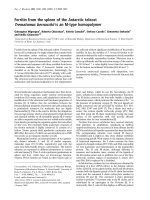
A key finding is the considerable freedom of movement
that exists at the ankle. For the frontal and transverse
planes, respectively, Lundgren et al [26] reported a mean
total range of motion of 8.1° and 7.9° during walking (n
= 5) (figure 1), Arndt et al [27] reported 12.2° and 8.7° in
slow running (n = 4), and using a dynamic cadaver model
of stance, Nester et al [9] reported a mean of 15.3°, and
10.0° (n = 13). Whilst in almost all cases the range of sag-
ittal plane motion was greater, the ankle is certainly not
limited to the role of a dorsi- and plantarflexion provider,
as was traditionally thought.
Furthermore, there is clear evidence that in some feet the
ankle displays more frontal and transverse plane motion
Journal of Foot and Ankle Research 2009, 2:18 />Page 3 of 7
(page number not for citation purposes)
than the subtalar joint, which was traditionally perceived
as the rearfoot joint most able to move in these planes. In
the case of transverse plane motion, Lundgren et al [26]
reported that the total range of ankle motion was greater
than the equivalent subtalar motion in 3 of 4 participants
(in walking). Nester et al [9] reported greater transverse
plane ankle motion compared to subtalar motion in 7 of
11 cadaver feet, and Arndt et al [27] reported the same in
all 3 of their participants (in slow running). In the case of
frontal plane motion, Arndt et al [27] found ankle motion
to be greater than the equivalent frontal plane subtalar
motion in 2 of 3 participants for which data was available
(slow running). Lundgren reported the same in 1 of 4 par-
ticipants in walking [26] as did Nester et al [9] in 8 of 11
cadavers. Based on these data, the subtalar joint is cer-
tainly not the sole 'torque converter' described in many
texts, and in fact the ankle and subtalar jonts share this
function, with each adopting different roles for different
individuals.
The inter-subject difference in how the ankle and subtalar
joints move is also evident in the pattern of movement
during stance. Lundgren et al's [26] subject-specific data
illustrates that some people display adduction of the talus
at the ankle (5–10°) in the first 20% of stance, with other
participants showing little motion at all (figure 1). Simi-
larly in slow running [27], 2 of 4 participants showed
eversion of the talus at the ankle (> 10° in first 40% of
stance), the other two showing little motion at all over the
same period. The variation between subjects in the frontal
and transverse plane 'role' of the ankle and subtalar joints
suggests they could work in tandem to provide the motion
required for each person. Certainly, we should never pre-
scribe distinctive roles to these two joints as has been the
case (ankle = sagittal plane, subtalar = torque converter)
and we might consider them to have quite similar func-
tional roles in the frontal and transverse planes.
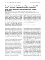
Published data consistently illustrate the significant free-
dom of movement at the talonavicular joint (figure 2),
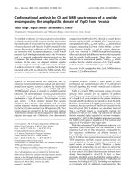
and to a lesser extent the calcaneocuboid joint (figure 3).
In the sagittal, frontal and transverse plane respectively,
Lundgren et al [26] reported 8.4° (1.1°), 14.9° (6.1°),
16.3° (6.5°) total range of motion at the talonavicular
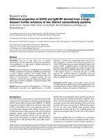
Ankle kinematics for 5 subjects during stance (0–100%)Figure 1
Ankle kinematics for 5 subjects during stance (0–
100%). Each band describes the mean +/- 1SD for each sub-
ject. +ve angles are eversion and adduction of the talus rela-
tive to tibia. Motion in degrees(°).
Talo-navicular kinematics for 5 subjects during stance (0–100%)Figure 2
Talo-navicular kinematics for 5 subjects during
stance (0–100%). Each band describes the mean +/- 1SD
for each subject. +ve angles are plantarflexion, eversion and
adduction of the talus relative to tibia. Motion in degrees(°).
Journal of Foot and Ankle Research 2009, 2:18 />Page 4 of 7
(page number not for citation purposes)
joint during walking. Arndt et al [27] reported similar sag-
ittal and frontal plane motion in slow running, but ~50%
less transverse plane motion. Given the more angular
articular facets it is no surprise that the calcaneocuboid
joint demonstrates less motion than the talonavicular
joint in most subjects studied. However, the mean total
range of calcaneocuboid motion in stance (7.8°, 6.3°,
6.9° respectively [26]) is greater than the equivalent sub-
talar joint motion in some in-vivo subjects [26,27] and
cadaver feet [9], reinforcing its important role in overall
foot function.
As with ankle and subtalar motion, there is no consistent
pattern between people in the range of motion the talona-
vicular and calcaneocuboid joints display. For one partic-
ipant of Lundgren et al [26] study, a total of 21° of motion
was observed in the frontal and transverse planes during
stance, yet only 5.2° and 6.0° in another participant.
Remarkably, despite these stark differences, in the sagittal
plane the same participants displayed 8.0° and 8.1° range
of sagittal plane motion, respectively. Quite how such
inter-subject variation is integrated into a clinical concep-
tual model of foot kinematics has yet to be determined.
However, given these data are from asymptomatic feet,
the data makes a mockery of any notion that a clinician
should seek to alter the foot biomechanics of all patients
such that their feet achieve some hypothetical mechanical
ideal (i.e. one foot model fits all feet). It is far from fitting
that in the year we celebrate the 150th anniversary of Dar-
win's 'discovery' of essential variations in nature, that foot
health professionals continue to use a clinical model of
foot function which seeks to eliminate all variation
between our patients. Furthermore, remaining as a 'varia-
tion' of nature rather than a clone of the hypothetical
'Root' foot type is likely to be central to a person remain-
ing symptom-free for most of their lives, since their own
body will have adapted to adequately cope with its own
variations.
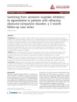
Many clinical models of foot biomechanics combine the
navicular and cuboid, but data from Lundgren et al [26]
indicates that motion between these bones is comparable
or greater than that at the subtalar joint (which we never
ignore) (figure 4). Identifying this capability, and the fact
that motion between the medial cuneiform and navicular
is equal to or greater than motion at the talonavicular
joint in some feet, is perhaps one of the most important
findings from the recent dynamic cadaver and invasive
foot kinematic studies. This is important because data
demonstrate that the tarsal bones are able to make a sig-
nificant contribution to the kinematics of the overall foot.
Motion that was previously attributed to the midtarsal
joint and rearfoot was most likely taking place between
the cuneiforms, the navicular, and cuboid. These move-
ments are invisible clinically due to overlying tissue and
consequently are completely absent from most if not all
clinical models of the foot.
For the forefoot, data have confirmed the greater stability
of the first, second and third metatarsals compared to
metatarsals four and five. The fourth and fifth metatarsals
are functionally distinct from the other three metatarsals,
in that they consistently displayed more motion during
stance. Using a dynamic cadaver model, Nester et al [9]
reported > 12° mean total range of motion in the sagittal
and frontal planes between the fifth metatarsal and
cuboid. These figures were broadly confirmed in subse-
quent invasive study (13.3° and 10.4° respectively [26])
(figure 5). Equivalent data for the other metatarsals was 5
to 8°.
Calcaneo-cuboid kinematics for 6 subjects during stance (0–100%)Figure 3
Calcaneo-cuboid kinematics for 6 subjects during
stance (0–100%). Each band describes the mean +/- 1SD
for each subject. +ve angles are plantarflexion, eversion and
adduction of the talus relative to tibia. Motion in degrees(°).
Journal of Foot and Ankle Research 2009, 2:18 />Page 5 of 7
(page number not for citation purposes)
Furthermore, the average total range of motion between
the first metatarsal and medial cuneiform reported by
Lundgren et al [26] was far less than the motion between
the equivalent fifth metatarsal and cuboid (5.3°, 5.4° and
6.1° in the sagittal, frontal, transverse planes compared to
13.3°, 10.4° and 9.8°). This mobility on the lateral side
of the foot is in addition to the motion between the
cuboid and calcaneus (9.7°, 11.3° and 8.1° respectively)
clearly demonstrating an infrequently discussed 'lower-
ing' of the lateral arch of the foot.
There is an important observation from the slow running
data reported by Arndt et al [27] and walking data from
Lundgren et al [26], which is even more valuable since the
data for the former study was collected on the same sub-
jects and in the same session (day) as the latter study. The
total range of motion at the subtalar, talonavicular, calca-
neo-cuboid, cuboid-navicular, medal cuneiform-navicu-
lar, metatarsal 1-cunieform, and metatarsal 5-cuboid, was
smaller in (slow) running than in walking. For the ankle,
the range of motion during walking was far greater in the
sagittal plane, and slightly greater in the frontal and trans-
verse planes. Less foot motion suggests a stiffer structure,
and given external forces are known to be greater during
running, this suggests that greater muscle forces would be
generated to control foot movements. One extrapolation
from this observation is that foot orthoses for running
need not be stiffer or have greater 'control' features (such
as high levels of medial heel wedging) compared to
orthoses for walking, since the motion taking place is
already less.
Cuboid-navicular kinematics for 6 subjects during stance (0–100%)Figure 4
Cuboid-navicular kinematics for 6 subjects during
stance (0–100%). Each band describes the mean +/- 1SD
for each subject. +ve angles are plantarflexion, eversion and
adduction of the talus relative to tibia. Motion in degrees(°).
5
th
Metatarsal-cuboid kinematics for 6 subjects during stance (0–100%)Figure 5
5
th
Metatarsal-cuboid kinematics for 6 subjects dur-
ing stance (0–100%). Each band describes the mean +/-
1SD for each subject. +ve angles are plantarflexion, eversion
and adduction of the talus relative to tibia. Motion in
degrees(°).
Journal of Foot and Ankle Research 2009, 2:18 />Page 6 of 7
(page number not for citation purposes)
Conclusion
Do we know how the foot really moves during gait?
Recent dynamic cadaver and invasive kinematic research
has provided some useful insights. The rearfoot plays only
a part of overall foot kinematics and we have consistently
undervalued the contribution from mid- and forefoot
articulations. This suggests that in order to control foot
pronation, orthoses need to provide support across the
entire rear- and mid foot and that the use of heel wedges
alone is unlikely to produce the desired biomechanical
effects on the foot. The forefoot undergoes a complex
series of rotations which must influence the action of the
intrinsic muscles of the foot, and researchers are only
recently being able to investigate some of their functions
[6].
Finally, variation between people in foot kinematics is
high and normal. This includes variation in how specific
joints move and how combinations of joints move. The
foot continues to demonstrate its flexibility in enabling us
to get from A to B via a large number of different kine-
matic solutions. Rather than continue to apply a poorly
founded model of foot type whose basis is to make all feet
meet criteria for the mechanical 'ideal' or 'normal' foot,
we should embrace variation between feet and identify it
as an opportunity to develop patient-specific clinical
models of foot function. Clinicians should consider foot
function in terms of the entire foot, and, given what we
know about the variation between subjects, the general
ranges of motion likely at specific joints, and what is
observable clinically, rationalise the most likely kinematic
solution for each patient. It is hoped that patient-specific
conceptual models for foot biomechanics will lead to
improved understanding of the role (if any) of foot bio-
mechanics in causation of foot and lower limb problems,
and improve our design of orthoses such that they have
more precise and predictable biomechanical effects. With
evidence of wide variation in foot kinematics from even
small samples of participants, and of patient-specific
response to orthoses [19], how can clinical practice con-
tinue to be so heavily based on the idea that one foot
model should fit all, and that orthosis design and pre-
scription is based on the ideal foot, rather than the
dynamics of the foot of each patient?
Competing interests
The author declares that they have no competing interests.
Authors' contributions
The author is the sole writer of this paper. Contributions
from prior research collaborations are identified under
acknowledgements.
Additional material
Acknowledgements
The author wishes to acknowledge important contributions from three
teams with whom the author collaborated to collectively produce the
research discussed in this paper. Dr's Erin Ward (DPM), Jay Cocheba
(DPM) and Tim Derrick (PhD), from and affiliated with Iowa State Univer-
sity USA. Dr's Toni Arndt (PhD) (University College of Physical Education
and Sport, Stockholm, Sweden), Arne Lundberg (PhD) and Paul Lundgren
(Karolinska Institute, Stockholm, Sweden). Dr Peter Wolf (PhD) and (late)
Dr Alex Stacoff (PhD) of Institute of Biomechanics, ETH Zurich, Switzer-
land. Prof David Howard, Richard Jones and Anmin Liu of the University of
Salford, UK.
References
1. Jenkyn TR, Anas K, Nichol A: Foot segment kinematics during
normal walking using a multisegment model of the foot and
ankle complex. J Biomech Eng 2009, 131(3):034504.
2. Ness ME, Long J, Marks R, Harris G: Foot and ankle kinematics in
patients with posterior tibial tendon dysfunction. Gait Posture
2008, 27(2):331-9.
3. Rao S, Saltzman C, Yack HJ: Segmental foot mobility in individ-
uals with and without diabetes and neuropathy. Clin Biomech
(Bristol, Avon) 2007, 22(4):464-71.
4. Stebbins J, Harrington M, Thompson N, Zavatsky A, Theologis T:
Repeatability of a model for measuring multi-segment foot
kinematics in children. Gait Posture 2006, 23(4):401-10.
5. Leardini A, Benedetti MG, Berti L, Bettinelli D, Nativo R, Giannini S:
Rear-foot, mid-foot and fore-foot motion during the stance
phase of gait. Gait Posture 2007, 25(3):453-62.
6. Kirane YM, Michelson JD, Sharkey NA: Contribution of the flexor
hallucis longus to loading of the first metatarsal and first
metatarsophalangeal joint. Foot Ankle Int 2008, 29(4):367-77.
7. Suckel A, Muller O, Herberts T, Langenstein P, Reize P, Wulker N:
Talonavicular arthrodesis or triple arthrodesis: peak pres-
sure in the adjacent joints measured in 8 cadaver specimens.
Acta Orthop 2007, 78(5):592-7.
8. Suckel A, Muller O, Herberts T, Wulker N: Changes in Chopart
joint load following tibiotalar arthrodesis: in vitro analysis of
8 cadaver specimens in a dynamic model. BMC Musculoskelet
Disord 2007, 8(8):80.
9. Nester CJ, Liu AM, Ward E, Howard D, Cocheba J, Derrick T, Pat-
terson P: In vitro study of foot kinematics using a dynamic
walking cadaver model. J Biomech 2007, 40(9):1927-37.
10. Erdemir A, Hamel AJ, Fauth AR, Piazza SJ, Sharkey NA: Dynamic
loading of the plantar aponeurosis in walking. J Bone Joint Surg
Am 2004, 86-A(3):546-52.
11. Ward ED, Smith KM, Cocheba JR, Patterson PE, Phillips RD, William
J: Stickel Gold Award. In-vivo forces in the plantar fascia dur-
ing the stance phase of gait: sequential release of the plantar
fascia. J Am Podiatr Med Assoc 2003, 93(6):429-42.
Additional file 1
Video 1 cadaver video. The video illustrates the performance of the
dynamic foot model.
Click here for file
[ />1146-2-18-S1.wmv]
Additional file 2
Video 2 bonepinvideo. The video illustrates walking with the bone pins
insitu.
Click here for file
[ />1146-2-18-S2.wmv]
Publish with Bio Med Central and every
scientist can read your work free of charge
"BioMed Central will be the most significant development for
disseminating the results of biomedical research in our lifetime."
Sir Paul Nurse, Cancer Research UK
Your research papers will be:
available free of charge to the entire biomedical community
peer reviewed and published immediately upon acceptance
cited in PubMed and archived on PubMed Central
yours — you keep the copyright
Submit your manuscript here:
/>BioMedcentral
Journal of Foot and Ankle Research 2009, 2:18 />Page 7 of 7
(page number not for citation purposes)
12. Michelson JD, Hamel AJ, Buczek FL, Sharkey NA: Kinematic behav-
ior of the ankle following malleolar fracture repair in a high-
fidelity cadaver model. J Bone Joint Surg Am 2002, 84-
A(11):2029-38.
13. Sharkey NA, Hamel AJ: A dynamic cadaver model of the stance
phase of gait: performance characteristics and kinetic valida-
tion. Clin Biomech 1998, 13(6):420-433.
14. Hurschler C, Emmerich J, Wülker N: In vitro simulation of stance
phase gait part I: Model verification. Foot Ankle Int 2003,
24(8):614-22.
15. Wülker N, Hurschler C, Emmerich J: In vitro simulation of stance
phase gait part II: Simulated anterior tibial tendon dysfunc-
tion and potential compensation. Foot Ankle Int 2003,
24(8):623-9.
16. Kim K, Kitaoka H, Luo Z, et al.: In vitro simulation of the stance
phase of human gait. J Musculoskeletal Research 2001, 5:113-121.
17. Lundberg A: The foot: block, gearbox, or cushion? Some con-
cepts in foot kinematics. J Orthop Sports Phys Ther 2004,
34(9):A6-7.
18. Arndt A, Westblad P, Ekenman I, Lundberg A: A comparison of
external plantar loading and in-vivo local metatarsal defor-
mation wearing two different military boots. Gait Posture 2003,
18(2):20-6.
19. Stacoff A, Reinschmidt C, Nigg BM, Bogert AJ van den, Lundberg A,
Denoth J, Stüssi E: Effects of foot orthoses on skeletal motion
during running. Clin Biomech 2000, 15(1):54-64.
20. Arndt A, Westblad P, Ekenman I, Halvorsen K, Lundberg A: An in
vitro comparison of bone deformation measured with sur-
face and staple mounted strain gauges. J Biomech 1999,
32(12):1359-63.
21. Lundberg A, Svensson OK, Bylund C, Selvik G: Kinematics of the
ankle/foot complex – Part 3: Influence of leg rotation. Foot
Ankle 1989, 9(6):304-9.
22. Lundberg A, Svensson OK, Bylund C, Goldie I, Selvik G: Kinematics
of the ankle/foot complex – Part 2: Pronation and supination.
Foot Ankle
1989, 9(5):248-53.
23. Lundberg A, Goldie I, Kalin B, Selvik G: Kinematics of the ankle/
foot complex: plantarflexion and dorsiflexion. Foot Ankle 1989,
9(4):194-200.
24. Lundberg A, Svensson OK, Németh G, Selvik G: The axis of rota-
tion of the ankle joint. J Bone Joint Surg Br 1989, 71(1):94-9.
25. Lundberg A: Kinematics of the ankle and foot. In-vivo roent-
gen stereophotogrammetry. Acta Orthop Scand Suppl 1989,
233:1-24.
26. Lundgren P, Nester C, Liu A, Arndt A, Jones R, Stacoff A, Wolf P, Lun-
dberg A: Invasive in-vivo measurement of rear-, mid- and
forefoot motion during walking. Gait Posture 2008,
28(1):93-100.
27. Arndt A, Wolf P, Liu A, Nester C, Stacoff A, Jones R, Lundgren P, Lun-
dberg A: Intrinsic foot kinematics measured in-vivo during
the stance phase of slow running. J Biomech 2007,
40(12):2672-2678.