Báo cáo y học: "Efficacy of customised foot orthoses in the treatment of Achilles tendinopathy: study protocol for a randomised trial" pps
Bạn đang xem bản rút gọn của tài liệu. Xem và tải ngay bản đầy đủ của tài liệu tại đây (886.19 KB, 13 trang )
BioMed Central
Page 1 of 13
(page number not for citation purposes)
Journal of Foot and Ankle Research
Open Access
Study protocol
Efficacy of customised foot orthoses in the treatment of Achilles
tendinopathy: study protocol for a randomised trial
Shannon E Munteanu*
1,2
, Karl B Landorf
1,2
, Hylton B Menz
1
, Jill L Cook
1,3
,
Tania Pizzari
1,4
and Lisa A Scott
2
Address:
1
Musculoskeletal Research Centre, Faculty of Health Sciences, La Trobe University, Bundoora 3086, Victoria, Australia,
2
Department of
Podiatry, Faculty of Health Sciences, La Trobe University, Bundoora 3086, Victoria, Australia,
3
School of Exercise and Nutrition Sciences, Faculty
of Health, Medicine, Nursing and Behavioural Sciences, Deakin University, Burwood 3125, Victoria, Australia and
4
School of Physiotherapy,
Faculty of Health Sciences, La Trobe University, Bundoora 3086, Victoria, Australia
Email: Shannon E Munteanu* - ; Karl B Landorf - ;
Hylton B Menz - ; Jill L Cook - ; Tania Pizzari - ;
Lisa A Scott -
* Corresponding author
Abstract
Background: Achilles tendinopathy is a common condition that can cause marked pain and disability. Numerous
non-surgical treatments have been proposed for the treatment of this condition, but many of these treatments
have a poor or non-existent evidence base. The exception to this is eccentric calf muscle exercises, which have
become a standard non-surgical intervention for Achilles tendinopathy. Foot orthoses have also been advocated
as a treatment for Achilles tendinopathy, but the long-term efficacy of foot orthoses for this condition is unknown.
This manuscript describes the design of a randomised trial to evaluate the efficacy of customised foot orthoses
to reduce pain and improve function in people with Achilles tendinopathy.
Methods: One hundred and forty community-dwelling men and women aged 18 to 55 years with Achilles
tendinopathy (who satisfy inclusion and exclusion criteria) will be recruited. Participants will be randomised, using
a computer-generated random number sequence, to either a control group (sham foot orthoses made from
compressible ethylene vinyl acetate foam) or an experimental group (customised foot orthoses made from semi-
rigid polypropylene). Both groups will be prescribed a calf muscle eccentric exercise program, however, the
primary difference between the groups will be that the experimental group receive customised foot orthoses,
while the control group receive sham foot orthoses. The participants will be instructed to perform eccentric
exercises 2 times per day, 7 days per week, for 12 weeks. The primary outcome measure will be the total score
of the Victorian Institute of Sport Assessment - Achilles (VISA-A) questionnaire. The secondary outcome
measures will be participant perception of treatment effect, comfort of the foot orthoses, use of co-interventions,
frequency and severity of adverse events, level of physical activity and health-related quality of life (assessed using
the Short-Form-36 questionnaire - Version two). Data will be collected at baseline, then at 1, 3, 6 and 12 months.
Data will be analysed using the intention to treat principle.
Discussion: This study is the first randomised trial to evaluate the long-term efficacy of customised foot orthoses
for the treatment of Achilles tendinopathy. The study has been pragmatically designed to ensure that the study
findings are generalisable to clinical practice.
Trial registration: Australian New Zealand Clinical Trials Registry Number: ACTRN12609000829213.
Published: 24 October 2009
Journal of Foot and Ankle Research 2009, 2:27 doi:10.1186/1757-1146-2-27
Received: 28 May 2009
Accepted: 24 October 2009
This article is available from: />© 2009 Munteanu et al; licensee BioMed Central Ltd.
This is an Open Access article distributed under the terms of the Creative Commons Attribution License ( />),
which permits unrestricted use, distribution, and reproduction in any medium, provided the original work is properly cited.
Journal of Foot and Ankle Research 2009, 2:27 />Page 2 of 13
(page number not for citation purposes)
Background
Achilles tendinopathy is a common musculoskeletal dis-
order, accounting for between 8-15% of all injuries in rec-
reational runners [1-3] and having a cumulative lifetime
incidence of approximately 6% in non-athletes and 24%
in athletes [4]. Interestingly, one-third of patients with
chronic Achilles tendinopathy are not physically active [5]
and Achilles tendinopathy is more common in those aged
35 years and over [6]. In some settings, approximately
30% of patients who present with this condition require
surgical treatment [7]. Since physical inactivity is a risk
factor for many multisystem diseases [8], Achilles tendin-
opathy may lead to poorer overall health and greater mor-
bidity, not just sporting inconvenience.
Numerous non-surgical treatments have been proposed
for the treatment of Achilles tendinopathy including:
footwear modification, activity modification and weight
reduction [9]; ultrasound and manual therapy techniques
[10]; flexibility and strengthening exercises [11]; extracor-
poreal shock wave therapy [12]; as well as various phar-
macological agents including corticosteroids, heparin,
dextrose, aprotinin, glyceryl trinitrate and sclerosing
agents [10]. However, many of these treatments have a
poor or a non-existent evidence base [10].
Eccentric calf muscle exercise is an emerging treatment
intervention for the management of tendinopathy, partic-
ularly for the Achilles tendon. Although the mechanism of
action [13] and optimum dosage (speed of contractions,
duration and frequency) for rehabilitation using eccentric
calf muscle exercises has yet to be clearly established, up
to three sets of fifteen repetitions, performed twice daily
for at least eleven weeks of a twelve week period has been
shown to be effective in high quality studies [14]. Recent
systematic reviews have concluded that eccentric calf mus-
cle exercise is a promising intervention and has the most
evidence to reduce pain in those with chronic Achilles
tendinopathy [15,16]. In a review of 9 clinical trials,
eccentric calf muscle exercise reduced pain by an average
of 60% [15]. However, eccentric calf muscle exercise alone
may not be effective in all people, as up to 40% of those
with Achilles tendinopathy do not improve with this
intervention [12], and eccentric calf muscle exercise has
been shown to be less effective in inactive people [17].
Also, major criticisms of current research in this area are
the lack of use of disease-specific functional outcome
measures and inadequately powered study designs
[15,18]. Nevertheless, eccentric calf muscle exercise is cur-
rently considered the best evidence-based intervention
available.
A further intervention that has been advocated for the
treatment of Achilles tendinopathy is foot orthoses
[11,19-21]. The classical theoretical mechanism for the
use of foot orthoses for this condition is that they align the
calcaneus to a more vertical position and reduce bending
stress applied to the Achilles tendon, particularly in a pro-
nated foot [22]. However, this theory has recently been
challenged by recent findings that a more laterally
directed force distribution during early stance followed by
a more medially directed force distribution during late
stance may be a risk factor for Achilles tendinopathy [2].
Further, recent studies indicate that the mechanical effects
of foot orthoses are non-specific and small, and that their
mechanism of action is likely to be more complicated,
possibly involving neuromotor effects [23-26]. Therefore,
there is currently a lack of evidence to explain the mecha-
nism by which foot orthoses exert their effects when used
to treat Achilles tendinopathy.
Despite the mechanism by which foot orthoses exert their
effects being unclear, there is evidence from a small
number of studies to suggest that they may reduce symp-
toms in those with Achilles tendinopathy [21,27,28].
Mayer and co-workers [27] performed a randomised clin-
ical trial comparing the effects of four weeks of physio-
therapy treatment (n = 11) (total of 10 treatments: deep
friction massage, ultrasound, ice and sensory motor train-
ing consisting of balance and eccentric exercises) versus
semi-rigid customised foot orthoses (n = 10) versus a no-
intervention control group (n = 10) in athletes with Achil-
les tendinopathy. Outcome measures for symptoms were
the 'Pain Disability Index' (PDI) and 'Pain Experience
Scale' (PES) scores. After four weeks, there were significant
differences between the groups. Both the physiotherapy
and customised foot orthoses groups, compared to the
control group, demonstrated significantly greater
improvements in pain, as measured by the PDI and PES
scores.
In a retrospective case-series study, Donoghue and col-
leagues [21] evaluated the effectiveness of customised
high-density ethylene vinyl acetate (EVA) foot orthoses to
alter lower limb kinematics and reduce pain in athletes
with chronic Achilles tendinopathy who displayed a pro-
nated foot type (n = 12). Participants reported a mean
improvement of 92 ± 16% in symptoms with the use of
the orthoses.
Whilst these studies suggest that customised foot orthoses
can reduce symptoms in those with Achilles tendinopa-
thy, they both have a number of limitations. First, the
sample sizes used were small. Second, the study by Dono-
ghue and colleagues [21] was retrospective and lacked a
control group for comparison. Third, neither study used
blinding of the participants or assessors which could have
lead to bias. It is therefore possible that the positive symp-
tom-modifying effects of the foot orthoses measured in
these studies may have been overestimated. Also, another
Journal of Foot and Ankle Research 2009, 2:27 />Page 3 of 13
(page number not for citation purposes)
criticism of current research in this area is the lack of use
of disease-specific functional outcome measures [15,17].
Finally, in the study by Mayer and co-workers [27], the
physiotherapy and custom foot orthoses interventions
were used mutually exclusive of one another. In clinical
practice, the two interventions are likely to be used con-
comitantly.
In light of the limitations of previous studies, the aim of
this project is to conduct a participant-blinded ran-
domised trial to determine the effectiveness of customised
foot orthoses on (i) pain, function and activity (using the
Victorian Institute of Sport Assessment - Achilles ques-
tionnaire) [29]; (ii) participant perception of change in
symptoms; (iii) comfort of the foot orthoses; (iv) use of
co-interventions; (v) frequency and severity of adverse
events; (vi) level of physical activity in previous week; and
(vii) health-related quality of life (using the Short-Form-
36 questionnaire) in people with Achilles tendinopathy.
The study protocol is presented in this paper, consistent
with the recommendations of Editorial Board of BioMed
Central [30].
Methods
Design
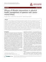
This study is a parallel group, participant blinded, ran-
domised controlled trial with a 12 month follow-up (Fig-
ure 1). Participants will be randomised to a control group
(sham foot orthoses) or an experimental group (custom-
ised foot orthoses). To ensure all participants, who will
have some level of pain and disability, receive some form
of intervention, both groups will be prescribed the same
eccentric calf muscle exercise program. This design covers
any ethical concerns of not treating participants in pain,
but will allow the effectiveness of customised foot
orthoses to be evaluated. Allocation to either of the inter-
vention groups will be achieved using a computer-gener-
ated random number sequence. The allocation sequence
will be generated and held by an external person not
directly involved in the trial. Concealment of the alloca-
tion sequence will be ensured as each participant's alloca-
tion will be contained in a sealed opaque envelope.
Envelopes will be made opaque by using a sheet of alu-
minium foil inside the envelope. In addition, a system
using carbon paper will be employed so the details (name
of participant and date of recruitment) are transferred
from the outside of the envelope to the paper inside the
envelope containing the allocation prior to opening the
seal.
Participants
The Human Studies Ethics Committee at La Trobe Univer-
sity (Human Ethics Committee Application No. 08-114)
has approved the study. Written informed consent will be
obtained from all participants prior to their participation.
People with Achilles tendinopathy will be recruited from
a number of sources:
(i) Advertisements in relevant Melbourne (Australia)
newspapers;
(ii) Mail-out advertisements to appropriate health
professionals in Melbourne;
(iii) Advertisements using relevant internet web-sites;
(iv) Posters displayed in local community centres,
sporting clubs and universities located in Melbourne.
Respondents will initially be screened by telephone inter-
view to ensure they are suitable for the study. Suitable
individuals will then be invited to participate in the study
and attend an initial assessment.
To be included in the study, participants must meet the
following inclusion criteria:
(i) Aged 18 to 55 years;
(ii) Have symptoms in the Achilles tendon of one lower
limb only for at least 3 months duration;
(iii) Be literate in English and able to complete the Victo-
rian Institute of Sport Assessment - Achilles (VISA-A)
questionnaire [29];
(iv) Score less than 80 on the VISA-A questionnaire [29];
(v) Regularly use footwear that can accommodate custom-
ised foot orthoses. This is defined as using footwear that
can accommodate foot orthoses for at least 90% of the
time during weightbearing activities [31];
(vi) Be willing to not receive any physical therapy on the
involved Achilles tendon(s) or trial of foot orthoses or
bracing (other than those allocated in the current study)
during the study period.
Achilles tendinopathy will be diagnosed from a clinical
assessment as well as from a musculoskeletal ultrasound
assessment using the following criteria [32-34]:
(i) Insidious onset of pain in the Achilles tendon region
that is aggravated by weightbearing activities and worse in
the morning, and/or during the initial stages of weight-
bearing activities;
(ii) Pain and swelling located 2-6 cm proximal to the
Achilles tendon insertion (as described by patient and pal-
pated by the investigator);
Journal of Foot and Ankle Research 2009, 2:27 />Page 4 of 13
(page number not for citation purposes)
(iii) Musculoskeletal ultrasound imaging of the Achilles
tendon showing local thickening (anterior-posterior)
and/or irregular fibre orientation and/or irregular tendon
structure with hypoechoic areas and/or vascularisation
within the mid-portion of the Achilles tendon.
Exclusion criteria for participants in this study will be
[12,17]:
(i) Previous Achilles tendon surgery in the symptomatic
lower limb;
(ii) Previous Achilles tendon rupture in the symptomatic
lower limb;
(iii) Previous lower limb trauma that has caused structural
imbalance (e.g. ankle fracture);
Design of studyFigure 1
Design of study.
Journal of Foot and Ankle Research 2009, 2:27 />Page 5 of 13
(page number not for citation purposes)
(iv) Osseous abnormality of the ankle (e.g. anterior or
posterior tibio-talar osteophytes);
(v) Inflammatory arthritis (e.g. ankylosing spondylitis);
(vi) Metabolic or endocrine disorders (e.g. type I or II dia-
betes);
(vii) Neurological disorders (e.g. Charcot-Marie-Tooth
disease);
(viii) Previous breast cancer and/or use of oestrogen
inhibitors;
(ix) Treatment with foot orthoses, heel lifts or eccentric
calf muscle exercises within the previous 3 months;
(x) Disorders of the Achilles tendon that are not mid-por-
tion tendinopathy (such as paratendinitis and insertional
Achilles tendon disorders);
(xi) Taken fluoroquinolones within the previous 2 years;
(xii) Injection of local anaesthetic, cortisone or other
pharmaceutical agents into the Achilles tendon or sur-
rounding area within the previous 3 months;
(xiii) Injury or pathology of the foot, knee, hip and/or
back or any condition that, in the opinion of the investi-
gators, may interfere with participation in the study.
Investigators will enquire about the above inclusion/
exclusion criteria during the initial telephone contact with
the potential participant and at the initial appointment.
Intervention
Participants will be randomised to one of two groups: an
intervention group (customised foot orthoses) or a con-
trol group (sham foot orthoses). Both groups will receive
eccentric calf muscle exercises. To maintain blinding of
participants, they will be advised that they will receive one
of two types of 'shoe inserts' during the study.
Data collection and the interventions will be adminis-
tered by 2 experienced qualified podiatrists. These podia-
trists will have attended two seminars for explanation and
discussion of the intervention protocols prior to the study
recruitment. During the seminars, the podiatrists will
receive further training regarding the administration of
the eccentric exercise program by qualified sports physio-
therapists (JLC and TP) who have extensive experience in
the management of Achilles tendinopathy. A detailed
manual outlining study procedures will be provided to all
project investigators.
An appointment will be given to all participants one
month after receiving their intervention to review the par-
ticipant's condition, assess compliance with the interven-
tion, ensure the foot orthoses are comfortable and
confirm that proper form and technique of the eccentric
calf muscle exercises is being adhered to.
Participants will be requested to refrain from other forms
of physical therapy intervention, not use any mechanical
interventions (apart from the foot orthoses provided as
part of this study), and not to consume non-steroidal anti-
inflammatory medications. They will be allowed to take
500 mg of paracetamol on an ad-hoc basis if the tendon is
painful.
The advice given to the participants with regard to the
amount of activity allowed during the study will be based
on the pain-monitoring model [35]. This approach allows
participants with Achilles tendinopathy to continue with
some level of activity during rehabilitation and shows
equivalent outcomes to programs that involve complete
rest from the aggravating activity with no negative effects
[35]. Using this approach, participants will be advised
that they can continue their activities after receiving their
intervention. However, Achilles tendon pain should not
be allowed to reach level 5 on the visual analogue scale
(VAS), where 0 is no pain and 10 is worst pain imagina-
ble, during the activity. The pain after the activity can
reach 5 on a VAS but should have subsided by the follow-
ing morning. Pain and stiffness in the Achilles tendon
should not increase from week to week [35].
Customised foot orthoses
Participants randomised to this intervention group will
receive customised foot orthoses for both feet. The basic
contour of the shell of all of the customised foot orthoses
will be based on the description of the modified Root style
of orthoses [36], and posted to vertical [36]. This style of
foot orthoses has been shown to be most commonly pre-
scribed by Australian and New Zealand podiatrists
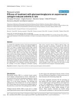
[37,38]. All customised foot orthoses will be manufac-
tured from polypropylene with a 400 kg/m
3
ethylene
vinyl acetate (EVA) rearfoot post and a shell-length cover-
ing fabric (Nora
®
Lunasoft SL 2 mm) (Figure 2a).
The foot orthoses will be further customised using the
information obtained from assessment of the foot posture
of each foot and the participants' body mass. The foot pos-
ture will be measured using the Foot Posture Index-6
(FPI), which is a valid and reliable tool [39]. The FPI con-
sists of six specific criteria: talar head palpation, supralat-
eral and infralateral malleolar curvature, calcaneal frontal
plane position, prominence in the region of the talonavic-
ular joint, medial arch height and abduction and adduc-
tion of the forefoot on the rearfoot. Each FPI criterion is
Journal of Foot and Ankle Research 2009, 2:27 />Page 6 of 13
(page number not for citation purposes)
scored on a 5-point scale (range, -2 to +2). The six scores
obtained are then summated to give an overall score of
foot posture. The summated score has the potential to
range from -12 (highly supinated) to +12 (highly pro-
nated) [39]. Feet that are assessed to have an FPI of (i) 0
or less will be considered to be supinated, (ii) +1 to +7
will be considered normal, and (iii) +8 or greater will be
considered to be pronated [40].
Those feet that are assessed to be pronated, defined as
obtaining an FPI summated score of +8 or greater [40],
will have a 4.0 mm Kirby medial heel skive (15 degree
varus heel wedge) incorporated into their orthosis [41].
This modification is thought to increase the anti-prona-
tion effect of the foot orthosis. The thickness of the poly-
propylene used for the customised foot orthoses will vary
depending on the body mass of the participant. For those
feet assessed as being normal or pronated, the thickness of
the polypropylene will be 4.0 mm for participants with a
body mass of less than 75 kg, and 4.5 mm for participants
with a body mass equal to or greater than 75 kg [42].
Those feet that are assessed to be supinated, defined as
obtaining an FPI summated score of 0 or less [40], will
receive an 'anti-supination' foot orthosis, based on the
description by Burns et al. [43] and Hertel et al. [44], but
with some modifications. This foot orthosis will have the
medial half of the rearfoot post removed [43,44] and be
fabricated from a relatively more flexible polypropylene
(Figure 2b) [43]. However, the thickness of the polypro-
pylene will be determined by the participant's body mass.
The thickness of the polypropylene will be 3.0 mm for
participants with a body mass of less than 75 kg, and 4.0
mm for participants with a body mass equal to or greater
than 75 kg. The Burns et al. [43] original description of
this device also used a padded full-length top cover as it
was designed to reduce excessive plantar pressures. In our
study, the anti-supination foot orthosis will not have a
padded top cover as the aim of our orthotic intervention
is to provide a pronatory force to the foot (resist supina-
tion), rather than reduce plantar pressures.
The customised foot orthoses will be manufactured and
donated by a commercial laboratory (Footwork Podiatric
Laboratory Pty Ltd, Victoria, Australia). Once fabricated,
the customised foot orthoses will be dispensed to partici-
pants two weeks after the initial appointment. Partici-
pants will be advised to remove any existing inner soles
from their shoes. The participant will also be given a
handout that provides instructions for using the orthoses,
including adjustment to them.
Sham foot orthoses
This condition will act as a control for the customised foot
orthoses intervention and be provided for both feet of
each participant. The sham foot orthoses will be made
from 4.0 mm thick ethylene vinyl acetate (EVA) with a
density of 90 kg/m
3
and have an identical covering fabric.
The shape of the sham foot orthoses will be derived from
being vacuum moulded over a standard cast which has
been obtained from replication of a prefabricated foot
orthosis (Prothotic S, Footech Orthotics™, The Orthotic
Laboratory Pty Ltd, Victoria, Australia). The sham foot
orthoses will have similar shape to the customised foot
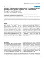
The customised foot orthoses for a foot with a normal foot posture (FPI score of +1 to +7) (A), a supinated foot posture (FPI score of 0 or less) (B) and sham foot orthoses (C) used in this studyFigure 2
The customised foot orthoses for a foot with a normal foot posture (FPI score of +1 to +7) (A), a supinated
foot posture (FPI score of 0 or less) (B) and sham foot orthoses (C) used in this study. Upper panels show poste-
rior-medial view and lower panels show plantar view.
Journal of Foot and Ankle Research 2009, 2:27 />Page 7 of 13
(page number not for citation purposes)
orthoses (Figure 2c), however, they will not provide any
mechanical support as the arch will flatten upon minimal
compressive force. This form of device has been used pre-
viously as a sham condition in a previous trial [38]. The
sham foot orthoses will be dispensed to the participant
two weeks after the initial appointment. The participant
will also be given the same handout that provides instruc-
tions for using the sham foot orthoses as participants that
receive the customised foot orthoses.
Eccentric calf muscle exercise program
The eccentric calf muscle exercise program will be per-
formed by all participants and is based on the method by
Alfredson et al. [45]. Participants will be given an infor-
mation package that includes a booklet and DVD with
instructions on performing the eccentric exercises for
Achilles tendinopathy [see Additional files 1, 2, 3, 4]. The
program is described below:
The participants will be instructed to do eccentric calf
muscle exercises 2 times daily, 7 days per week, for 12
weeks. Two types of eccentric calf muscle exercises will be
used. The calf muscle will be eccentrically loaded both
with the knee straight to maximise the activation of the
gastrocnemius muscle, and also with the knee bent to
maximise the activation of the soleus muscle. Each of the
two exercises will include 15 repetitions done in 3 sets
(i.e. 3 sets of 15 repetitions). The participants will be told
that muscle and tendon soreness during the first 4 weeks
of training is to be expected. After 12 weeks, the partici-
pants will be required to perform the exercises once daily,
3 days per week for the remainder of the study (12
months).
In the beginning, the loading will consist of bodyweight
and the participants will be standing with all their body-
weight on the injured leg. Participants will stand with
their heels over the edge of a step. From an upright body
position and standing with all bodyweight on the forefoot
and the ankle joint in plantar flexion, the calf muscle will
be loaded by having the participant lower the heel
beneath the forefoot. Only eccentric loading the calf mus-
cle will be allowed: minimal concentric loading will be
performed. Instead, the non-injured leg will be used to
return to the start position. If participants are unable to
load their injured leg with all of their bodyweight, they
will be advised to use their non-injured leg to assist until
they are able to load their injured leg with all of their bod-
yweight. Participants will be advised to perform the exer-
cise even if they experience pain. However, they will be
warned to stop the exercise if the pain becomes disabling.
When the exercise can be completed with no pain or dis-
comfort, participants will progress to performing the exer-
cise with a weighted back-pack containing 5 kg of mass
(bricks, books etc). They will be advised to continue to
add mass in multiples of 5 kg, up to a maximum of 20 kg,
if they do not experience pain in the Achilles tendon by
the end of the third set of the eccentric calf muscle exer-
cises. Participants will be advised to apply ice on the
affected area of the Achilles tendon for 15 minutes after
completion of an exercise session.
Assessments
Initial assessments
An initial assessment will be performed to determine the
eligibility of participants for this study. Participants will
complete a questionnaire to obtain data concerning the
presentation of symptoms (lower limb affected, location,
characteristics and duration of symptoms). Demographic
and anthropometric data will also be collected including
the age, gender, waist and hip circumference [46], height
and mass of participants. Data concerning the partici-
pants' sporting activities (including type, frequency and
duration) will also be obtained.
Foot posture will be determined for both feet of each par-
ticipant during the initial assessment. The foot posture
will be measured using the Foot Posture Index-6 (FPI),
which has been previously described [39].
A pair of neutral suspension plaster casts of both feet with
participants positioned non-weightbearing (prone) will
be taken to allow fabrication of the customised foot
orthoses. Plaster casts will be taken as previously
described [47]. To maintain blinding of participants, all
participants will have plaster casts taken of their feet.
To confirm that participants have Achilles tendinopathy,
an ultrasound assessment will also be performed, as
described by Leung and Griffith [34]. A qualified sonogra-
pher, who will be aware of the clinical status of the partic-
ipants, will perform the examinations using grey scale
settings of an ultrasound machine with a 13.5 MHz linear
transducer (Siemens Anatares, Siemens, Germany). The
participant will be positioned prone with the feet hanging
free in a neutral position over the end of the examination
table. Tendon and paratendinous structures will be
imaged in both transverse and longitudinal planes [34].
Assessments of tendon dimensions, echogenicity, echo-
texture, and presence of calcifications will be performed.
Paratendinous structures (subcutaneous tissue, para-
tenon, Kager's fat pad, retrocalcaneal and Achilles/precal-
caneal bursae, calcaneal cortical outline) will also be
assessed. The dimensions of the Achilles tendon (both
maximum antero-posterior diameter and cross-sectional
area) will be measured at 3 sites: the musculotendinous
junction, just proximal to the calcaneal insertion, and at
the midpoint between the previous two sites [34]. Trans-
verse sections will be used to measure tendon thickness
(with the electronic calipers) and cross-sectional area (by
Journal of Foot and Ankle Research 2009, 2:27 />Page 8 of 13
(page number not for citation purposes)
tracing of the tendon's outline) [33,34]. After assessment
of grey-scale characteristics, colour Doppler assessment of
the entire tendon will be performed in both transverse
and longitudinal planes (to assess for neovascularisation).
All images will be recorded for subsequent review by one
of the study investigators.
Participants who have local thickening [33] and/or irreg-
ular fibre orientation and/or irregular tendon structure
with hypoechoic areas and/or neovascularisation within
the mid-portion of the Achilles tendon (1 or more vessels
visible within the Achilles tendon) [48]) will be deemed
to have Achilles tendinopathy [12]. Participants will not
be excluded if they have any of the aforementioned sono-
graphic features accompanied by fluid in the retrocalca-
neal bursae (up to 4.0 mm), focal calcifications,
paratenon thickening (considered to be present if the
paratenon measures more than 2.0 mm in thickness [34]),
or calcaneal cortical anomalies (such as spurring). These
features have been shown to concomitantly exist in those
with Achilles tendinopathy, and may also exist in asymp-
tomatic people [34].
Baseline assessments and outcome measures
Participants who are eligible for the study will be invited
to attend a baseline assessment. During the baseline
assessment, participants will undergo primary and sec-
ondary outcome measurements prior to receiving their
intervention. Outcome measurements (primary and sec-
ondary) will occur at five time-points at baseline, 1, 3, 6
and 12 months. The outcome measurements at 6 and 12
months will occur via questionnaires mailed to partici-
pants at these times. A pre-paid envelope will be included
to facilitate the return of these questionnaires. Partici-
pants will be free to contact the researchers at any time
during the study. The researchers involved in the data
entry phases of this study will be blinded as to the inter-
vention the participants have been allocated to.
Participant compliance with the eccentric calf muscle
exercise will be measured by daily registration in the form
of a diary which will be returned to the investigators at 1
and 3 months. Participants will be required to document
the number of repetitions, sets and load performed for
each day of the exercise program (12 weeks). Compliance
at 3 months will be determined by the number of exercise
sessions performed per week (e.g., 100% compliance = 14
sessions per week) [49]. Compliance will be classified into
four categories. When <25% of the exercises are per-
formed, participant compliance will be classified as poor,
between 25 and 50% it will be moderate, between 50 and
75% will be classified as good and >75% will be classified
as excellent. The number of participants classified as dem-
onstrating 'poor or moderate', 'good', and 'excellent' com-
pliance will be documented for each intervention group
[50]. The compliance with the customised foot orthoses
or sham orthoses will be assessed at 1, 3, 6 and 12
months. Participants will provide information concerning
the number of hours per day and number of days they
have worn their foot orthoses during the past week. The
use of the foot orthoses for sports and exercise will also be
determined using a 5-point Likert scale. The scale will ask
"How much of the time have you worn the shoe inserts
during your sport or other physical activity in the previous
week?", and have the following five responses: "all of the
time", "most of the time", "some of the time", "a little of
the time" and "none of the time". For the purpose of anal-
ysis, this scale will then be dichotomised according to
compliance for exercise, where 'compliance for exercise' is
defined as most or all of the time on this scale.
Primary outcome measures
The primary outcome measure will be the total score of
the Victorian Institute of Sport Assessment - Achilles
(VISA-A) questionnaire. The VISA-A questionnaire has
been developed primarily to assess the clinical severity of
Achilles tendinopathy [29]. The VISA-A questionnaire
evaluates three domains that are clinically relevant to
patients: pain, function and activity. The VISA-A question-
naire has been validated (construct validity), and shows
good test-retest reliability [29]. Other strengths of the
VISA-A questionnaire are that it can be self-administered,
is likely to be sensitive to small changes occurring over a
medium duration of time and has previously been used to
monitor the clinical severity of Achilles tendinopathy in
response to treatments [12,17,29,35].
The VISA-A questionnaire contains 8 questions that cover
3 domains of pain (questions 1 to 3), function (questions
4 to 6), and activity (questions 7 and 8). Questions 1 to 7
are scored out of 10, and question 8 has a maximum score
of 30. Scores are summated to give a total score out of 100.
Higher scores indicate less severe Achilles tendinopathy.
Therefore, an asymptomatic person would score 100 [29].
Secondary outcome measures
The secondary outcome measures will be:
(i) Participant perception of treatment effect
The perception of treatment effect will be assessed using a
5-point Likert scale. The scale will ask "How has the pain
in your Achilles tendon(s) changed since you received
treatment?", and have the following five responses:
"marked worsening", "moderate worsening", "same",
"moderate improvement", and "marked improvement".
For the purpose of analysis, this scale will then be dichot-
omised according to success, where 'success' is defined as
marked or moderate improvement on this scale [51,52].
Journal of Foot and Ankle Research 2009, 2:27 />Page 9 of 13
(page number not for citation purposes)
(ii) Comfort of the interventions (customised foot orthoses and sham
foot orthoses)
The comfort of the customised foot orthoses and sham
foot orthoses will be assessed using a 150 mm visual ana-
logue scale with the left end of the scale (0 mm) labelled
"not comfortable at all" and the right end of the scale (150
mm) labelled "most comfortable imaginable". Partici-
pants will be asked "Please indicate the comfort of your
shoe inserts compared to when they are not in your shoes,
the further the right the more comfortable the shoe
inserts". The reliability of this scale has been shown to be
good (ICC = 0.799) when a protocol including a control
condition is used [53].
(iii) Use of co-interventions to relieve pain at the Achilles tendon(s)
The number of participants who consume rescue medica-
tion (i.e., paracetamol) and mean consumption of rescue
medication to relieve pain at the Achilles tendon(s)
(mean grams of paracetamol/participant/month] will be
assessed using a medications diary that participants will
self-complete [54-56]. The diary will be returned to the
investigators at monthly intervals for analysis.
A questionnaire regarding the use of other treatments to
relieve pain at the Achilles tendon(s) by participants will
be completed at 1, 3, 6 and 12 months. Other treatments
will include oral non-steroidal anti-inflammatory medica-
tion, visits to health-care practitioners (general practition-
ers, specialists and allied health professionals such as
physiotherapists and podiatrists), changes to foot
orthoses or wedging, massage, acupuncture, complemen-
tary medicine (such as osteopaths and naturopaths), top-
ical medicaments (such as rubefacients or topical non-
steroidal anti-inflammatory medication), taping or brac-
ing [57]. Participants will also be questioned to determine
if they have changed their footwear they normally wear
(worn for everyday or sporting activities) to accommodate
their foot orthoses.
(iv) Frequency and severity of adverse events
The frequency (number of participants affected and
number of cases), types (including rubbing or blistering
of the feet or ankles, pain in the feet, lower limbs or other
part(s) of the body) and severity (mild, moderate or
severe as rated by the participant) of adverse events in
each intervention group during the trial will be recorded
using a questionnaire that participants will complete at 1,
3, 6 and 12 months. An open-response type format will
also be available for participant responses.
(v) Level of physical activity in the previous week
The level of physical activity in the previous week will be
evaluated with a questionnaire, the 7-day Recall Physical
Activity Questionnaire [58]. This questionnaire records all
physical activities (work as well as leisure and household
activities) during the preceding week. The questionnaire
involves quantifying the time (hours) spent in moderate,
hard and very hard activities during the preceding 7 days.
The time (hours) spent in each activity is then multiplied
by its metabolic equivalent (MET) where 1 MET is the
energy expended by a person while sitting at rest (equal to
1 kilocalorie per kilogram per hour). The total calories
(kilocalories) of energy expended per kilogram of body
weight can then be calculated. Kilocalories per day (for the
participant) can then be derived by multiplying the kilo-
calories per kilogram by the participant's body weight and
dividing this by 7. This questionnaire has been shown to
have good reliability and validity [58] and has been used
previously in studies investigating the effects of interven-
tions for lower limb musculoskeletal pathology [51,52].
(vi) Health-related quality of life
The Short-Form-36 (Version two) (SF-36) questionnaire
will be used to assess health-related quality of life. The SF-
36 is a 36 question survey that measures eight health con-
cepts most affected by disease and treatment. The eight
health concepts can then be used to form two summary
measures: physical health and mental health. The SF-36 has
been extensively validated and is one of the most widely
used instruments to measure health status. The SF-36 has
sound reliability and validity [59-62].
Sample size
The sample size for the study has been pre-specified using
an a priori power analysis using the primary outcome
measure of the total score of the VISA-A questionnaire
[29]. One hundred and forty participants (i.e. 70 per
group) would provide power of over 80% to detect an
effect size of 10-points on the VISA-A questionnaire with
the significance level set at p < 0.05. An effect size of 10
points was determined to be a clinically significant differ-
ence worth detecting [17] and a standard deviation of 20
was derived from previous reports (i.e. standard devia-
tions derived from the VISA-A questionnaire)
[12,17,29,35]. This calculation includes a 10% drop-out
rate [12]. Further, we have conservatively ignored the extra
precision provided by covariate analysis when estimating
the sample size.
Statistical analysis
Statistical analysis will be undertaken using SPSS version
14.0 (SPSS Corp, Chicago, IL, USA) statistical software. All
analyses will be conducted on an intention-to-treat prin-
ciple using all randomised participants in the groups they
were originally randomised to [63-65]. Missing data will
be replaced with the last score carried forward; although
the authors reserve the right to review this if a significantly
larger number of participants drop out of one group (15%
difference between groups) [66] as this technique may
falsely affect the results [67]. Standard tests for normal dis-
Journal of Foot and Ankle Research 2009, 2:27 />Page 10 of 13
(page number not for citation purposes)
tribution will be used and transformation carried out if
required.
Demographic and anthropometric characteristics (gender,
age, mass, height, body mass index, waist-to-hip circum-
ference ratio, sporting activities, foot posture using the
FPI) will be determined at the baseline visit for each treat-
ment group. Summary statistics will be calculated for
duration of symptoms, side affected (left, right), sono-
graphic measurements (antero-posterior thickness, cross-
sectional area, presence of vascularisation, presence of
irregular tendon structure with hypo-echoicity) of the
Achilles tendon, as well as all primary and secondary out-
come measurements for each treatment group.
Analyses will be conducted on 1, 3, 6 and 12 month out-
come measures. However, the primary end-point will be
change in the total score of the VISA-A questionnaire at 3
months. The continuously scored outcome measures at 1,
3, 6 and 12 months will be compared using analysis of
covariance with baseline scores and intervention group
entered as independent variables [68,69]. The exception
to this will be the comfort of the foot orthoses interven-
tions and compliance with the foot orthoses interventions
which will be analysed using independent t-tests. Nomi-
nal and ordinal scaled data will be compared using chi-
square analyses (or Fisher's exact test where appropriate)
and Mann-Whitney U-tests, respectively. Effect sizes will
be determined using Cohen's d (continuous scaled data)
or odds ratios (nominal and ordinal scaled data) as
appropriate. Hypothesis tests will be considered signifi-
cant if p < 0.05.
Discussion
This study is a randomised controlled trial designed to
investigate the efficacy of customised foot orthoses to
reduce pain and improve function in people with Achilles
tendinopathy. Two studies have previously investigated
the efficacy of customised foot orthoses for the treatment
of pain associated with Achilles tendinopathy [21,27].
However, these studies had limitations in that the sample
sizes used were small, the study protocols did not blind
participants and they also lacked the use of disease-spe-
cific functional outcome measures such as the VISA-A
questionnaire.
The study protocol described here will overcome these
limitations. It has been designed using recognised criteria
for quality assessment of randomised clinical trials [70].
The primary outcome measure will be the Victorian Insti-
tute of Sport Assessment - Achilles (VISA-A) questionnaire
[29]. The secondary outcome measures will be the partic-
ipant perception of change in symptoms, comfort of the
foot orthoses, use of co-interventions, frequency and
severity of adverse events, level of physical activity in pre-
vious week, and health-related quality of life (using the
SF-36 questionnaire). Previous studies investigating the
usefulness of foot orthoses for lower limb musculoskele-
tal pathologies have shown that the short- versus long-
term symptom-modifying effects of foot orthoses may dif-
fer [38,57,71]. Thus, the use of follow-up assessments at
multiple time points, up to 12 months, will allow us to
more comprehensively determine the effects of the cus-
tomised foot orthoses.
We have chosen to evaluate the effectiveness of custom-
ised foot orthoses in participants with Achilles tendinop-
athy who are undergoing an eccentric calf muscle exercise
program. Eccentric calf muscle exercises have become the
accepted treatment for Achilles tendinopathy. As such,
other interventions such as foot orthoses would be more
likely to be used in conjunction with an eccentric exercise
program rather than in isolation [50,72]. Hence, our
study protocol using this approach is more likely to be
clinically valid. Further, as all participants have some level
of pain and disability, including a calf muscle eccentric
exercise program in both study groups will overcome any
ethical concerns of not treating participants in pain.
At present, there are no empirically-proven guidelines for
the prescription of customised foot orthoses. In light of
this limitation, our customised foot orthoses prescription
protocol has been developed by consensus using 3 podia-
trists (SEM, KBL and HBM), all with at least 10 years clin-
ical experience. The customised foot orthoses need to
reflect what is commonly prescribed in clinical practice.
As such, the technique for obtaining the impressions of
the participants' feet (neutral suspension casting) and the
basic design of the customised foot orthoses (modified
Root style made from polypropylene for the orthotic shell
material and EVA for the rearfoot posting material) have
been shown to be most commonly prescribed by Austral-
ian and New Zealand podiatrists [37]. Several other varia-
tions to the basic design of the orthoses shell can also be
used to improve the customisation of the orthoses [37].
We have included the medial heel skive technique (15
degree varus heel wedge) as a means of further increasing
the ability of the custom foot orthoses to control prona-
tory forces at the foot in those feet that are pronated [41].
In contrast, those feet that are assessed as being supinated
will receive foot orthoses that are modified (modified
rearfoot heel post, flexible shell material) to exert an anti-
supinatory force to the foot [43,44]. We considered add-
ing heel lifts to the customised foot orthoses as this is a
commonly recommended intervention for reducing
Achilles tendon loading in those with Achilles tendinopa-
thy [9]. However, we did not use this intervention as bio-
mechanical analyses have shown that heel lifts may
increase Achilles tendon loading [73].
Journal of Foot and Ankle Research 2009, 2:27 />Page 11 of 13
(page number not for citation purposes)
In summary, this project is the first randomised controlled
trial to be conducted to evaluate the efficacy of customised
foot orthoses for reducing pain and improving function
and disability in people with Achilles tendinopathy
undergoing an eccentric calf muscle exercise program. The
study protocol, including interventions, has been prag-
matically designed to ensure that the study findings are
generaliseable to clinical practice. Recruitment for the
study will commence in June 2009, and we expect final
results to be available in late 2010.
Competing interests
HBM, KBL and SEM are Editor-in-Chief, Deputy Editor-in-
Chief and Assistant Editor, respectively, of Journal of Foot
and Ankle Research. It is journal policy that editors are
removed from the peer review and editorial decision mak-
ing processes for papers they have co-authored.
Authors' contributions
SEM, KBL, HBM and JLC conceived the idea and obtained
funding for the study. All authors designed the trial proto-
col and drafted the manuscript. All authors have read and
approved the final manuscript.
Additional material
Acknowledgements
This study is funded by the Prescription Foot Orthotic Laboratory Associ-
ation (PFOLA). HBM is currently a National Health and Medical Research
Council fellow (Clinical Career Development Award, ID: 433049). Foot-
work Podiatric Laboratory Pty Ltd is donating the customised foot
orthoses for this study. George Murley provided technical support for pro-
duction of the eccentric calf muscle exercise DVD.
References
1. Lysholm J, Wiklander J: Injuries in runners. Am J Sports Med 1987,
15(2):168-171.
2. Van Ginckel A, Thijs Y, Hesar NGZ, Mahieu N, De Clerq D, Roosen
P, Witvrouw E: Intrinsic gait-related risk factors for Achilles
tendinopathy in novice runners: A prospective study. Gait Pos-
ture 2008, 29(3):387-391.
3. Johansson C: Injuries in elite orienteers. Am J Sports Med 1986,
14(5):410-415.
4. Kujala U, Sarna S, Kaprio J: Cumulative incidence of Achilles ten-
don rupture and tendinopathy in male former elite athletes.
Clin J Sports Med 2005, 15(3):133-135.
5. Rolf C, Movin T: Etiology, histopathology, and outcome of sur-
gery in achillodynia. Foot Ankle Int 1997, 18(9):565-569.
6. Kvist M: Achilles tendon injuries in athletes. Sports Med 1994,
18(3):173-201.
7. Paavola M, Kannus P, Paakkala T, Pasanen M, Jarvinen M: Long-term
prognosis of patients with Achilles tendinopathy: An obser-
vational 8-year follow-up study. Am J Sports Med 2000,
28(5):634-642.
8. Booth FW, Gordon SE, Carlson CJ, Hamilton MT: Waging war on
modern chronic diseases: primary prevention through exer-
cise biology. J Appl Physiol 2000, 88(2):774-787.
9. Mazzone MF, McCue T: Common conditions of the Achilles
tendon. Am Fam Physician 2002, 65(9):1805-1810.
10. Rees JD, Wilson AM, Wolman RL: Current concepts in the man-
agement of tendon disorders. Rheumatology 2006,
45(5):508-521.
11. Kountouris A, Cook J: Rehabilitation of Achilles and patellar
tendinopathies. Best Practice Res Clin Rheumatol 2007,
21(2):295-316.
12. Rompe JD, Nafe B, Furia JP, Maffulli N: Eccentric loading, shock-
wave treatment, or a wait-and-see policy for tendinopathy of
the main body of tendo Achillis: A randomized controlled
trial. Am J Sports Med
2007, 35(3):374-383.
13. Knobloch K: Eccentric training and the science behind. Med Sci
Sports Exerc 2009, 41(1):251.
14. Meyer A, Tumilty S, Baxter G: Eccentric exercise protocols for
chronic non-insertional Achilles tendinopathy: how much is
enough? Scand J Med Sci Sports 2009, 19(5):609-615.
15. Kingma JJ, de Knikker R, Wittink HM, Takken T: Eccentric over-
load training in patients with chronic Achilles tendinopathy:
a systematic review. Br J Sports Med 2007, 41(6):e3.
16. Magnussen RA, Dunn WR, Thomson AB: Nonoperative treat-
ment of midportion Achilles tendinopathy: A systematic
review. Clin J Sport Med 2009, 19(1):54-64.
17. Sayana MK, Maffulli N: Eccentric calf muscle training in non-ath-
letic patients with Achilles tendinopathy. J Sci Med Sport 2007,
10(1):52-58.
18. Woodley BL, Newsham-West RJ, Baxter GD, Kjaer M, Koehle MS:
Chronic tendinopathy: effectiveness of eccentric exercise. Br
J Sports Med 2007, 41(4):188-198.
19. Alfredson H, Cook J: A treatment algorithm for managing
Achilles tendinopathy: new treatment options. Br J Sports Med
2007, 41(4):211-216.
20. Wilson JJ, Best TM: Common overuse tendon problems: A
review and recommendations for treatment. Am Fam Physician
2005, 72(5):811-818.
21. Donoghue OA, Harrison AJ, Laxton P, Jones RK: Orthotic control
of rear foot and lower limb motion during running in partic-
ipants with chronic Achilles tendon injury. Sports Biomech 2008,
7(2):194-205.
22. Schepsis AA, Jones H, Haas AL: Achilles tendon disorders in ath-
letes. Am J Sports Med 2002, 30(2):287-305.
Additional file 1
The eccentric calf muscle exercise instructional DVD: part 1 of 4. The
video shows the eccentric calf muscle exercise instructional DVD provided
to participants: part 1 of 4.
Click here for file
[ />1146-2-27-S1.M4V]
Additional file 2
The eccentric calf muscle exercise instructional DVD: part 2 of 4. The
video shows the eccentric calf muscle exercise instructional DVD provided
to participants: part 2 of 4.
Click here for file
[ />1146-2-27-S2.M4V]
Additional file 3
The eccentric calf muscle exercise instructional DVD: part 3 of 4. The
video shows the eccentric calf muscle exercise instructional DVD provided
to participants: part 3 of 4.
Click here for file
[ />1146-2-27-S3.M4V]
Additional file 4
The eccentric calf muscle exercise instructional DVD: part 4 of 4. The
video shows the eccentric calf muscle exercise instructional DVD provided
to participants: part 4 of 4.
Click here for file
[ />1146-2-27-S4.M4V]
Journal of Foot and Ankle Research 2009, 2:27 />Page 12 of 13
(page number not for citation purposes)
23. Stacoff A, Reinschmidt C, Nigg BM, Bogert AJ van den, Lundberg A,
Denoth J, Stussi E: Effects of foot orthoses on skeletal motion
during running. Clin Biomech 2000, 15(1):54-64.
24. Mundermann A, Nigg BM, Neil Humble R, Stefanyshyn DJ: Orthotic
comfort is related to kinematics, kinetics, and EMG in recre-
ational runners. Med Sci Sports Exerc 2003, 35(10):1710-1719.
25. Nawoczenski DA, Ludewig PM: Electromyographic effects of
foot orthotics on selected lower extremity muscles during
running. Arch Phys Med Rehabil 1999, 80(5):540-544.
26. Donoghue O, Harrison A, Coffey N, Hayes K: Functional data
analysis of running kinematics in chronic Achilles tendon
injury. Med Sci Sports Exerc 2008, 40(7):1323-1335.
27. Mayer F, Hirschmuller A, Muller S, Schuberth M, Baur H: Effects of
short-term treatment strategies over 4 weeks in Achilles
tendinopathy. Br J Sports Med 2007, 41(7):e6.
28. Gross M, Davlin L, Evanski P: Effectiveness of orthotic shoe
inserts in the long-distance runner. Am J Sports Med 1991,
19(4):409-412.
29. Robinson JM, Cook JL, Purdam C, Visentini PJ, Ross J, Maffulli N,
Taunton JE, Khan KM: The VISA-A questionnaire: a valid and
reliable index of the clinical severity of Achilles tendinopa-
thy. Br J Sports Med 2001, 35(5):335-341.
30. Godlee F: Publishing study protocols: Making them visible will
improve registration, reporting and recruitment. BMC News
and Views 2001, 2:4 [ />].
31. Kulig K, Reischl SF, Pomrantz AB, Burnfield JM, Mais-Requejo S,
Thordarson DB, Smith RW: Nonsurgical management of poste-
rior tibial tendon dysfunction with orthoses and resistive
exercise: A randomised controlled trial. Phys Ther 2009,
89(11-12 [ />].
32. Kader D, Saxena A, Movin T, Maffulli N: Achilles tendinopathy:
some aspects of basic science and clinical management. Br J
Sports Med 2002, 36(4):239-249.
33. Khan KM, Forster BB, Robinson J, Cheong Y, Louis L, Maclean L,
Taunton JE: Are ultrasound and magnetic resonance imaging
of value in assessment of Achilles tendon disorders? A two
year prospective study. Br J Sports Med 2003, 37(2):149-153.
34. Leung JL, Griffith JF: Sonography of chronic Achilles tendinopa-
thy: a case-control study. J Clin Ultrasound 2008, 36(1):27-32.
35. Silbernagel KG, Thomee R, Eriksson BI, Karlsson J: Continued
sports activity, using a pain-monitoring model, during reha-
bilitation in patients with Achilles tendinopathy: A rand-
omized controlled study. Am J Sports Med 2007, 35(6):897-906.
36. Root ML: Development of the functional foot orthosis. Clin
Podiatr Med Surg 1994, 11(2):183-210.
37. Landorf K, Keenan A-M, Rushworth RL: Foot orthosis prescrip-
tion habits of Australian and New Zealand podiatric physi-
cians. J Am Podiatr Med Assoc 2001, 91(4):174-183.
38. Landorf KB, Keenan A-M, Herbert RD: Effectiveness of foot
orthoses to treat plantar fasciitis: A randomized trial. Arch
Intern Med 2006, 166(12):1305-1310.
39. Redmond AC, Crosbie J, Ouvrier RA: Development and valida-
tion of a novel rating system for scoring standing foot pos-
ture: The Foot Posture Index. Clin Biomech 2006, 21(1):89-98.
40. Redmond A, Crane Y, Menz H: Normative values for the Foot
Posture Index. J Foot Ankle Res 2008, 1(1):6.
41. Kirby K: The medial heel skive technique. Improving prona-
tion control in foot orthoses. J Am Podiatr Med Assoc 1992,
82(4):177-188.
42. Philps JW: The Functional Foot Orthosis. Edinburgh: Churchill
Livingstone; 1995.
43. Burns J, Crosbie J, Ouvrier R, Hunt A: Effective orthotic therapy
for the painful cavus foot: A randomized controlled trial.
J
Am Podiatr Med Assoc 2006, 96(3):205-211.
44. Hertel J, Sloss BR, Earl JE: Effect of foot orthotics on quadriceps
and gluteus medius electromyographic activity during
selected exercises. Arch Phys Med Rehabil 2005, 86(1):26-30.
45. Alfredson H, Pietila T, Jonsson P, Lorentzon R: Heavy-load eccen-
tric calf muscle training for the treatment of chronic Achilles
tendinosis. Am J Sports Med 1998, 26(3):360-366.
46. Malliaras P, Cook JL, Kent PM, Alfredson H: Anthropometric risk
factors for patellar tendon injury among volleyball players.
Br J Sports Med 2007, 41(4):259-263.
47. Root M, Weed J, Orien W: Neutral Position Casting Tech-
niques. Clinical Biomechanics Corporation, Los Angeles; 1971.
48. de Vos RJ, Weir A, Cobben LP, Tol JL: The value of power Dop-
pler ultrasonography in Achilles tendinopathy: a prospective
study. Am J Sports Med 2007, 35(10):1696-1701.
49. Roos EM, Engström M, Lagerquist A, Söderberg B: Clinical
improvement after 6 weeks of eccentric exercise in patients
with mid-portion Achilles tendinopathy: a randomized trial
with 1-year follow-up. Scand J Med Sci Sports 2004, 14(5):286-295.
50. de Vos RJ, Weir A, Visser RJA, de Winter T, Tol JL: The additional
value of a night splint to eccentric exercises in chronic mid-
portion Achilles tendinopathy: a randomised controlled
trial. Br J Sports Med 2007, 41(7):e5.
51. Crossley K, Bennell K, Green S, Cowan S, McConnell J: Physical
therapy for patellofemoral pain: a randomized, double-
blinded, placebo-controlled trial. Am J Sports Med 2002,
30(6):857-865.
52. Vicenzino B, Collins N, Crossley K, Beller E, Darnell R, McPoil T:
Foot orthoses and physiotherapy in the treatment of patel-
lofemoral pain syndrome: a randomised clinical trial. BMC
Musculoskelet Disord 2008, 27(9):27.
53. Mundermann A, Nigg BM, Stefanyshyn DJ, Humble RN: Develop-
ment of a reliable method to assess footwear comfort during
running. Gait Posture 2002,
16(1):38-45.
54. Sun SF, Chou YJ, Hsu CW, Hwang CW, Hsu PT, Wang JL, Hsu YW,
Chou MC: Efficacy of intra-articular hyaluronic acid in
patients with osteoarthritis of the ankle: a prospective study.
Osteoarthritis Cartilage 2006, 14(9):867-874.
55. Lee PB, Kim YC, Lim YJ, Lee CJ, Sim WS, Ha CW, Bin SI, Lim KB, Choi
SS, Lee SC: Comparison between high and low molecular
weight hyaluronates in knee osteoarthritis patients: Open-
label, randomized, multicentre clinical trial. J Int Med Res 2006,
34(1):77-87.
56. Munteanu SE, Menz HB, Zammit GV, Landorf KB, Handley CJ, Elzarka
A, Deluca J: Efficacy of intra-articular hyaluronan (Synvisc) for
the treatment of osteoarthritis affecting the first metatar-
sophalangeal joint of the foot (hallux limitus): study protocol
for a randomised placebo controlled trial. J Foot Ankle Res 2009,
2(1):2.
57. Collins N, Crossley K, Beller E, Darnell R, McPoil T, Vicenzino B:
Foot orthoses and physiotherapy in the treatment of patel-
lofemoral pain syndrome: randomised clinical trial. BMJ 2008,
337:a1735.
58. Sallis JF, Haskell WL, Wood PD, Fortmann SP, Rogers T, Blair SN,
Paffenbarger RS Jr: Physical activity assessment methodology
in the five-city project. Am J Epidemiol 1985, 121(1):91-106.
59. Ware J, Sherbourne C: The MOS 36-item short-form health
survey (SF-36). I. Conceptual framework and item selection.
Med Care 1992, 30(6):473-483.
60. McHorney CA, Ware JE Jr, Lu JF, Sherbourne CD: The MOS 36-
item Short-Form Health Survey (SF-36): III. Tests of data
quality, scaling assumptions, and reliability across diverse
patient groups. Med Care 1994, 32(1):40-66.
61. McHorney CA, Ware JE Jr, Raczek AE: The MOS 36-Item Short-
Form Health Survey (SF-36): II. Psychometric and clinical
tests of validity in measuring physical and mental health con-
structs. Med Care 1993, 31(3):247-263.
62. McHorney CA, Ware JE Jr, Rogers W, Raczek AE, Lu JF: The validity
and relative precision of MOS short- and long-form health
status scales and Dartmouth COOP charts. Results from the
Medical Outcomes Study. Med Care 1992,
30(5
Suppl):MS253-265.
63. Gibaldi M, Sullivan S: Intention-to-treat analysis in randomized
trials: who gets counted? J Clin Pharmacol 1997, 37(8):667-672.
64. Sheiner LB, Rubin DB: Intention-to-treat analysis and the goals
of clinical trials. Clin Pharmacol Ther 1995, 57:6-15 [http://
www.nature.com/clpt/journal/v57/n1/abs/clpt19952a.html].
65. Newell DJ: Intention-to-treat analysis: Implications for quan-
titative and qualitative research. Int J Epidemiol 1992,
21(5):837-841.
66. Lang TA, Secic M: How to Report Medical Statistics in Medi-
cine. Philadelphia: American College of Physicians; 1997.
67. Peat JK, Barton B: Medical Statistics: A Guide to Data Analysis
and Critical Appraisal. Malden, Massachusetts: Blackwell; 2005.
68. Vickers AJ, Altman DG: Statistics Notes: Analysing controlled
trials with baseline and follow up measurements. BMJ 2001,
323(7321):1123-1124.
Publish with BioMed Central and every
scientist can read your work free of charge
"BioMed Central will be the most significant development for
disseminating the results of biomedical research in our lifetime."
Sir Paul Nurse, Cancer Research UK
Your research papers will be:
available free of charge to the entire biomedical community
peer reviewed and published immediately upon acceptance
cited in PubMed and archived on PubMed Central
yours — you keep the copyright
Submit your manuscript here:
/>BioMedcentral
Journal of Foot and Ankle Research 2009, 2:27 />Page 13 of 13
(page number not for citation purposes)
69. Twisk J, Proper K: Evaluation of the results of a randomized
controlled trial: how to define changes between baseline and
follow-up. J Clin Epidemiol 2004, 57(3):223-228.
70. Verhagen AP, de Vet HCW, de Bie RA, Kessels AGH, Boers M,
Bouter LM, Knipschild PG: The Delphi list: A criteria list for
quality assessment of randomized clinical trials for conduct-
ing systematic reviews developed by Delphi consensus. J Clin
Epidemiol 1998, 51(12):1235-1241.
71. Torkki M, Malmivaara A, Seitsalo S, Hoikka V, Laippala P, Paavolainen
P: Surgery vs orthosis vs watchful waiting for hallux valgus: A
randomized controlled trial. JAMA 2001, 285(19):2474-2480.
72. Brown R, Orchard J, Kinchington M, Hooper A, Nalder G: Aprotinin
in the management of Achilles tendinopathy: a randomised
controlled trial. Br J Sports Med 2006, 40(3):275-279.
73. Reinschmidt C, Nigg BM: Influence of heel height on ankle joint
moments in running. Med Sci Sports Exerc 1995, 27(3):410-416.