Báo cáo y học: " Foot posture influences the electromyographic activity of selected lower limb muscles during gait" doc
Bạn đang xem bản rút gọn của tài liệu. Xem và tải ngay bản đầy đủ của tài liệu tại đây (824.78 KB, 9 trang )
BioMed Central
Page 1 of 9
(page number not for citation purposes)
Journal of Foot and Ankle Research
Open Access
Research
Foot posture influences the electromyographic activity of selected
lower limb muscles during gait
George S Murley*
1,2
, Hylton B Menz
2
and Karl B Landorf
1,2
Address:
1
Department of Podiatry, Faculty of Health Sciences, La Trobe University, Bundoora, Australia and
2
Musculoskeletal Research Centre,
Faculty of Health Sciences, La Trobe University, Bundoora, Australia
Email: George S Murley* - ; Hylton B Menz - ; Karl B Landorf -
* Corresponding author
Abstract
Background: Some studies have found that flat-arched foot posture is related to altered lower
limb muscle function compared to normal- or high-arched feet. However, the results from these
studies were based on highly selected populations such as those with rheumatoid arthritis.
Therefore, the objective of this study was to compare lower limb muscle function of normal and
flat-arched feet in people without pain or disease.
Methods: Sixty adults aged 18 to 47 years were recruited to this study. Of these, 30 had normal-
arched feet (15 male and 15 female) and 30 had flat-arched feet (15 male and 15 female). Foot
posture was classified using two clinical measurements (the arch index and navicular height) and
four skeletal alignment measurements from weightbearing foot x-rays. Intramuscular fine-wire
electrodes were inserted into tibialis posterior and peroneus longus under ultrasound guidance,
and surface EMG activity was recorded from tibialis anterior and medial gastrocnemius while
participants walked barefoot at their self-selected comfortable walking speed. Time of peak
amplitude, peak and root mean square (RMS) amplitude were assessed from stance phase EMG
data. Independent samples t-tests were performed to assess for significant differences between the
normal- and flat-arched foot posture groups.
Results: During contact phase, the flat-arched group exhibited increased activity of tibialis anterior
(peak amplitude; 65 versus 46% of maximum voluntary isometric contraction) and decreased
activity of peroneus longus (peak amplitude; 24 versus 37% of maximum voluntary isometric
contraction). During midstance/propulsion, the flat-arched group exhibited increased activity of
tibialis posterior (peak amplitude; 86 versus 60% of maximum voluntary isometric contraction) and
decreased activity of peroneus longus (RMS amplitude; 25 versus 39% of maximum voluntary
isometric contraction). Effect sizes for these significant findings ranged from 0.48 to 1.3,
representing moderate to large differences in muscle activity between normal-arched and flat-
arched feet.
Conclusion: Differences in muscle activity in people with flat-arched feet may reflect
neuromuscular compensation to reduce overload of the medial longitudinal arch. Further research
is required to determine whether these differences in muscle function are associated with injury.
Published: 26 November 2009
Journal of Foot and Ankle Research 2009, 2:35 doi:10.1186/1757-1146-2-35
Received: 24 June 2009
Accepted: 26 November 2009
This article is available from: />© 2009 Murley et al; licensee BioMed Central Ltd.
This is an Open Access article distributed under the terms of the Creative Commons Attribution License ( />),
which permits unrestricted use, distribution, and reproduction in any medium, provided the original work is properly cited.
Journal of Foot and Ankle Research 2009, 2:35 />Page 2 of 9
(page number not for citation purposes)
Background
Human foot posture is highly variable among healthy
individuals and ranges from flat- to high-arched [1].
While foot posture is strongly influenced by some sys-
temic conditions, such as neurological and rheumatolog-
ical diseases, there is emerging evidence that variations in
foot posture among healthy individuals are associated
with changes in lower limb motion [2,3], and in some
cases, increased risk of lower limb injury [4,5]. The link
between variations in foot posture and increased risk of
lower limb injury may arise from abnormal muscle activ-
ity. For example, it has been suggested that the flat-arched
foot relies on additional muscular support during gait [2],
and that fatigue of these controlling muscles with exercise
can result in the development of various injuries such as
tibial stress fractures [6].
With this mind, we recently conducted a systematic review
of studies that investigated the effect of foot posture on
lower limb muscle activity during walking or running [7].
The review concluded that there is some evidence to indi-
cate that pronated foot posture is associated with greater
electromyography (EMG) amplitude for invertor muscles,
such as tibialis posterior, and lower EMG amplitude for
evertor muscles, such as peroneus longus, when com-
pared to normal or supinated foot posture. However,
these findings may not be generaliseable to the wider pop-
ulation because of highly selected samples. For instance,
the best evidence to date that indicates tibialis posterior
muscle activation is greater in flat-arched foot posture was
reported by a study comprising older adults with long-
standing rheumatoid arthritis [8]. Therefore, other than
the early descriptive work of Gray and Basmajian in 1968
[9], it is unknown whether foot posture influences tibialis
posterior muscle activation in adults without pain or dys-
function.
Another issue with previous studies is that strategies for
classifying foot posture have infrequently included valid
and reliable measurements. Several methods of classifying
foot posture have been employed, including: the arch
index [10], the arch ratio [11], radiographic alignment [8],
two-dimensional video analysis [12] and subjective clini-
cal observation [2,9]. Furthermore, only in the last decade
has normative foot posture data for various clinical and
radiological measurements been published [3,13-16].
Utilising these data, we recently developed a protocol for
classifying foot posture based on both clinical and radio-
graphic measurements [16]. We hypothesised that adopt-
ing a more systematic approach to classifying foot posture
would assist in the identification of functional differences
in EMG activity between foot types.
With these issues in mind, the objective of this study was
to investigate EMG activity of tibialis posterior, peroneus
longus, tibialis anterior and medial gastrocnemius in
healthy adults with normal- and flat-arched foot posture.
Methods
Participants
Sixty adults aged 18 to 47 years were recruited to this
study. Of these, 30 had normal-arched feet (15 male and
15 female) and 30 had flat-arched feet (15 male and 15
female). Participant characteristics are presented in Table
1. A foot screening protocol that included both clinical
and radiographic measures to classify foot posture was
used to recruit participants with normal- and flat-arched
feet [16]. This protocol was derived from normative foot
posture values for two clinical measurements (the arch
index and navicular height) and four angular measure-
ments obtained from antero-posterior and lateral x-rays
(talus-second metatarsal angle, talonavicular coverage
angle, calcaneal inclination angle and calcaneal-first met-
atarsal angle) [16]. To qualify for the normal-arched foot
group, participants had either a normal arch index or
navicular height measurement, and their four radio-
Table 1: Participant characteristics
Foot posture groups
Flat-arch
n = 30
Normal-arch
n = 30
General anthropometric
Gender ratio (female/male) 15/15 15/15
Age mean ± SD (years) 21.8 ± 4.3 23.6 ± 5.9
Height mean ± SD (cm) 171.0 ± 10.0 169.7 ± 9.7
Weight mean ± SD (Kg) 73.3 ± 15.50 69.9 ± 13.6
Left or right foot count
FC
13 right/17 left 13 right/17 left
Clinical measurements
AI mean ± SD 0.30 ± 0.07* 0.24 ± 0.04*
NNHt mean ± SD 0.18 ± 0.04
†
0.27 ± 0.03
†
Radiographic measurements
CIA mean ± SD (degrees) 15.7 ± 4.5
#
20.8 ± 3.5
#
C1MA mean ± SD (degrees) 142.3 ± 6.0
‡
132.8 ± 4.1
‡
TNCA mean ± SD (degrees) 27.6 ± 9.0^ 11.9 ± 8.1^
T2MA mean ± SD (degrees) 27.1 ± 10.1
¥
13.0 ± 6.4
¥
Walking velocity 1.21 ± 0.13** 1.10 ± 0.11**
AI arch index, NNHt normalised navicular height truncated, CIA
calcaneal inclination angle, C1MA calcaneal first metatarsal angle,
TNCA talo-navicular coverage angle, T2MA talus-second
metatarsal angle.
FC
denotes the number of participants whose left or
right foot was suitable for inclusion in their respective group (i.e.
normal-arch or flat-arch).
Mean differences and 95% confidence interval (CI) expressed relative
to normal-arch.
Statistically significant findings for comparisons listed below (p <
0.001):
* AI: mean difference 0.06, 95% CI 0.03 to 0.09
†
NNHt: mean difference -0.09, 95% CI -0.11 to -0.08
#
CIA: mean difference -5.13°, 95% CI -7.21 to -3.05°
‡
C1MA: mean difference 9.47°, 95% CI 6.8 to 12.14°
^TNCA: mean difference 15.70°, 95% CI 11.28 to 20.12°
¥
T2MA: mean difference 14.08°, 95% CI 9.73 to 18.44°
** Walking speed: mean difference 0.11 ms, 95% CI 0.05 to 0.17 ms
Journal of Foot and Ankle Research 2009, 2:35 />Page 3 of 9
(page number not for citation purposes)
graphic measurements were within a normal range. To
qualify for the flat-arched group, participants had an arch
index or navicular height measurement greater than two
standard deviations from mean values obtained for the
normal-arched group. Furthermore, their radiographic
measurements were greater than 1 standard deviation
from the mean values obtained for the normal-arched
group for either the sagittal and or transverse plane meas-
urements. Figures 1, 2 and 3 illustrate the clinical and
radiographic measurements.
The participants were without symptoms of macrovascu-
lar disease (e.g. angina, stroke, peripheral vascular dis-
ease), neuromuscular disease, or any biomechanical
abnormalities that affected their ability to walk. Ethical
approval was obtained for the study from the La Trobe
University Human Ethics Committee (Ethics ID:
FHEC06/205) and the study was registered with the Radi-
ation Safety Committee of the Victorian Department of
Human Services. The x-rays were performed in accordance
with the Australian Radiation Protection and Nuclear
Safety Agency Code of Practice for the Exposure of
Humans to Ionizing Radiation for Research Purposes
(2005) [17].
Experimental protocol
Bipolar fine-wire intramuscular electrodes were used to
record the EMG signal from tibialis posterior and per-
oneus longus. The electrodes were fabricated from 75 μm
Teflon
®
coated stainless steel wire (A-M Systems, Washing-
ton, USA) with 1 mm of insulation stripped to form the
recording surface of the two wires. The electrode wires
were inserted into a 23 gauge sterilized single use hypo-
dermic needle with the exposed electrode tips bent 3 mm
and 5 mm to prevent the contact areas from touching dur-
ing recording. The process of fine-wire electrode construc-
tion and positioning of wires in vivo was undertaken in
accordance with previous work [14] (Additional file 1).
Tibialis anterior and medial gastrocnemius EMG was
recorded with the use of DE-3.1 surface electrodes (Delsys
Inc., Boston, USA). The electrodes featured a double dif-
ferential 3-bar type configuration with a 99.9% silver con-
tact material and an inter-electrode distance of 10 mm.
The application of surface electrodes followed the recom-
mendations of SENIAM [18].
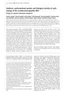
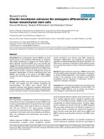
Footprint with reference lines for calculating the arch indexFigure 1
Footprint with reference lines for calculating the
arch index. The length of the foot (excluding the toes) is
divided into equal thirds to give three regions: A forefoot;
B midfoot; and C heel. The arch index is then calculated
by dividing the midfoot region (B) by the entire footprint
area (i.e. Arch index = B/[A+B+C]).
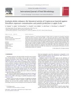
Calculating normalised navicular height truncatedFigure 2
Calculating normalised navicular height truncated.
The distance between the supporting surface and the navicu-
lar tuberosity is measured. Foot length is truncated by meas-
uring the perpendicular distance from the 1
st
metatarsophalangeal joint to the most posterior aspect of
the heel. Normalised navicular height truncated is calculated
by dividing the height of the navicular tuberosity from the
ground (H) by the truncated foot length (L) (i.e. Normalised
navicular height truncated = H/L).
Journal of Foot and Ankle Research 2009, 2:35 />Page 4 of 9
(page number not for citation purposes)
The temporal characteristics of the walking cycle were
measured using circular force sensitive resistors (foots-
witches) with a diameter of 13 mm (Model: 402, Interlink
Electronics, California, USA). These were placed on the
plantar surface of the interphalangeal joint of the hallux
and the most posterior plantar aspect of the calcaneus to
record the timing of heel contact, toe contact, heel off and
toe off.
During testing, participants were instructed to walk at
their self-selected walking speed, which was established
following a warm-up period from two trials along a 9 m
walkway. Six trials were recorded during testing, with any
trial exceeding ± 5% of the average warm-up speed
excluded and subsequently repeated.
Maximum voluntary isometric contractions (MVIC) were
used for normalising EMG amplitude parameters. At the
completion of each testing session, three MVICs for each
muscle were undertaken comprised of a gradual and con-
tinuous 2 s build-up followed by a maximum 2 s effort.
Each participant was instructed to perform a maximum
contraction against the resistance of the tester and was
given verbal encouragement while doing so. The resisted
movements included; supination - tibialis posterior, pro-
nation - peroneus longus, dorsiflexion - tibialis anterior,
plantarflexion (knee extended) - medial gastrocnemius.
The participant sat on a bench while performing the
MVICs for tibialis posterior, tibialis anterior and the pero-
neal muscles. For the medial gastrocnemius MVICs, the
participant sat on the floor with their back against a wall,
to ensure the participant did not slide backward during
the contraction.
Three consecutive maximum efforts were separated by a 1
min recovery period. A 600 ms window in the middle of
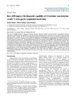
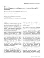
Traces from two representative participants illustrate x-ray angular measurements from normal (left) and flat-arched (right) foot postureFigure 3
Traces from two representative participants illustrate x-ray angular measurements from normal (left) and
flat-arched (right) foot posture. Lateral views (top) show: calcaneal inclination angle; calcaneal-first metatarsal angle; ante-
rior posterior views (bottom) show: talonavicular coverage angle; talus second metatarsal angle. A - calcaneal inclination angle,
B - calcaneal-first metatarsal angle, C - talo-navicular coverage angle, D - talus-second metatarsal angle. Angle A decreases with
flat-arched foot posture; angle B, C and D increase with flat-arched foot posture, compared to the normal-arched foot posture.
Journal of Foot and Ankle Research 2009, 2:35 />Page 5 of 9
(page number not for citation purposes)
the 2 s recording period was used to calculate average root
mean square (RMS) from three trials.
Data processing
During the gait trials, the raw EMG signal was passed
through a differential amplifier at a gain of 1000 with a
sampling frequency of 2 kHz. A band pass filter (built into
the amplifier; Delsys Inc., Boston, USA) of 20-2000 Hz
was applied to the intramuscular electrodes and 20-450
Hz for the surface electrodes.
EMG data from the MVICs and walking trials were full
wave rectified and low pass filtered at a cut off frequency
of 6 Hz through a 4
th
order Butterworth filter with phase
lag. Data were analysed from the third or fourth stride
depending on the quality of the footswitch signal. Two
consecutive strides were analysed for each trial and aver-
aged from the last four of six trials for each speed (i.e. four
average gait cycles derived from eight ipsilateral steps).
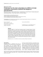
Three EMG parameters were analysed for each muscle,
including: (i) time of peak amplitude; (ii) root mean
square (RMS); and (iii) peak amplitude (Figure 4). These
parameters have been utilised in previous single-session
investigations [14,19,20]. The following phases of the gait
cycle were assessed based on when these muscles are most
active in normal-arched feet [14]: tibialis posterior and
peroneus longus - contact and combined midstance/pro-
pulsion phase; tibialis anterior - contact phase; and
medial gastrocnemius - combined midstance/propulsion
phase.
Statistical analysis
The distribution of data was evaluated from skewness and
kurtosis values and Levene's test for equality of variances.
Independent samples t-tests were performed to assess for
significant differences between the normal- and flat-
arched groups for anthropometric characteristics, walking
speed and EMG parameters with p values less than 0.05
considered significant.
Results
Participant characteristics
The normal- and flat-arched foot posture groups were
matched for age, gender, height and weight, with no sig-
nificant differences for any of these characteristics except
for the clinical and radiographic measures of foot posture
(Table 1). However, the self-selected comfortable walking
speed of the flat-arched group was slightly greater than the
normal-arched group (mean difference: 0.11 ms, 95% CI:
0.05 to 0.17, p < 0.001).
A single gait cycle showing raw and processed EMG for tibialis posterior from a single participantFigure 4
A single gait cycle showing raw and processed EMG for tibialis posterior from a single participant. Time of peak
amplitude, peak amplitude and RMS amplitude (root mean square) were derived from the linear envelope (processed curve).
Journal of Foot and Ankle Research 2009, 2:35 />Page 6 of 9
(page number not for citation purposes)
Effect of foot posture on muscle EMG activation
Comparisons of EMG variables between the normal- and
flat-arched foot groups are presented in Table 2. Statisti-
cally significant differences in peak and RMS EMG ampli-
tude were detected for tibialis posterior, peroneus longus
and tibialis anterior. There were no significant differences
in EMG time of peak amplitude.
Contact phase - heel contact to toe contact
For tibialis anterior, the flat-arched group exhibited
increased peak EMG amplitude (mean difference: 19.0%;
95% CI: 11.2 to 26.9; d = 1.3; p < 0.001) and RMS ampli-
tude (mean difference: 10.4%; 95% CI: 4.0 to 16.8; d =
0.87; p = 0.002), compared to the normal-arched group.
For peroneus longus, the flat-arched foot group exhibited
decreased peak EMG amplitude (mean difference: -
12.8%; 95% CI: -25.1 to -0.5; d = 0.48; p = 0.041), com-
pared to the normal-arched group (Figure 5). For tibialis
posterior, the flat-arched foot group exhibited decreased
peak EMG amplitude (mean difference: -14.3%; 95% CI:
-29.1 to 0.4; d = 0.51; p = 0.058) compared to the normal
arched group, although this finding did not reach statisti-
cal significance (Figure 5).
Midstance/propulsion phase - toe contact to toe-off
For peroneus longus, the flat-arched foot group exhibited
decreased peak EMG (mean difference: -13.7%; 95% CI: -
26.1 to -1.4; d = 0.58; p = 0.030), compared to the normal-
arched group (Figure 5). For tibialis posterior, the flat-
arched group exhibited increased peak EMG amplitude
(mean difference: 26.5%; 95% CI: 4.2 to 48.7; d = 0.69; p
= 0.021) and RMS amplitude (mean difference: 16.4%;
95% CI: 3.6 to 29.1; d = 0.68; p = 0.013), compared to the
normal-arched group (Figure 5). No significant differ-
ences between groups were detected for medial gastrocne-
mius.
Discussion
The objective of this study was to investigate the effect of
flat-arched foot posture on the EMG activity of selected leg
muscles. During comfortable walking, participants in the
flat-arched foot group functioned at a significantly greater
percentage of their maximum amplitude for tibialis poste-
rior during midstance/propulsion phase, compared to
participants in the normal-arched group (peak amplitude,
86 versus 60% of MVIC; RMS amplitude, 48 versus 31%
of MVIC). Similar trends have been reported by earlier
studies comparing these foot types [8,9], however these
studies did not report 95% confidence intervals for the
percentage difference or effect size calculations, making it
difficult to assess the precision and the magnitude of the
differences observed [7]. Effect sizes for the differences
observed in peak and RMS for tibialis posterior amplitude
were 0.68 and 0.69 respectively, representing moderate
Table 2: Effect of foot posture on all EMG variables
Muscle Phase of gait cycle EMG
parameter
% mean difference ^ 95% CI Effect size
#
p value
(2-tailed)
Tibialis posterior Contact TimePeak 0.1 0.0 to 1.7 0.52 0.051
PeakAmp -14.3 -29.1 to 0.4 0.51 0.058
RMS -7.8 -18.4 to 2.7 0.39 0.144
Mid/Prop TimePeak 0.0 -3.8 to 3.7 0.01 0.980
PeakAmp 26.5* 4.2 to 48.7 0.69 0.021*
RMS 16.4* 3.6 to 29.1 0.68 0.013*
Peroneus longus Contact TimePeak 1.6 0.0 to 3.2 0.51 0.057
PeakAmp -12.8* -25.1 to -0.5 0.48 0.041*
RMS -6.6 -13.9 to 0.6 0.48 0.075
Mid/Prop TimePeak 3.3 -0.3 to 6.9 0.47 0.079
PeakAmp -20.0 -42.9 to 2.9 0.46 0.086
RMS -13.7* -26.1 to -1.4 0.58 0.030*
Tibialis anterior Contact TimePeak 0.1 -0.7 to 0.9 0.09 0.737
PeakAmp 19.0* 11.2 to 26.9 1.3 <0.001*
RMS 10.4* 4.0 to 16.8 0.87 0.002*
Medial oastrocnemius Mid/Prop TimePeak 0.4 -1.8 to 2.7 0.10 0.715
PeakAmp 2.7 -15.4 to 20.7 0.12 0.766
RMS 7.2 -12.3 to 16.9 0.22 0.753
Contact contact period of gait cycle; Mid/Prop combined midstance and propulsion period of gait cycle; TimePeak time of peak amplitude;
PeakAmp peak EMG amplitude; RMS root mean square amplitude; ^ relative to normal-arch foot group; CI confidence interval;
#
Cohen's d
calculation;
* statistically significant independent sample t-test (p < 0.05)
Journal of Foot and Ankle Research 2009, 2:35 />Page 7 of 9
(page number not for citation purposes)
differences in muscle activity. Despite the issue of random
variability for tibialis posterior EMG amplitude during
gait [14,20], our results provide strong evidence to indi-
cate that tibialis posterior is working harder (i.e. as a per-
centage of a maximum contraction) during midstance/
propulsion in participants with flat-arched feet, compared
to those with normal-arched feet.
One explanation for our findings is that the medial longi-
tudinal arch and supportive structures (e.g. ligaments) of
a flat-arched foot may undergo greater loading during
walking, compared to the normal-arched foot. Greater
loading of the medial arch would require greater work
from tibialis posterior to protect the arch structures from
excessive tissue stress and injury. While cadaveric research
has shown an increased loading of the foot's medial struc-
tures with simulated tibialis posterior tendon dysfunction
[21], it is also possible that these events can occur in
reverse, that is, the flat-arched foot may place a greater
demand on tibialis posterior. This mechanism is further
supported by our findings for peroneus longus.
In contrast to tibialis posterior, participants in the flat-
arched group functioned at a significantly lower percent-
age of their maximum amplitude for peroneus longus
during contact phase and midstance/propulsion phase,
compared to participants in the normal-arched group
(peak amplitude - contact phase, 24 versus 37% MVIC;
RMS amplitude - midstance/propulsion, 25 versus 39%
MVIC). These findings indicate that peroneus longus is
working less during the contact and midstance/propul-
sion phases in participants with flat-arched feet, com-
pared to those with normal-arched feet. Effect sizes for
these differences were 0.48 and 0.58 for peak amplitude
(contact phase) and RMS (midstance/propulsion phase)
amplitude respectively, representing moderate differences
in muscle activity. These functional differences between
foot types may reflect a compensatory lack of activity in
peroneus longus to avoid further overloading the medial
arch. Alternatively, this finding may occur as a result of
flat-arched feet being less laterally unstable, therefore
requiring less peroneus longus activity.
A further significant finding was that participants in the
flat-arched group functioned at a significantly greater per-
centage of their maximum amplitude for tibialis anterior
during contact phase, compared to participants in the nor-
mal-arched group (peak amplitude, 65 versus 46% MVIC;
RMS amplitude, 43 versus 32% MVIC). Effect sizes for
these differences were 1.3 and 0.87 for peak and RMS
amplitude respectively, representing large differences in
muscle activity. During contact phase of the gait cycle,
tibialis anterior is thought to decelerate ankle joint
plantarflexion and resist foot pronation [22]. Interest-
Ensemble averaged EMG curves for tibialis posterior, per-oneus longus and tibialis anterior for 30 participants with normal-arch and 30 participants with flat-arch feetFigure 5
Ensemble averaged EMG curves for tibialis posterior,
peroneus longus and tibialis anterior for 30 partici-
pants with normal-arch and 30 participants with flat-
arch feet. The curves differ slightly to the actual results
(Table 2), as these curves are derived from a single gait cycle
for each participant to illustrate the main findings. Solid lines
mean amplitude; shaded area surrounding solid line 95%
confidence interval. Significant differences are generally indi-
cated where 95% confidence intervals separate between
groups. HC - heel contact.
Journal of Foot and Ankle Research 2009, 2:35 />Page 8 of 9
(page number not for citation purposes)
ingly, the role of tibialis anterior in resisting pronation of
the foot during the contact phase was not assisted via
strong co-activation of tibialis posterior. In fact, tibialis
posterior functioned at a lower percentage amplitude dur-
ing contact phase compared to the normal arched group,
although this finding did not reach statistical significance
(p = 0.058).
There were no differences in medial gastrocnemius timing
or amplitude EMG parameters comparing normal- and
flat-ached feet. This finding adds to the growing body of
evidence that medial gastrocnemius muscle activation is
not affected by differences in foot posture [7]. Further-
more, this indicates that medial gastrocnemius is unlikely
to have a significant function as an inverter of the hind-
foot, since deviations in hindfoot alignment have not
been shown to cause changes in the activity of this muscle
[7].
The finding that participants in the flat-arched foot group
walked slightly faster than those in the normal-arched
group (mean difference, 0.11 ms) was unexpected and
may have influenced some results in this study. It should
be noted that both foot posture groups were instructed to
walk at their normal comfortable walking speed and data
collection was carried out under identical conditions. This
difference in walking speed required some consideration,
as numerous studies investigating the influence of walk-
ing speed on EMG amplitude have indicated that EMG
amplitude increases linearly with walking speed [23-25].
There may be a biological or compensatory reason why
participants with flat-arched feet walked faster than those
with normal-arched feet, such as a means of increasing
stability of the foot and lower limb during walking. In this
case, the independent variable (flat-arch foot posture)
may have influenced the covariate (walking speed), and
this poses a conceptual issue preventing us from adopting
an analysis of co-variance approach to adjust for walking
speed [26]. However, we believe that the differences in
muscle activity observed between the groups are unlikely
to have been caused by differences in walking speed. Par-
ticipants in the flat-arch group functioned at a signifi-
cantly lower percentage of their maximum amplitude for
peroneus longus during contact phase and midstance/
propulsion phase, despite walking faster. Furthermore,
den Otter and colleagues [23] have shown that negative
amplitude gains (i.e. increased amplitude with reduced
walking speed) of peroneus longus only occur at very slow
speeds. Therefore, it is unlikely that the normal-arched
group displayed a relative 'negative gain' compared to the
flat-arched group.
The results presented here may have implications for the
management of lower extremity overuse conditions.
Although it is still unknown whether these functional dif-
ferences in muscle activation are beneficial or detrimental
in relation to injury, preliminary evidence indicates that
these differences may be reversible with intervention [27].
In a recent study, Franettovich and colleagues [27] inves-
tigated the effect of an anti-pronation taping technique on
lower limb EMG muscle activation in four adults with
pronated foot posture. They reported that the anti-prona-
tion tape significantly reduced the EMG amplitude of the
tibialis posterior and tibialis anterior muscles during
walking. While this indicates that anti-pronation tape
may bring muscle function in a flat-arched foot closer to
that observed in a normal-arched foot, further research is
required to ascertain whether these changes are associated
with clinical outcomes.
This study has several strengths, including the use of a rig-
orous protocol to classify foot posture, the use of in-dwell-
ing needle electrodes to assess tibialis posterior and
peronus longus, and a relatively large sample size (n = 60
compared to 17 to 43 in previous studies [2,7-10,12]).
However, the results of this study also need to be inter-
preted in light of two limitations. Firstly, we did not
simultaneously record other kinematic and kinetic varia-
bles, thus we can only speculate as to the mechanical
effects of the EMG differences. Secondly, the participants
in this study were relatively homogenous as they were
mostly young, healthy and without musculoskeletal
injury. Therefore, caution should be taken in generalising
these results to symptomatic or clinical populations. A
further limitation was that we used MVICs to normalise
the EMG amplitude parameters. It is difficult to control
and monitor the participants' effort or output with MVICs
which may be a factor that leads to greater between-partic-
ipant variability compared to other normalisation proto-
cols [20].
Conclusion
Lower limb muscle function is affected by foot posture.
The flat-arched group functioned at a greater percentage of
their maximum EMG amplitude during contact phase for
tibialis anterior and during midstance/propulsion for tibi-
alis posterior, compared to normal-arched feet. The flat-
arched foot group also functioned at a lower percentage of
their maximum EMG amplitude throughout stance phase
for peroneus longus, compared to normal-arched feet.
These differences in muscle activity may reflect neuromus-
cular compensation to reduce overload of the medial lon-
gitudinal arch in people with flat-arched feet. Further
research is required to determine whether these differ-
ences in muscle function are associated with injury.
Competing interests
HBM and KBL are Editor-in-Chief and Deputy Editor-in-
Chief, respectively, of Journal of Foot and Ankle Research. It
is journal policy that editors are removed from the peer
Publish with Bio Med Central and every
scientist can read your work free of charge
"BioMed Central will be the most significant development for
disseminating the results of biomedical research in our lifetime."
Sir Paul Nurse, Cancer Research UK
Your research papers will be:
available free of charge to the entire biomedical community
peer reviewed and published immediately upon acceptance
cited in PubMed and archived on PubMed Central
yours — you keep the copyright
Submit your manuscript here:
/>BioMedcentral
Journal of Foot and Ankle Research 2009, 2:35 />Page 9 of 9
(page number not for citation purposes)
review and editorial decision-making processes for papers
they have co-authored.
Authors' contributions
GSM, HBM and KBL conceived the idea and obtained
funding for the study. GSM, HBM and KBL designed the
study protocol. GSM recruited participants, conducted the
laboratory testing and processed data. GSM, HBM and
KBL drafted the manuscript. All authors have read and
approved the final manuscript.
Additional material
Acknowledgements
This project was supported by a research grant from the Australian Podia-
try Education and Research Foundation (APERF). We thank Mark White-
side, Lisa Scott and Bianca David for assisting with participant recruitment
and testing; and Southern Cross Medical Imaging at La Trobe University
Medical Centre. We also thank Monika Buljan and Paul Kabaila (La Trobe
University) for statistical support relating to this study. HBM is currently a
National Health and Medical Research Council fellow (Clinical Career
Development Award, ID: 433049).
References
1. Redmond AC, Crane YZ, Menz HB: Normative values for the
Foot Posture Index. J Foot Ankle Res 2008, 1:6.
2. Hunt AE, Smith RM: Mechanics and control of the flat versus
normal foot during the stance phase of walking. Clin Biomech
2004, 19:391-397.
3. Redmond AC, Crosbie J, Ouvrier RA: Development and valida-
tion of a novel rating system for scoring standing foot pos-
ture: The Foot Posture Index. Clin Biomech 2006, 21:89-98.
4. Burns J, Keenan A-M, Redmond A: Foot Type and overuse onjury
in triathletes. J Am Podiatr Med Assoc 2005, 95:235-241.
5. Yates B, White S: The incidence and risk factors in the devel-
opment of medial tibial stress syndrome among naval
recruits. Am J Sports Med 2004, 32:772-780.
6. Milgrom C, Radeva-Petrova DR, Finestone A, Nyska M, Mendelson S,
Benjuya N, Simkin A, Burr D: The effect of muscle fatigue on in
vivo tibial strains. J Biomech 2007, 40:845-850.
7. Murley GS, Landorf KB, Menz HB, Bird AR: Effect of foot posture,
foot orthoses and footwear on lower limb muscle activity
during walking and running: a systematic review. Gait Posture
2009, 29:172-187.
8. Keenan MA, Peabody TD, Gronley JK, Perry J: Valgus deformities
of the feet and characteristics of gait in patients who have
rheumatoid arthritis. J Bone Joint Surg Am 1991, 73:237-247.
9. Gray EG, Basmajian JV: Electromyography and cinematography
of leg and foot ("normal" and flat) during walking. Anat Rec
1968, 161:1-15.
10. Backmann CK: The effect of treadmill compliance and foot
type on electromyography of lower extremity muscles dur-
ing running. Western Washington University; 1997.
11. Williams DS III, McClay IS, Hamill J: Arch structure and injury pat-
terns in runners. Clin Biomech 2001, 16:341-347.
12. Cornwall MW, McPoil TG: The influence of tibialis anterior
muscle activity on rearfoot motion during walking. Foot Ankle
Int
1994, 15:75-79.
13. McPoil TG, Vicenzino B, Cornwall MW, Collins N, Warren M: Reli-
ability and normative values for the foot mobility magnitude:
a composite measure of vertical and medial-lateral mobility
of the midfoot. J Foot Ankle Res 2009, 2:6.
14. Murley GS, Buldt AK, Trump PJ, Wickham JB: Tibialis posterior
EMG activity during barefoot walking in people with neutral
foot posture. J Electromyogr Kinesiol 2009, 19:e69-e77.
15. Scott G, Menz HB, Newcombe L: Age-related differences in foot
structure and function. Gait Posture 2007, 26:68-75.
16. Murley GS, Menz HB, Landorf KB: A protocol for classifying nor-
mal- and flat-arched foot posture for research studies using
clinical and radiographic measurements. J Foot Ankle Res 2009,
2:22.
17. Exposure of Humans to Ionizing Radiation for Research Pur-
poses [ />]
18. Hermens HJ, Freriks B, Merletti R, Stegeman D, Blok J, Rau G, Dissel-
horst-Klug C, Hägg G: SENIAM 8 - European Recommendations for Sur-
face ElectroMyoGraphy 2nd edition. Enschede: Roessingh Research and
Development; 1999.
19. Murley GS, Bird AR: The effect of three levels of foot orthotic
wedging on the surface electromyographic activity of
selected lower limb muscles during gait. Clin Biomech 2006,
21:1074-1080.
20. Murley GS, Menz HB, Landorf KB, Bird AR: Reliability of lower
limb electromyography during overground walking: a com-
parison of maximal- and sub-maximal normalisation tech-
niques. J Biomech 2009 in press. doi:10.1016/j.jbiomech.2009.10.014
21. Imhauser CW, Siegler S, Abidi NA, Frankel DZ: The effect of pos-
terior tibialis tendon dysfunction on the plantar pressure
characteristics and the kinematics of the arch and the hind-
foot. Clin Biomech 2004, 19:161-169.
22. Hunt AE, Smith RM, Torode M: Extrinsic muscle activity, foot
motion and ankle joint moments during the stance phase of
walking. Foot Ankle Int 2001, 22:31-41.
23. den Otter AR, Geurts AC, Mulder T, Duysens J: Speed related
changes in muscle activity from normal to very slow walking
speeds. Gait Posture 2004, 19:270-278.
24. van Hedel HJ, Tomatis L, Muller R: Modulation of leg muscle
activity and gait kinematics by walking speed and body-
weight unloading. Gait Posture 2006, 24:35-45.
25. Warren GL, Maher RM, Higbie EJ: Temporal patterns of plantar
pressures and lower-leg muscle activity during walking:
effect of speed. Gait Posture 2004, 19:91-100.
26. Tabachnick BG, Fidell LS: Using multivariate statistics 5th edition. Bos-
ton: Pearson/Allyn & Bacon; 2007.
27. Franettovich M, Chapman A, Vicenzino B: Tape that increases
medial longitudinal arch height also reduces leg muscle
activity: a preliminary study. Med Sci Sports Exerc 2008,
40:593-600.
Additional file 1
A video demonstration of the insertion of an intramuscular electrode
into tibialis posterior via the posterior approach
Click here for file
[ />1146-2-35-S1.m4v]