Báo cáo y học: "Changes in joint coupling and variability during walking following tibialis posterior muscle fatigue" ppt
Bạn đang xem bản rút gọn của tài liệu. Xem và tải ngay bản đầy đủ của tài liệu tại đây (584.07 KB, 8 trang )
RESEARCH Open Access
Changes in joint coupling and variability during
walking following tibialis posterior muscle fatigue
Reed Ferber
1,2*†
, Michael B Pohl
1†
Abstract
Background: The tibialis posterior muscle is believed to play a key role in controlling foot mechanics during the
stance phase of gait. However, an experiment involving localised tibialis posterior muscle fatigue, and analysis of
discrete rearfoot and forefoot kinematic variables, indicated that reduced force output of the tibialis posterior
muscle did not alter rearfoot and forefoot motion during gait. Thus, to better understand how muscle fatigue
affects foot kinematics and injury potential, the purpose of this study was to reanalyze the data and investigate
shank, rearfoot and forefoot joint coupling and coupling variability during walking.
Methods: Twenty-nine participants underwent an exercise fatigue protocol aimed at reducing the force output of
tibialis posterior. An eight camera motion analysis system was used to evaluate 3 D shank and foot joint coupling
and coupling variability during treadmill walking both pre- and post-fatigue.
Results: The fatigue protocol was successful in reducing the maximal isometric force by over 30% and a
concomitant increase in coupling motion of the shank in the transverse plan e and forefoot in the sagittal and
transverse planes relative to frontal plane motion of the rearfoot. In addition, an increase in joint coupling
variability was measured between the shank and rearfoot and between the rearfoot and forefoot during the
fatigue condition.
Conclusions: The reduced function of the tibialis posterior muscle following fatigue resulted in a disruption in
typical shank and foot joint coupling patterns and an increased variability in joint coupling. These results could
help explain tibialis posterior injury aetiology.
Background
Although runners often sustain acute injuries such as
ankle sprains and muscle strains, a vast majority of
running injuries could be classified as cumulative
micro-trauma (overuse) injuries [1-4]. The aetiology of
an overuse running injury is multifactorial but muscle
fatigue and/or weakness has been discussed as a pri-
mary contributing factor [5-9]. Indeed, many lower
extremity overuse injuries have been attributed to aty-
pical foot mechanics during gait [10-13]. The tibialis
posterior is believ ed to play a key role in controlling
rearfoot eversion [14,15] and providing dynamic sup-
port across the midfoot and forefoot during the stance
phase of gait [15-17].
The proximal origin of tibi alis posterior lies o n the
interosseous membrane and posterior surfaces of the
tibia and fibula. The muscle has multiple distal inser-
tions including the navicular tubercle, the plantar sur-
face of the cuneiforms and cuboid, and bases of the
second, third and fourth metatarsals [18]. Biomechanical
research conducted on patients with posterior tibialis
tendon dysfunction (PTTD) highlights the importance
of this muscle in controlling rearfoot, midfoot, and fore-
foot mechanics dur ing gait [19-21]. However, these stu-
dies involved patients with moderate- to advanced-stage
PTTD and may not provide adequate information to
help us understand the contribution that the tibialis
posterior muscle plays in controlling foot pronation in
healthy individuals.
One method of assessing a muscle’s contribution to a
specific movement pattern is via fatigue-inducing exer-
cise of that muscle. Christina et al. [ 22] showed that
localised fatigue of the ankle invertors resulted in a
* Correspondence:
† Contributed equally
1
Faculty of Kinesiology, University of Calgary, Calgary, AB, Canada
Full list of author information is available at the end of the article
Ferber and Pohl Journal of Foot and Ankle Research 2011, 4:6
/>JOURNAL OF FOOT
AND ANKLE RESEARCH
© 2011 Ferber and Pohl ; licensee BioMed Centra l Ltd. This is an Open Access article distributed under the terms of the Creative
Commons Attribution License ( which permits unrestricted use, distribution, and
reproduction in any medium, provided the original work is properly cited.
trend towards greater rearfoot eversion during running.
However, fatigue of the invertor musculature was
achieved using open chain resisted supination exercises,
which would result in the recruitment of all invertor
muscles including tibialis posterior. Kulig et al. [23]
investigated which exercise most selectively and effec-
tively activates tibialis posterior: 1) closed chain resisted
foot adduction, 2) unil ateral heel raise, and 3) ope n
chain resisted foot supination using MRI to quantify
changes in pre- to post-exercise signal intensity. These
authors reported that isolated activation of tibialis pos-
terior is best achieved using closed chain resisted foot
adduction as opposed to open chain supi nation. In addi-
tion, they reported the greatest tibialis posterior increase
occurred with closed c hain resisted foot adduction,
whereas the mean increase in the other muscles was less
than 5%. Therefore, to better understand the role of
tibialis posterior fatigue on foot mechanics it seems pru-
dent to use an exercise that more selectively activates
and subsequently fatigues this muscle.
Pohl et al. [15] recently conducted a study investigat-
ing the eff ect of localised tibialis posterior muscle fati-
gue on foot kinematics during walking. These authors
reported that following a 30% reduction in tibialis pos-
terior maximal isometric force production, no changes
in rearfoot or forefoot kinematics were measured. Spe ci-
fically, a 0.7 degree increase in peak rearfoot eversion
was reported as statistically signif icant but this change
was smaller than the precision error of a within-day gait
analysis (0.9 degree). Therefore, these authors postulated
the results were not clinically relevant and that it was
possible that other muscles, such as tibialis anterior,
may have compensated for the lack of tibialis posterior
force production thereby resulting in no change in dis-
crete kinematic variables. However, inspection of the
data also revealed that 24 out of 29 participants demon-
strated an increase in peak rearfoot angle following fati-
gue (ranging from 0.5 - 2.0 degrees). Since such a
consiste nt change was observed, it raises the question of
what other mechanisms and potential explanations can
account for these systematic changes. Thus, In light of
these findings, it may be worthwhile to investigate the
effect of localised muscle fatigue u sing a joint coupling
and coupling variability approach.
The timing or coupling of joint movements has been
shown to be a useful tool for understanding injury
aetiology based on the notion that asynchrony in joint
coupling and changes in joint coupling variability of
movement may result in injury [24]. Some researchers
have subsequently investigated changes in joint coupling
for both injured and healthy partic ipants and reported
that, overall, non-injurious coupling involves an in-
phase relationship and injurious coupling involves more
out-of-phase joint coupling relationship throughout
stance [5,25-27]. However, these studies have focused
primarily on thigh:shank or rearfoot:shank couplings in
an effor t to understand knee-rel ated injuries . Moreover,
these studies have utilised a cross-sectional approach
and compared the joint coupling and /or coupling varia-
bility patterns between injured and non-injured groups.
Few studies have investigated the complexities of the
multiple foot segments using a joint coupling approach
or by investigating joint coupling variability.
Variability in joint coupling has been suggested to play
a role in the aetiology of injury. Hamill et al. [24] pro-
posed that injured runner s exhibit reduced jo int cou-
pling variability thereby reducing the flexibility in the
system a nd increasing the potential for musculoskeletal
injury. Other studies have supported this finding for
patients with iliotibial band syndrome [28] and for
female runners as a possible mechanism to explain the
higher incidence of ACL injuries compared to males
[29]. More over, Miller et al. [ 28] suggested that muscle
dysfunction and/or weakness may be a possible explana-
tion for the reduced joint coupling variability measured
after an exhaustive run for a group of injured runners.
However, these authors did not measure changes in
muscle strength following the run and the aforemen-
tioned studies [28,29] utilised a cross-sectional approach
and/or extrinsic perturbations to investigate changes in
joint coupling variability. To our knowledge, no study
has utilised a muscle fatigue protocol (an intrinsic per-
turbation) to better understand potential changes in
joint coupling and/or joint coupling variability to shed
light on injury aetiology.
Therefore, the purpose of this study was to examine
the effect of localised tibialis posterior muscle fatigue on
shank, rearfoot and forefoot joint coupling and coupling
variability during walking. It was hypothesised that fol-
lowing a bout of fatigue-inducing exercise participants
would demonstrate altered and non-synchronous joint
coupling between the respective segments. Since no
study has specifically investigated changes in joint cou-
pling for the ankle and foot segments in such a manner,
we chose to leave this hypothesis non-d irectional. Since
several other muscles, specifically tibialis anterior, flexor
hallucis longus, flexor digitorumlongus,andperoneus
longus, also serve to control foot and a nkle kinematics,
it is reasonable to assume that localised fatigue of one
muscle would force the supporting musculature to
increase their role i n maintaining a normal mechanical
pattern. Since fewer muscles are now functioning to
perform a given task, we hypothesised that following
fatigue a reduction in coupling variability would be mea-
sured. We also hypothesised that the greatest changes in
joint coupling and variability would occur at or near
midstance when loading to the foot and shank would be
greatest.
Ferber and Pohl Journal of Foot and Ankle Research 2011, 4:6
/>Page 2 of 8
Methods
Participants
Twenty-nine (11 males, 18 females) recreationally active
participants (age = 27.3 ± 8.1 years; mass = 68.8 ± 13.5 kg;
height = 172.8 ± 13.5 cm) volunteered to participate in the
study. All participants were currently free from lower
extremity injury, had no prior history of surgery, and were
familiar with treadmill walking. The study was approved
by the institutional ethics board, and written informed
consent was obtained from all participants.
Procedures
More in depth explanations of the procedures and
methods can be found in a previous publication [15]. In
brief, three-dimensional kin ematic data were collected
for all participants walking barefoot on a treadmill both
prior to, and following, fatigue-inducing exercise of the
tibialis posterior muscle of the right limb. Seventeen
reflective markers (9 mm diameter) were attached to the
skin of the forefoot, rearfoot and shank as described
previously [15,30]. Kinematic walking data were col-
lected at 120 Hz using an eight-camera motion analysis
system (Vicon Motion Systems Ltd, Oxford, UK)
arranged around a treadmill (StarTrac, Irvine, USA).
A standing static calibration trial was recorded followed
by walking on a treadmill at 1.1 ms
-1
. Subjects were pro-
vided 2-3 minutes to accommodate to the treadmill and
the speed chosen. Once accommodated and comfortab le
on the treadmill, ten footfalls of kinematic data were
collected to represent the “pre-fatigue” (PRE) condition.
Upon completion of the fatigue exercise protocol, parti-
cipants immediately completed the “ post-fatigue”
(POST) walk and another 10 footfalls were collected.
Muscle fatigue was defined as a reduction in the capa-
city of the muscle to perform work or generate force
[15,22]. Participants were seated in a chair while their
right foot was placed in a custom built device containing
a dynamometer (Lafayette Instrument, L afayette, USA:
Model 01163) that 1) allowed participants to perform
concentric/eccentric foot adduction contractions with
adjustable resistance and 2) enabled the measurement of
a maximal voluntary isometric contraction (MVIC) dur-
ing foot adduction. The mean of three MVIC trials was
taken t o represent baseline strength. Then, participants
performed sets of 50 concentric/eccentric contractions
at 50% MVIC through a 30° range of motion with
10 seconds of rest between each set. MVICs were
repeate d after every four sets and exerc ises were contin-
ued until participants MVICs had dropped below 70%
of the pre-fatigue values or they were unable to com-
plete two consecutive s ets. A final set of MVICs were
taken immediately follo wing the post-fatigue walk
(within 2 minutes) to determine whether participants
had recovered in strength during the walking trial.
Data processing
Ten foot falls for the PRE and POST kinema tic walking
data were selected for analysis. Raw kinematic data were
filtered using a fourth order low-pass Butterworth filter
at 12 Hz. Anatomical co-ordinate systems for the shank,
rearfoot and forefoot, along with three-dimensional seg-
ment angles were calculated using Visual 3 D software
(C-motion Inc, Rockville, USA) [15,31]. All segment
angles we re defined as motion measured relative to the
next most proximal segment [19,21] and the segment
angles during walking were expressed relative to the
standing calibration trial. All kinematic data w ere ana-
lysed for the stance phase and normalised to 101 data
points. Initial contact (IC) and toe off (TO) were identi-
fied using a kinematic velocity-based algorithm [32]
applied to the posterior calcaneal and dorsal phalanx
markers respectively. Custom Labview (National Instru-
ments Corp, Austin, USA) software was used to extract
the kinematic coupling variables of in terest for each
subject. Specifically, the following joint coupling and
coupling variability relationships were investigated:
1) tibia internal/external rotation:rearfoot inversion/ever-
sion (TIBrot:RFi/ev), 2) rearfoot inversion/eversion:fore-
foot dorsi/plantarflexion (RFi/ev:FFd/pf), and 3) rearfoot
inversion/eversion:forefoot abd/adduction(RFi/ev:FFab/d).
We chose these joint coupling relationships to compare
the results with previous studies [5,26,28,30,31].
Angle-angle plots of proximal and distal segments for
each trial were created. The coupling angle was deter-
mined using a modification of a vector coding technique
suggested by Heiderscheit et al. [33]. The absolute resul-
tant vector between two adjacent data points during the
stance phase of running was calculated (equation 1)
and, following conversion from radians to degrees, the
resulting range of values for coupling angle was 0-90°.
Ø
i
1
11
abs tan y y x x=
−
++
[(–/–)]
iiii
(1)
where i = 1,2, and n
Thus, with the distal segment motion plotted on the
abscissa and proximal segment motion plotted on the
ordinate, a coupling angle of 45° would indicate equal
amounts of segmental motion. An angle greater than
45° indicates greater proximal segment motion relative
to distal segment. Similar to previous studies [24,26], for
the purpose of analyzing the coupling angles and varia-
bility within specific regions of stance, each relative
motion plot was first normalized to 100% of stance and
then divided into 4 phases. Phase 1 ranged from heel
strike to 25% of stance, phase 2 from 25-50% of stance,
phase 3 from 50-75% of stance, and phase 4 from
75-100% of stance. To calculate the average coupling
angle values for each phase of stance, each da ta point
Ferber and Pohl Journal of Foot and Ankle Research 2011, 4:6
/>Page 3 of 8
was averaged on a point-by-point basis across the ten
trials resulting in an average trace. From the aver age
trace, the average coupling angle for each phase of
stance was calculate d over time. The standard deviation
was calculated on a point-by-po int basis across the 10
trials and the between-trial, within-subject joint coupling
variability for each phase of stance was calculated across
time for each phase of stance.
Data analysis
Group descriptive statistics were calculated for each vari-
able for both PRE and POST fatigue conditions. Paired
sample t-tests (two-tailed) were conducted for the va ri-
ables of interest for between-condition statistical compar-
ison. Since we hypothesised that the greatest changes in
coupling and variabi lity would occur at or near mid-
stance, a priori t-tests were performed on phase 2 and
phase 3 data and significance for these tests was set at an
alpha level of P < 0.05. If necessary, analysis of phases 1
or 4 were performed to help better understand our
results and an alpha level of P < 0.01 was established to
minimize type I error. All analyses were undertaken
using SPSS 15.0 (SPSS Inc, Chicago, USA).
Results
Strength
As reported previously, following the fatigue exercise
protocol the MVIC strength dropped to 67% of the
baseline values (p = 0.001; PRE = 66.2 N; POST = 44.6).
Eight participants did not drop below the predetermined
threshold of 70% base line MVIC but were sti ll included
in the analysis since they were unable to complet e two
additional sets of 50 repetitions due to muscle fatigue
and also exhibited a 21% reduction in force output.
Immediately following the p ost-fatigue walk, the MVIC
strength was 80% of the baseline.
Joint Coupling
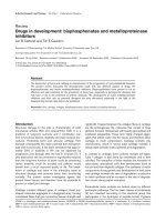
A summary of pre- and post-fatigue changes in TIBrot:
RFi/ev, RFi/ev:FFd/pf, and RFi/ev:FFab/d joint coupling
angle is provided in Figure 1 and Table 1. For TIBrot:
RFi/ev, a significant increase in joint coupling angle dur-
ing Phase 2 (p = 0.05; PRE = 42.42°; POST = 44.71°) was
measured following the f atigue protocol. RFi/ev:FFd/pf
significantly decreased during Phase 2 (p = 0.04; PRE =
45.91°; POST = 40.58°) and Phase 3 (p = 0.01; PRE =
48.19°; POST = 43.42°) and RFi/ev:FFab/d, also significantly
decreased during Phase 2 (p = 0.01; PRE = 54.62°; POST =
52.06°) and Phase 3 (p = 0.01; PRE = 59.09°; POST =
53.94°) compared to pre-fatigue values.
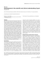
Coupling Variability
A s ummary of pre- post-fatigue changes in TIBrot:RFi/
ev, RFi/ev:FFd/pf, and RFi/ev:FFab/d joint coupling
variability is provided in Figure 2 and Table 1. TIBrot:
RFi/ev significantly increased during Phase 2 (p = 0.01;
PRE = 20.66°; POST = 22.34°) and Phase 3 (p = 0.01; PRE
= 18.71°; POST = 20.84°) following the fatigue protocol. A
significant increase in RFi/ev:FFab/d joint coupling varia-
bility was measured for Phase 2 ( p = 0.01; PRE = 21.34°;
POST = 23.37°) and Phase 3(p = 0.01; PRE = 20.64°; POST
= 22.94°) compared to pre-fatigue values. No changes in
RFi/ev:FFd/pf variability were measured across any phase
compared to pre-fatigue va lues (Table 1).
Figure 1 Joint coupling angle prior to and following the
fatigue protocol and across phase of stance. Note, * indicates
P < 0.05.
Ferber and Pohl Journal of Foot and Ankle Research 2011, 4:6
/>Page 4 of 8
Discussion
The purpose of this study was to examine the effect of
localised tibialis posterior muscle fatigue on shank, rear-
foot, and forefoot joint coupling and coupling variability
during walking. Two main hypotheses were put forth:
following a bout of fatigue-inducing exercise participants
would demo nstrate 1) altered an d non-synchrono us
joint coupling between the respective segments as well
as 2) reduced coupling variability. To test these hypoth-
eses, a unique approach was utilised to selectively fati-
gue the tibialis posterior muscle.
The protocol for reducing the force output of this
muscle was developed based on Kulig et al. [24] who
showed that isolated activation of tibialis posterior is
best achieved using closed chain resisted foot adduction.
The results of the present study indicate that this fatigue
protocol was successful in reducing the MVIC force by
over 30%. Although eight participants did not achie ve
the targeted 30% reduction in force production, they did
all achieve at least a 21% reduction and were unable to
complete 2 consecutive sets of the 50-repetition exer-
cise. Furthermore, there was no evidence that these
eight participants differed systematically from the rest of
the sample in terms of kinematic changes following fati-
gue based on the results of the current study and pre-
vious study [15]. Finally, the reduction in strength was
still apparent following the POST data collection indi-
cating that the fatigue protocol was effective.
The decrements in isometric force are similar to pre-
vious f atigue studies and studies involving healthy run-
ners and PTTD patients. Cheung and Ng [34] reported
similar findings for fatigue of the invertor muscles in
healthy runners following an exhaustive run. Moreover,
Alvarez et al. [35] reported a 40% reductio n in con-
centric ankle invertor strength for PTTD patients prior
to a 16-week rehabili tation program. However, it should
be recognized that the PTTD patients in this study
included advance-stage PTTD patients who had symp-
toms for approximately 16.5 weeks prior to treatment.
Pilot data from our laboratory shows that early-stage
PTTD patients exhibit a 17% reduction in ankle invertor
MVIC strength compared to healthy controls. Thus, we
are confident that our fatigue protocol and a priori cri-
teria for localised muscle fatigue is sufficient t o induce
tibi alis posterior muscle fatigue and concomitant reduc-
tions in force output during a dynamic task such as
walking.
In support of t he first hypothesis, a change in joint
coupling angle between 2.3° and 5.3° was measured dur-
ing Phase 2 and 3. Moreover, the tibia and forefoot all
increased their respective motions relative to the rear-
foot. While we are not aware of another study that h as
investigated changes in foot and ankle joint coupling fol-
lowing a fatigue protocol, the pre- and post-fatigue cou-
pling angle data in the current study are similar to Pohl
et al. [31] who also reported joint coupling angles at or
near 45° for the same coupling relationships while walk-
ing. Specifically, the pre-fatigue values for the TIBrot:
RFi/ev coupling angle indicate a near 1:1 ratio in cou-
pling for Phase 1 then greater overall motion of the
rearfoot throughout the remainder of stance which is
similar to previous studies [25,26].
Post-fatigue, and during Phase 2 of stance, inc reased
tibial motion was measured, relative to the rearfoot,
Table 1 Summary of shank, rearfoot, and forefoot joint coupling and coupling variability (Mean, (SD)) prior to (PRE)
and following fatigue (POST)
Coupling Angle (deg) Phase 1 Phase 2 Phase 3 Phase 4
PRE POST PRE POST PRE POST PRE POST
TIBrot:RFi/ev 45.63 45.54 42.42 44.71* 41.59 42.36 41.06 42.82
(5.74) (5.68) (9.58) (6.66) 7.78 8.73 6.11 6.11
RFi/ev:FFd/pf 44.34 45.92 45.91 40.58* 48.19 43.42* 45.32 41.17*
(7.12) (6.47) (11.50) (8.35) 6.63 11.14 7.02 9.55
RFi/ev:FFab/d 51.30 51.72 54.62 52.06* 59.09 53.94* 52.76 49.97
(6.22) (6.38) (8.47) (6.72) 8.68 9.24 7.05 7.48
Coupling Variability (deg) Phase 1 Phase 2 Phase 3 Phase 4
PRE POST PRE POST PRE POST PRE POST
TIBrot:RFi/ev 23.81 23.41 20.66 22.34* 18.71 20.84* 16.88 20.11*
(2.78) (1.55) (3.10) (2.99) 3.89 4.15 3.15 3.35
RFi/ev:FFd/pf 25.11 24.57 23.04 23.58 22.63 22.88 22.24 22.62
(1.79) (2.15) (3.22) (2.57) 4.48 3.42 4.16 2.40
RFi/ev:FFab/d 24.15 24.16 21.34 23.37* 20.64 22.94* 19.89 21.16
(2.32) (1.96) (4.01) (2.50) 4.93 3.84 4.38 2.48
* indicates p < 0.05.
Ferber and Pohl Journal of Foot and Ankle Research 2011, 4:6
/>Page 5 of 8
suggesting that fatigue of the tibialis posterior disrupted
the typical coupling mechanics b etween these two seg-
ments. This same relationship was observed for the RFi/
ev:FFd/pf coupling relationship wherein a 1:1 coupling
relationship is measured pre-fatigue and post-fatigue
shows a change in forefoot motion relative to the rear-
foot for Phases 2 and 3. Finally, greater overall rearfoot
motion, relative of the fore foot (RFi/ev:FFab/d), was
measured pre-fatigue, which is consistent w ith previous
studies [31], and fatigue of the tibialis posterior dis-
rupted this relationship resulting in greater motion of
the forefoot relative to the rearfoot for Phases 2 and 3
of stance. Thus, it can be concluded that when the tibia-
lis posterior is unable to produce sufficient force, there
are significant alterations in coupling patterns for the
shank and foot.
We postulate that the overall greater motion of the
tibia and forefoot (relative to the rearfoot) following fati-
gue is the result of the functional anatomy of the tibialis
posterior muscle itself. The tibialis posterior muscle ori-
ginates from the tibia and the tendon does not attach
directly to the rearfoot (calcaneus), but has several
attachment points to the midfoot and forefoot. Thus, it
is reasonable to speculate that greater relative motion of
these segments is the result of the inab ility of t he mus-
cle, via fatigue and reduced force output, to control the
individual motions of these foot segments.
In contrast to the sec ond hypothesis, an increase in
joint coupling variability was measured fo llowing the
fatigue protocol for TIBrot:RFi/ev and RFi/ev:FFab/d
during Phase 2 and 3. These results are in contrast to
several other studies investigating joint coupling varia-
bility. Ferb er et al. [26] studied different types of ortho-
tics during running and reported no significant changes
in TIBrot:RFi/ev variability across orthotic conditions or
compared to a con trol group. Hamill et al. [24] studied
patients with patellofemoral pain syndrome (PFPS) and
reported overall reduced joint coupling variability for
thigh and shank coupling variability compared to the
uninjured leg and a control group. However, these
authors measured thigh and shank c oupling patterns or
variability so it is difficult to compare their results with
those of the present study. Also in contrast to the
results of the present study, Miller et al. [36] reported
that runners with a history of iliotibial band syndrome
(ITBS) demonstrated reduced TIBrot:RFi/ev coupling
variability while running on a treadmill compared to a
control group. However, it is important to note that
these studies involved runners who were either injured
at the time of testing or had a long history of running-
related injuries. The participants in the current study
were healthy athletes with nohistoryofchronicinjury
and involved an intrinsic perturba tion rather than a
cross-sectional comparison. In addition, these authors
[24,36] used a different measure for coordination (con-
tinuous relative phase), which may no t directly compare
to the present vector coding method and could explain
the different findings of the present study. Thus, com-
parisons to previous studies must be made with caution.
While we are no t aware of another study utilising a
muscle-fatigue protocol to measure changes in either
joint coupling or coupling variability, two studies have
investigated changes in movement variability following
an intervention of some type. Ferber et al . [37] reported
that following a 3-week strengthening protocol, a
Figure 2 Joint coupling variability prior to and following the
fatigue protocol and across phase of stance. Note, * indicates
P < 0.05.
Ferber and Pohl Journal of Foot and Ankle Research 2011, 4:6
/>Page 6 of 8
reduced variability in st ride-to-stride knee joint kine-
matic patterns was adopted by the PFPS group. These
authors suggested that, from a clinical perspective,
restoration of a more consistent and predictable move-
ment pattern is expected with the increases in muscle
strength and reductions in pain. Thus, the increase in
coupling variability following fatigue in the present
study is consistent with these authors [37]. Further
research involving changes in coupling variability follow-
ing successful rehabilitation from a musculoskeletal
injury is, however, warranted.
Few studies have investigated the effect of fatigue o n
changes in joint coupling. Miller et al. [28] studied
changes in joint coupling variability during an exhaus-
tive run for runners who had previously experienced
ITBS. Compared w ith the control group, the ITBS run-
ners were more variable in knee flex/extension-foot add/
abduction at the start o f the run, less variable in thigh
add/abduction-foot inv/eversion at the end of the run,
tended to be less variable in thigh add/abduction-tibia
rotation at the end of the r un but showed no change in
TIBrot:RFi/ev coupling variability either during the
entire stride cycle, swing, or stance phase. It is possible
that since a variety of changes in coupling variability
were reported, albeit the majority of joint coupling rela-
tionships showing reduced variability, that increased
coupling variability for the shank and foot is a possible
mechanism to explain injury aetiology similar to the
results of the present study.
Based on the redundancy of the various muscles that
serve to control frontal plane rearfoot and transverse
plane tibial motion, a potential strategy for the foot may
be to increase coupling variability to avoid injury. We
postulate that a diminished ability of the muscle to pro-
duce a vigorous contraction, a concomitant reduction in
joint contact force, and a resulting increase in joint cou-
pling variability could result. In other words, the
reduced function of the tibialis posterior muscle follow-
ing fatigue would result in less control of joint m ove-
ment since fewer muscles are functioning to achieve a
desired movement pattern. Moreo ver, since tibiali s pos-
terior is a major invertor of the foot, and we successfully
fatigued this muscle, other muscles must compensate to
control foot pronation. Given these muscles are not as
accustomed to localised fatigue conditions, this might
also contribute to the increase d variability that was
observed. Finally, it is p ossible that reduced posterior
tibialis function lead to increased activation levels of
other inverters with the goal of compensating for the
loss of force. Future studies are needed to improve our
understanding of the lower extremity as a dynamical
system in healthy and injured runners and how kine-
matic coupling and variability patt erns may change for
patients with chronic and more advanced PTTD.
There are factors that may have influenced the results
of this study. While closed-chain foot adduction has
been shown to be the best exercise at selectively activat-
ing tibialis posterior [23], as previously discussed, other
muscles also p lay a role in this movement. Therefore,
this study was limited in its ability to specifically quan-
tify the degree of fatigue that was achieved in the tibialis
posterior muscle. An alternative approach would be to
quantify changes in muscle activity and fatigue via the
use of e lectromyography [38]. Subsequent E MG studies
would also enable greater understanding of the compen-
sation strategies employed by other muscles. Second,
the order of conditions w as the same for all subjects
and ideally the order would be balanced to minimize
the changes of a presentation bias. However, in a fatigue
study, it is admittedly difficu lt to achieve randomization
oforderunlessEMG,MRI,orsomeothervalidmea-
surement technique was used to ensure that participants
recovered from the fatigue protocol prior to post-fatigue
testing. Third, we chose to investigate potential changes
in joint coupling and coupling v ariabilit y using a vector
coding technique. How ever, other t echniques, such as
continuous relative phase (CRP) are also available. Per-
haps using a method such as CRP would yield different
results but Miller et al. [39] stated that both vector cod-
ing and CRP methods seem to be valid metrics for
assessing variability. However, future research using dif-
ferent methods of assessing joint coupling is w arra nted.
Finally, our analysis wa s restricted to the stance phase
of gait and did not include the swing phase. Previous
studies h ave reported differences in coordination varia-
bility during swing or during the transitions between
swing to stance [24,28,33]. However, since the ankle
invertor muscles exhibit minimal or no activity during
the swing phase of gait [16], we chose not to analyze
these data.
Conclusions
Following a repeated bout of exercise, a fatigue protocol
was successful in reducing the MVIC force of the tibialis
posterior muscle by over 30%. Concomitant with t he
reduction in force output was a change in joint coupling
patterns and increase in coupling v ariability. We con-
cludethatoncethetibialisposteriormusclewasfati-
gued, fewer muscles are functioning to achieve a desired
movement pattern and alterations in joint coupling and
coupling variability result. These changes could help
explain tibialis posterior injury aetiology and serve to
optimize injury rehabilitation.
Acknowledgements
This work was supported in part by research grants from the Alberta
Innovates: Health Solutions (funded by the Alberta Heritage Foundation for
Medical Research endowment fund) and the Olympic Oval High
Ferber and Pohl Journal of Foot and Ankle Research 2011, 4:6
/>Page 7 of 8
Performance Fund at the University of Calgary, and through a charitable
donation from SOLE Inc. The authors gratefully acknowledge the help of
Chandra Lloyd, Melissa Rabbito, Lindsay Farr, and Andrea Bachand for their
assistance with the project.
Author details
1
Faculty of Kinesiology, University of Calgary, Calgary, AB, Canada.
2
Faculty of
Nursing, University of Calgary, Calgary, AB, Canada.
Authors’ contributions
MBP and RF developed the rationale for the study, designed the study
protocol, conducted the data collections, processed the data, and drafted
the manuscript. All authors have read and approved the final manuscript.
Competing interests
The authors declare that they have no competing interests.
Received: 24 August 2010 Accepted: 4 February 2011
Published: 4 February 2011
References
1. Ferber R, Hreljac A, Kendall KD: Suspected mechanisms in the cause of
overuse running injuries: a clinical review. Sports Health: A Multidisciplinary
Approach 2009, 1(3):242-246.
2. Stanish WD: Overuse injuries in athletes: A perspective. Med Sci Sports
Exerc 1984, 16(1):1-7.
3. Taunton JE, Ryan MB, Clement DB, McKenzie DC, Lloyd-Smith DR,
Zumbo BD: A retrospective case-control analysis of 2002 running
injuries. Br J Sports Med 2002, 36(2):95-101.
4. van Mechelen W: Can running injuries be effectively prevented? Sports
Med 1995, 19(3):161-165.
5. Dierks TA, Davis I: Discrete and continuous joint coupling relationships in
uninjured recreational runners. Clin Biomech 2009, 22(5):581-591.
6. Ireland ML, Willson JD, Ballantyne BT, Davis IM: Hip strength in females
with and without patellofemoral pain. J Orthop Sports Phys Ther 2003,
33(11):671-676.
7. Kendall KD, Ferber R, Louro M: Proximal and distal clinical measures
related to patellofemoral pain syndrome in runners. J Ath Training 2007,
42(2):S114.
8. Snyder KR, Earl JE, O’Connor KM, Ebersole KT: Resistance training is
accompanied by increases in hip strength and changes in lower
extremity biomechanics during running. Clin Biomech 2009, 24(1):26-34.
9. Thijs Y, Van Tiggelen D, Roosen P, De Clercq D, Witvrouw E: A prospective
study on gait-related intrinsic risk factors for patellofemoral pain. Clin J
Sport Med 2007, 17(6):437-445.
10. Levinger P, Gilleard W: Tibia and rearfoot motion and ground reaction
forces in subjects with patellofemoral pain syndrome during walking.
Gait Posture 2007, 25(1):2-8.
11. Messier SP, Pittala KA: Etiologic factors associated with selected running
injuries. Med Sci Sports Exerc 1988, 20(5):501-505.
12. Pohl MB, Mullineaux DR, Milner CE, Hamill J, Davis IS: Biomechanical
predictors of retrospective tibial stress fractures in runners. J Biomech
2008, 41(6):1160-1165.
13. Willems TM, De Clercq D, Delbaere K, Vanderstraeten G, De Cock A,
Witvrouw E: A prospective study of gait related risk factors for exercise-
related lower leg pain. Gait Posture 2006, 23(1):91-98.
14. O’Connor KM, Hamill J:
The role of selected extrinsic foot muscles during
running. Clin Biomech 2004, 19(1):71-77.
15. Pohl MB, Rabbito M, Ferber R: The role of tibialis posterior fatigue on foot
kinematics during walking. J Foot Ankle Res 2010, 3(6):1-8.
16. Kitaoka HB, Luo ZP, An KN: Effect of posterior tibial tendon on the arch
of the foot during simulated weightbearing: biomechanical analysis.
Foot Ankle Int 1997, 18(1):43-46.
17. Thordarson DB, Schmotzer H, Chon J, Peters J: Dynamic support of the
human longitudinal arch: a biomechanical evaluation. Clin Orthop Relat
Res 1995, 316:165-172.
18. Moore KL, Dalley AF: Clinically Oriented Anatomy. 5 edition. Baltimore MD:
Lippincott Williams & Wilkins; 2005.
19. Ness ME, Long J, Marks R, Harris G: Foot and ankle kinematics in patients
with posterior tibial tendon dysfunction. Gait Posture 2008, 27(2):331-339.
20. Rattanaprasert U, Smith R, Sullivan M, Gilleard W: Three-dimensional
kinematics of the forefoot, rearfoot, and leg without the function of
tibialis posterior in comparison with normals during stance phase of
walking. Clin Biomech 1999, 14(1):14-23.
21. Tome J, Nawoczenski DA, Flemister A, Houck J: Comparison of foot
kinematics between subjects with posterior tibialis tendon dysfunction
and healthy controls. J Orthop Sports Phys Ther 2006, 36(9):635-644.
22. Christina KA, White SC, Gilchrist LA: Effect of localised muscle fatigue on
vertical ground reaction forces and ankle joint motion during running.
Hum Mov Sci 2001, 20(3):257-276.
23. Kulig K, Burnfield JM, Requejo SM, Sperry M, Terk M: Selective activation of
tibialis posterior: evaluation by magnetic resonance imaging. Med Sci
Sports Exerc 2004, 36(5):862-867.
24. Hamill J, van Emmerik RE, Heiderscheit BC, Li L: A dynamical systems
approach to lower extremity running injuries. Clin Biomech 1999,
14(5):297-308.
25. DeLeo AT, Dierks TA, Ferber R, Davis IS: Lower extremity joint coupling
during running: a current update. Clin Biomech 2004, 19(10):983-991.
26. Ferber R, Davis IM, Williams DS: Effect of foot orthotics on rearfoot and
tibia joint coupling patterns and variability. J Biomech 2005,
38(3):477-483.
27. Stergiou N, Scholten SD, Jensen JL, Blanke D: Intralimb coordination
following obstable clearance during running: the effect of obstacle
height. Gait Posture 2001, 13(3):210-220.
28. Miller RH, Meardon SA, Derrick TR, Gillette JC: Continuous relative phase
variability during an exhaustive run in runner with a history of iliotibial
band syndrome. J Appl Biomech 2008, 24(3)
:262-270.
29. Pollard CD, Heiderscheit BC, van Emmerik RE, Hamill J: Gender differences
in lower extremity coupling variability during an unanticipated cutting
maneuver. J Appl Biomech 2005, 21(2):143-152.
30. Pohl MB, Messenger N, Buckley JG: Changes in foot and lower limb
coupling due to systematic variations in step width. Clin Biomech 2006,
21(2):175-183.
31. Pohl MB, Messenger N, Buckley JG: Forefoot, rearfoot and shank coupling:
effect of speed and mode of gait. Gait Posture 2007, 25(2):295-302.
32. Zeni JA, Richards JG, Higginson JS: Two simple methods for determining
gait events during treadmill and overground walking using kinematic
data. Gait Posture 2008, 27(4):710-714.
33. Heiderscheit BC, Hamill J, Van Emmerik RE: Q-angle influences on the
variability of lower extremity coordination during running. Med Sci Sports
Exerc 1999, 31(9):1313-1319.
34. Cheung RTH, Ng GYF: Efficacy of motion control shoes for reducing
excessive rearfoot motion in fatigued runners. Phys Ther Sport 2007,
8:75-81.
35. Alvarez RG, Marini A, Schmitt C, Saltzman CL: Stage I and II posterior tibial
tendon dysfunction treated by a structured nonoperative management
protocol: an orthosis and exercise program. Foot Ankle Int 2006, 27(1):2-8.
36. Miller RH, Meardon SA, Derrick TR, Gillette JC: Continuous relative phase
variability during an exhaustive run in runners with a history of iliotibial
band syndrome. J Appl Biomech 2008, 24(3):262-70.
37. Ferber R, Kendall KD, Farr L: Changes in knee biomechanics following a
hip abductor strengthening protocol for runners with patellofemoral
pain syndrome. J Ath Training .
38. Ringleb SI, Kavros SJ, Kotajarvi BR, Hansen DK, Kitaoka HB, Kaufman KR:
Changes in gait associated with acute stage II posterior tibial tendon
dysfunction. Gait Posture 2007, 25(4):555-564.
39. Miller RH, Chang R, Baird JL, Van Emmerik RE, Hamill J: Variability in
kinematic coupling assessed by vector coding and continuous relative
phase. J Biomech 2010, 43(13):2554-2560.
doi:10.1186/1757-1146-4-6
Cite this article as: Ferber and Pohl: Changes in joint coupling and
variability during walking following tibialis posterior muscle fatigue.
Journal of Foot and Ankle Research 2011 4:6.
Ferber and Pohl Journal of Foot and Ankle Research 2011, 4:6
/>Page 8 of 8