Báo cáo y học: "The clinical assessment study of the foot (CASF): study protocol for a prospective observational study of foot pain and foot osteoarthritis in the general population" docx
Bạn đang xem bản rút gọn của tài liệu. Xem và tải ngay bản đầy đủ của tài liệu tại đây (385.58 KB, 16 trang )
STUDY PROT O C O L Open Access
The clinical assessment study of the foot (CASF):
study protocol for a prospective observational
study of foot pain and foot osteoarthritis in the
general population
Edward Roddy
1*
, Helen Myers
1
, Martin J Thomas
1
, Michelle Marshall
1
, Deborah D’Cruz
1
, Hylton B Menz
1,2
,
John Belcher
1
, Sara Muller
1
and George Peat
1
Abstract
Background: Symptomatic osteoarthritis (OA) affects approximately 10% of adults aged over 60 years. The foot
joint compl ex is commonly affected by OA, yet there is relatively little research into OA of the foot, compared with
other frequently affected sites such as the knee and hand. Existing epidemiological studies of foot OA have
focussed predominantly on the first metatarsophalangeal joint at the expense of other joints. This three-year
prospective population-based observational cohort study will describe the prevalence of symptomatic radiographic
foot OA, relate its occurrence to symptoms, examination findings and life-style-factors, describe the natural history
of foot OA, and examine how it presents to, and is diagnosed and managed in primary care.
Methods: All adults aged 50 years and over registered with four general practices in North Staffordshi re, UK, will
be invited to participate in a postal Health Survey questionnaire. Respondents to the questionnaire who indicate
that they have experienced foot pain in the preceding twelve months will be invited to attend a research clinic for
a detailed clinical assessment. This assessment will consist of: clinical interview; physical examination; digital
photography of both feet and ankles; plain x-rays of both feet, ankles and hands; ultrasound examination of the
plantar fascia; anthropometric measurement; and a further self-complete questionnaire. Follow-up will be
undertaken in consenting participants by postal questionnaire at 18 month s (clinic attenders only) and three y ears
(clinic attenders and survey participants), and also by review of medical records.
Discussion: This three-year prospective epidemiological study will combine survey data, comprehensive clinical, x-
ray and ultrasound assessment, and review of primary care records to identify radiographic phenotypes of foot OA
in a population of community-dwelling older adults, and describe their impact on symptoms, function and clinical
examination findings, and their presentation, diagnosis and management in primary care.
Background
Symptomatic osteoarthritis (OA) is common in the gen-
eral population, affecting the daily lives of an estimated
10% of people aged over 60 years [1]. It has a major
impact on the quality of later life (O A is one of the ten
leading causes of disability-adjusted life years [2]), on
health care systems and costs (e.g. annual GP consulta-
tion rate of 250 pe r 10,000 persons aged 15 years and
over [3]), and on economic productivity [4]. An ageing
population and the rising prevalence of important
causes of OA (e.g. obesity) ensure that it is an increasing
challenge for the future [5].
The foot is the least studied joint complex affected by
OA [6]. The prevalence of foot pain, problems and
deformities (hallux valgus, arch deformities, hind-foot
valgus) is high in community-dwelling older adults
[7-12] and these contribute to locomotor disability
[13-16], poor balance and risk of falling [17-19]. How-
ever, the contribution of foot OA within this is unclear.
The first metatarsophalangeal joint (1
st
MTPJ) was
* Correspondence:
1
Arthritis Research UK Primary Care Centre, Primary Care Sciences, Keele
University, Staffordshire, ST5 5BG, UK
Full list of author information is available at the end of the article
Roddy et al . Journal of Foot and Ankle Research 2011, 4:22
/>JOURNAL OF FOOT
AND ANKLE RESEARCH
© 2011 Ro ddy et al; licensee BioMed Central Ltd. This is an Open Access article distributed under the terms of the Creative Commons
Attribu tion License (h ttp://creativecommons.org/licenses/by/2.0), which permits unrestr icted use, distribution, and reproduction in
any medium, provided the original work is properly cite d.
included in early descriptions of primary generalised OA
[20], where it was shown to be relatively strongly asso-
ciated with symptoms [21]. However, there are few
examples internationally of epidemiological research
that will extend our understanding of foot OA [6,22,23].
The recent publication of a validated atlas for scoring
OA not only at the 1
st
MTPJ but also at the 1
st
and 2
nd
cuneo-metatarsal joints (CMJ), the navicular-1
st
cunei-
form joint (NCJ) and the talo-n avicular joint (TNJ) [24]
now provides a basis for investigating patterns of radio-
graphic f oot OA, and their relation to impairment (e.g.
pain and deformity), activity limitation and participation
restriction.
The majority of ongoing formal healthcare for people
with OA is provided in primary care. Peripheral joint
pain is a common presentation to the primary care phy-
sician by older adults [25] and OA is one of the most
frequently made diagnoses [26], yet there have been few
systematic attempts to link defined clinical phenotypes
with the diagnosis of OA in primary care [27]. Such
research is needed to understand which phenotypes are
seen by general practitioners, which are recognised as
OA, and at what stage of development they are pre-
sented and recognised. Such research could form the
basis for improved recognition, assessment and manage-
ment of OA in primary care.
In additio n to the questions o f what phenotypes pre-
senttoprimarycareandhowtheyaremanaged,acru-
cially important issue is what effect primary care
management has on outcome. Non-consultation for per-
ipheral joint problems is common. Approximately 80%
of those with musculoskeletal foot problems do not
appear to consult their GP over prolonged periods of
time (three years) [28]. Part of this is likely be related to
the belief, pervasive among both the public and practi-
tioners, that “ nothing can be done” . Furthermore,
despite randomised controlled trial evidence about the
short-term efficacy of prima ry care treatment for some
peripheral joint problems [29], there are few investiga-
tions of the long-term effect of primary care consulta-
tion or OA management on impairment, activities or
participation.
In summary, there is a paucity of research evidence
concerning the radiographic phenotypes of foot OA and
their impact on symptoms, clinical fea tures, activity lim-
itation and participation restriction. Important questions
concerning how clinical phenotypes relate to the diagno-
sis of OA in primary care, and how the outcome from
foot pain and OA is influenced by primary care consul-
tation have been under-researched in relation to the
foot but also at other joint sites. This prospective, obser-
vational, c ohort study will combine unselected popula-
tion sampling of older adults, self-reported surve y data,
comprehensive clinical and radiographic assessment, and
linkage to computerised primary care records, to address
these issues over a three-year period. It is designed to
complement earlier studies of knee pain and OA [30]
and hand pain, problems and OA [31] and permit com-
bining of data across all three cohorts as well as direct
comparison between them.
The aims of the study are to:
(i) Describe the frequency and pa ttern of co-occur-
rence of radiographic features of symptomatic OA in
the following foot joints: the 1
st
MTPJ, the 1
st
and 2
nd
CMJs, the NCJ and the TNJ.
(ii) Relate the occurrence of radiographic OA,
described above, cross-sectionally to foot pain and dis-
ability, foot deformities, and soft tissue problems on
physical examination. The a ssociations between foot
OA, foot pain, disability and footwear will also be
examined.
(iii) Determine pros pectively the factors that predict
clinical deterioration, for example, radiographic OA,
footwear characteristics, pain/OA at other sites, and psy-
chosocial factors.
(iv) Identify which foot pain phenotypes present to
primary care and are diagnosed in this setting.
(v) Describe the patterns of self-care and primary
health care use for foot OA.
(vi) Model the effects of care on the outcome of
severe foot pain.
Methods
Study design
The study is a three-year pop ulation-based prospective
observational cohort study . Ethical approval for all
phases of the study has been obtained from Coventry
Research Ethics Committ ee (REC reference number: 10/
H1210/5). Adults aged 50 years and over registered with
four separate local general practices will be invited to
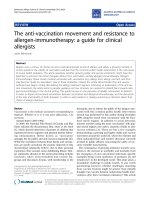
participate in the study, irrespective of consultation (Fig-
ure 1). Data collection will be in five phases:
Phase 1: Baseline postal Health Survey questionnaire
Phase 2: Baseline Clinical Assessment Study of the
Foot (CASF)
Phase 3: Review of general practice medical records
Phase 4: Follow-up mailed survey at 18 months (Phase
2 participants only)
Phase 5: Follow-up mailed survey at 3 years (Phase 1
and Phase 2 participants)
Phase 1: Baseline postal Health Survey questionnaire
Alladultsaged50yearsandoverregisteredwithfour
local general practices (ma iled population approximately
9000 adults) will be mailed a letter of invitation from
their general pra ctitioner, a Participant Information
Sheet, a Health Survey questionnaire, and a pre-paid
return envelope. The lead general practitioner (GP) at
Roddy et al . Journal of Foot and Ankle Research 2011, 4:22
/>Page 2 of 16
each practice will be invited to identify potentially vulner-
able patients (e.g. dementia, severe or terminal illnesses)
they feel should be excluded from the study. Practice lists
will be screened prior to mailing to ensure that addresses
are up to date and exclude any recent deaths or depar-
tures from the practice list. Health Survey questionnaires
will be mailed in batches (n = 500) to ensure regular
recruit ment to research clinics (Phase 2) and to limit the
interval between questionna ire completion and clinic
attendance. Pilot cognitive interviews have been underta-
ken with members of the R esearch Centre’ sResearch
User Group to test the Health Survey questionnaire’slay-
out, readability, conten t, language and length. The ques-
tionnaire will be divided into five main sections: (i)
general health (including generic measures of physical
function, psychosocial factors and lifestyle [32-36] (Addi-
tional File 1: Appendix 1)); (ii) specific health problems
including musculoskel etal co-morbidity and pain [37,38];
(iii) questions concerning the presence [39], duration,
location [14], severity [40], and impact [41,42] of foot
pain; (iv) demographic and socioeconomic characteristics
[43,44]; and (v) employment (Table 1). Non-responders
to the questionnaire will be sent a reminder postcard
after two weeks. Those who do not respond to the remin-
der postcard will be sent a repeat questionnaire and Par-
ticipant Information Sheet with a f urther covering letter
four weeks after the initial mailing. Quest ionnaires will
ask for consent (i) to contact participants again by post
and /or (ii) to review medical records. Responders will be
given the option of providing their telephone number for
further contact.
Phase 2: Baseline Clinical Assessment Study of the Foot
(CASF)
Responders to the Health Survey questionnaire who
report experiencing pain in or around the foot within
the last twelve months and who provide written consent
to further contact will be sent a letter of invitation to
attend a research clinic. The letter of invitation will be
accompanied by a Par ticipant Information Sheet provid-
ing details of the study. Participants will be ask ed to tel-
ephonetheResearchCentreiftheyareinterestedin
Data collection
p
oints are in bold
All adults aged 50 years and over registered with 4 general
practices in North Staffordshire
Phase 1: Mailed Health Survey questionnaire
Exclusions
Respondents to Health Survey questionnaire
Non-respondents
Foot pain in the last 12 months
No foot pain in the last 12 months
Phase 2: Clinical Assessment Study of the Foot (CASF)
“Clinic” population
Non-respondents/declined to make an appointment
Did not/unable to attend appointment
Non-consent to participate in CASF
Losses to follow-up
Phase 4: Mailed 18-month Follow-up Survey
Losses to follow-up
Phase 5: Mailed 3-year Follow-up Survey
Phase 3: Medical
record review
including 18-months
prior to clinic
attendance
“Survey”
population
Figure 1 Flowchart of study procedures.
Roddy et al . Journal of Foot and Ankle Research 2011, 4:22
/>Page 3 of 16
Table 1 Content of baseline postal Health Survey questionnaire
Concept Measurement method Detail
Section A: General health
Perceived
general health
MOS SF12 [33] Physical and mental component summary scores
Physical
function
MOS SF36 [32] Physical functioning sub-scale
Anxiety and
depression
Hospital anxiety and depression scale [34] Anxiety and depression sub-scales
Participation Keele Assessment of Participation (KAP) [35,73] 5-items assessing person-perceived, performance-based
participation
Support Emotional support: single question Yes, no, no need
Physical support: single question Yes, no, no need
Life-style Smoking status Current, previous, never
Anthropometric
characteristics
Self-reported height
Self-reported weight
Footwear Toe-box breadth line drawings (Additional File 1: Appendix 1) Type most frequently worn by decade
Heel height line drawings (females only) (Additional File 1:
Appendix 1)
Type most frequently worn by decade
Physical activity Short-Form International Physical Activity Questionnaire (IPAQ)
[36]
Frequency and duration of 4 activities performed during
previous 7 days
Section B: Specific health problems
Hallux valgus Self-completed line drawings [37] 5 line-drawings for each foot depicting increasing severity of
hallux valgus
Co-morbidities Falls, fractures, chest problems, heart problems, deafness,
problem with eyesight, raised blood pressure, diabetes, stroke,
cancer, liver disease, kidney disease, poor circulation, rheumatoid
arthritis
Yes, for any that apply
Intermittent
claudication
Edinburgh Claudication Questionnaire [38] Pain or discomfort in legs when walking, pain characteristics,
pain location (leg manikin)
Bodily pain Self-completed body manikin In the past 4 weeks, have you had pain that has lasted for one
day or longer in any part of your body? If yes, shade pain
location on manikin
Site-specific questions Have you had any problems with your hands or pain in your
hands/hips/knees/feet in the last year?
Section C: Foot pain
Foot pain
characteristics
Side of pain Both, right, left
Duration in past 12 months < 7 days, 1-4 weeks, 1-3 months, 3+ months
Foot injury: Have you ever injured your foot badly enough to
see a doctor about it?
No, right foot only, left foot only, both feet
Foot pain, aching, stiffness in last month [39] No days, few days, some days, most days, all days
Location: self-completed foot manikin [14] In the past month, have you had any ache or pain that has
lasted for one day or longer in your feet? If yes, shade pain
location on foot manikin
Foot pain intensity in last month [40] 0-10 NRS with verbal anchors (no pain, pain as bad as can be)
Complaint-
specific
functioning
Manchester Foot Pain and Disability Index [41] 19-items across four constructs: pain, function, appearance,
work/leisure
Coping
strategies for
foot pain
Foot-related fatigue: single item
Single-item coping strategies questionnaire [42]
None of the time, on some days, on most/every day(s)
0-6 NRS with verbal anchors (never do that, always do that)
Healthcare use Medication use in last month For foot pain, for other pain
Consultation in last 12 months for foot pain General practitioner, physiotherapist, podiatrist, chiropodist
(NHS and private)
Section D: Demographic/socioeconomic characteristics
Demographic
characteristics
Date of birth
Gender
Marital status Married, separated, divorced, widowed, cohabiting, single
Roddy et al . Journal of Foot and Ankle Research 2011, 4:22
/>Page 4 of 16
taking part in order to book an appointment. Non-
responders to this initial invitation letter will be sent a
reminder invitation approximately two weeks later.
Those willing to take part in the study will be
booked into the n ext convenient a ppointment and, if
necessary, travel arrangements (taxi) made. Postal con-
firmation of the appointment will be made by letter
and then by a reminder postcard shortly prior to the
appointment. The postcard will be mailed in an envel-
ope to maintain confidentiality about the n ature of the
appointment. Participants who do not attend the c linic
for their specified appointment will be sent another
letter asking them to re-contact the Research Centre
and book another appointment if they still wish to
participate.
Assessment clinics for the study will be conducted
twice-weekly in a local NHS Trust community rheuma-
tology hospital. A maximum of 12 appointments per
week are scheduled. Each clinic is to be staffed by a
Clinic Co-ordinator, a Clinic Suppo rt Worker, two
trained H ealth Professionals (podiatrist or physiothera-
pist) acting as Research Assessors, one trained Research
Assessor (physiotherapist, radiographer or nurse) acting
as an Ultrasonographer, and two Radiographers.
On ar riving at clinic, participants will be issued with a
file containing all assessment documentation marked
with their unique study number. Prior to commencing
the assessment, the procedures outlined in the Partici-
pant Information Sheet will be discussed with each par-
ticipant. Participants will be given the opportunity to
ask questions. Written informed consent to take part in
the study will be obtained from all participants. Appro-
priate clothing (shorts) for the assessment will be
provided.
Participants will undertake the following standardised
ass essment: digital photography of both feet and ankles;
plain radiography of both feet, ankles and hands; ultra-
sound of the plantar fa scia in both feet; clinical inter-
view; physical assessment o f the feet, lower limb and
hands; simple anthropometric measurement and self-
complete questionnaire (Table 2). Each participant’s visit
is expected to last approximately 2 hours.
Digital photography
Each participant will have three photographs taken by a
Research Assessor us ing a digital camera (Canon Digital
IXUS 75: Resolution 7.1 mega pixels, 3× zoom). Each
foot will be imaged separately with the participant
standing in a specially designed mirror-box that enables
images of the dorsum, medial and lateral aspects of
their foot to be captured in a single photograph. An
additional posterior view photograph of both feet will be
taken with the participant positioned in a self-selected
relaxed bipedal stance on a gym step using a separate
camera (Canon PowerShot A480: Resolution 10.0 mega
pix els 3.3× zoom) mounted on a tripod to the height of
the step. The photograph will be taken at a distance of
40 cm and will capture the heels, ankles and lower limb.
To preserve anonymity partic ipants’ faces will not be
included in any of the photographs: their unique study
number will b e placed in each frame. Permission to use
anonymised digital images for educati onal purposes will
be sought in the written consent form. Digital photogra-
phy w ill t ake approximately 5 minutes to complete for
each participant.
Plain radiography of the feet and hands
Digital radiographs of both feet, ankles and hands will
be obtained for all participants. Weight-bearing dorso-
plantar and later al views of each foot will be obtained
Table 1 Content of baseline postal Health Survey questionnaire (Continued)
Living arrangements Alone, not alone
Socioeconomic
characteristics
Current employment status Employed, not working due to ill-health, retired, unemployed/
seeking work, housewife, other
Current/recent job title Free text
Current/recent job title of spouse Free text
Adequacy of income [43] Find it a strain to get by from week to week, have to be careful
with money, able to manage without much difficulty, quite
comfortably off
Higher education Yes/no (If yes, age finished full-time education)
Ethnicity White UK/European, Afro Caribbean, Chinese, Asian, African,
Other
Section E: Work
Work status Working full-time, part time, or off work due to ill-health
Work performance 0-10 NRS with verbal anchors (worst performance, best
performance)
Work limitation due to a health problem or physical limitation Not affected, changed the way I do the job, reduced the
number of hours, currently off work
Job lock Would like to leave work but can’t due to financial needs
MOS SF 12 = Medical Outcomes Study Short Form 12; MOS SF 36 = Medical Outcomes Study Short F orm 36; NRS = numerical rating scale
Roddy et al . Journal of Foot and Ankle Research 2011, 4:22
/>Page 5 of 16
Table 2 Content of clinical assessment: clinical interview, physical examination and self-complete questionnaire
Concept Measurement method Detail
Clinical Interview
Pre-assessment
screening:
Screen for
clinical “red
flags”
Recent significant foot or hand injury
Recent sudden change in foot symptoms
Screen for
joint
surgery
History of joint operations
Foot pain
characteristics
Side of pain
Comparative severity of bilateral symptoms
Duration Within 12 months, 1-5 years, 5-10 years, 10+ years (for each foot)
Preceding accident/injury Yes/no
Foot pain/aching/discomfort in last month Yes/no
Foot pain
quality
Short-form McGill Pain Questionnaire [47] 15 sensory and affective descriptors
Sleep
disturbance
Self-report Yes/no
Sensory
disturbance
Self-reported tingling/numbness/pins and needles Yes/no (for each foot)
Causal
attribution
What do you think has caused the problem with your
foot/feet?
Recorded verbatim
Diagnostic
attribution
What do you think is the matter with your foot/feet
now?
Recorded verbatim
Foot surgery Details of any foot surgery Nature of surgery
Right/left
< 1 year, 1- < 5 years, 5- < 10 years, 10+ years ago
Foot/ankle
injury
Details of foot/ankle injury Sprain, fracture, other
Right/left; forefoot, mid-foot, heel, ankle
< 1 year, 1- < 5 years, 5- < 10 years, 10+ years ago
Planned
treatment
Are you waiting for any appointments or treatments for
this foot or ankle problem?
Yes/no (free text comments for yes)
Importance of
health problems
What would you consider to be your two most
important health problems at the moment? [Includes
foot problem]
Recorded verbatim
Physical examination
Screen for
clinical “red
flags”
Acutely, swollen, hot, painful feet or hands Yes/no (free text for comments)
Observation Skin lesions Bunionette, hyperkeratotic lesions, ulcers (plantar and dorsal aspect)
Toe deformity MTPJ and interphalangeal joint hyperextension
Mallet toe, hammer toe, claw toe, retracted toe
Present/absent (great toe)
Present/absent (lesser toes)
Palpation Mid-foot bony exostosis
Plantar fascia tenderness
Present/absent
Present/absent (insertion and mid-arch)
Foot posture Foot Posture Index [50] Six-criterion scoring system
Navicular Height [49] Millimetres
Foot Length [51] Millimetres
Arch index [48,49] Weightbearing footprint. Length of footprint excluding toes is divided
into equal thirds. Arch index = area of middle third divided by area of
entire footprint
Range of
movement
(foot/ankle)
Ankle dorsiflexion (with knee flexed and extended) [53] Degrees
Subtalar inversion [52] Degrees
Subtalar eversion [52] Degrees
1
st
MTP joint dorsiflexion [54] Degrees
Roddy et al . Journal of Foot and Ankle Research 2011, 4:22
/>Page 6 of 16
Table 2 Content of clinical assessment: clinical intervi ew, physical examination and self-complete questionnaire
(Continued)
Knee valgus/
varus deformity
Intercondylar distance Centimetres
Intermalleolar distance Centimetres
Anthropometric
measurements
Height Metres
Weight Kilograms
Lower limb
physical
function
Short physical performance battery (SPPB) [57] Standing balance test, timed repeated chair stand test, 4-metre gait
speed test
Hand
osteoarthritis
Deformity, enlargement, swelling, nodes [55] Observation and palpation: swelling (MCPJ), nodes (PIPJ and DIPJ),
deformity and enlargement (1
st
CMCJ, PIPJ and DIPJ)
Hand function Power grip strength (Jamar dynanometer) [56] Pounds
Pinch grip strength (B&L pinch gauge) [56] Pounds
Self-complete questionnaire
Section A: Foot
Pain
Foot pain
chronicity
Chronic Pain Grade [59] 6 questions (0-10 NRS) and 1 question (4 response options) giving
grade I-IV
Complaint-
specific
functioning
Symptom satisfaction [64] 5-point Likert scale (Very dissatisfied to Very satisfied)
Section B: Hand pain and problems
Hand pain
characteristics
Hand pain in last 12 months
Side of pain
Duration in past 12 months
Hand pain, aching, stiffness in last month [55]
Present/absent
< 7 days, 1-4 weeks, 1-3 months, 3+ months
No days, few days, some days, most days, all days
Hand pain intensity in last month [40] 0-10 NRS with verbal anchors (no pain, pain as bad as could be)
Location: self-completed hand manikin [60]
AUSCAN [65,66]
In the past month, have you had any ache or pain that has lasted for
one day or longer in your hand? If yes, shade location on hand manikin
Pain and stiffness sub-scales
Complaint-
specific
functioning
AUSCAN [65,66] Physical function sub-scale
Hand
dominance
Self-report Right, left, both
Healthcare use GP consultation within last 12 months for hand
problem
Section C: Hip
pain
Hip pain
characteristics
Side of pain Both, right, left
Duration in past 12 months < 7 days, 1-4 weeks, 1-3 months, 3+ months
Hip pain, aching, stiffness in last month [61] No days, few days, some days, most days, all days.
WOMAC (hip) [62] Pain and stiffness sub-scales
Complaint-
specific
functioning
WOMAC (hip) [62] Physical function sub-scale
Healthcare use GP consultation within last 12 months for hip pain
Section D: Knee
pain
Knee pain
characteristics
Side of pain Both, right, left
Duration in past 12 months < 7 days, 1-4 weeks, 1-3 months, 3+ months
Knee pain, aching, stiffness in last month [63] No days, few days, some days, most days, all days.
WOMAC (knee) [62] Pain and stiffness sub-scales
Roddy et al . Journal of Foot and Ankle Research 2011, 4:22
/>Page 7 of 16
according to a defined protocol [24] and stored on di sc.
The participa nt will stand in a relaxed position with the
weight of the participant’s body distributed equally. A
relaxed position will be achieved by asking the partici-
pant to walk on the spot for a few steps and then stand
relaxed. For the dorso-plantar view the participant will
stand with the plantar aspect of both feet on the detec-
tor. The x-ray tube will be angled 15° cranially with a
vertical central ray centred at the base of the third
metatarsal [24]. For lateral projections the participant
will stand on a low platform with the detector posi-
tioned at the side of the participant’ s foot. The x-ray
tube will be angled at 90° with a horizontal central ray
centred on the base on the base of the first metatar sal
[24]. Weight-bearing antero-posterior views of both
ankle joints will also be obtained with the participant
standing on the low platform. The detector will be posi-
tioned behind the participant. The x-ray tube will be
angled 90° with a horizontal central ray centred midway
between the malleoli [45]. Dorso-palmar views of both
hands are to be performed. The palmar aspect of the
hand will be placed on the detector with the fingers
extended, separated slightly and spaced evenly [31]. A
vertical central ray will be centred on the head of the
third metacarpal [45]. Each foot, ankle and hand will be
imaged separately and the film focus distance will be set
at 110 cm f or all projections. X-rays will take approxi-
mately 20 minutes to complete for each participant.
Ultrasound of the plantar fascia
The ultrasound examination will be performed using a
variable frequency 8-13 MHz linear transducer with a
Logiq-e ultrasound system (GE Healthcare). The partici-
pant will be positioned in a self-selected half-lying posi-
tion, or sitting position if the half-lying position cannot
be assumed by the participant, on a couch with their
feet hanging over the end of the couch and ankles dorsi-
flexed to 90 degrees. Real-time sagittal (longitudinal)
imaging of t he plantar apo neurosis will be performed
with the f ocus adjusted to the depth o f the fascia for
each participant. Plantar fa scia thickness will be mea-
sured at a standard reference point where the plantar
fascia crosses the anterior aspect of the inferior border
of the calcaneus on the longitudinal view but at its
thickest point in the transverse plane [46]. Three
measurements will be taken and recorded on a paper
proforma. The Research Assessor performing the ultra-
sound w ill be blind t o the results of the clinical assess-
ment. The scan will take approximately 10 minutes for
each participant.
Ultrasound images will be retained and digitally stored
at the Research Centre for quality control purposes.
Consent will be sought in the clinic consent process for
the use of anonymised images for educational purposes
and in presentations.
Clinical interview and physical examination
Participants will be interviewed and examined by a
trained Research A ssessor who will be blind to the
radiographic and sonographic findings. This procedure
will comprise three components. Firstly, a standardised
clinical interview w ill be conducted to gather quantita-
tive data relating to foot pain and symptoms in older
adults [47], causal and diagnostic attribution, previous
injury or su rgery, and planned treatment (Table 2). Sec-
ondly, a detailed, standardised, examination of both feet
will be conducted. This will include assessment of skin
lesions; common deformities; foot posture including sta-
tic arch index [48,49], Foot Posture Index [50], foot
length [51], navicular height [49,51]; and range of move-
ment of subtalar inversion and eversion [52], ankle dor-
siflexion [53], and 1
st
MTPJ dorsiflexion [54] (Table 2).
Thirdly, a brief standardised physical examination of
both hands, and both knees will be conducted (Table 2).
This will include assessment of presence of deformity,
enlargement, swelling and nodes in both hands [55];
maximal power and pinch grip strength using a Jamar
dynamometer and B&L pinch gauge respectively [56];
and presence of varus and valgus deformities at the
both knees. Lower extremity physical performance will
be also assessed [57].
Plantar pressures from both feet will be recorded dur-
ing level bar efoot walking using a pressure platform (RS
Scan
®
International, Olen, Bel gium). This syste m con-
sists of a 12 mm thick floor mat (578 mm × 418 mm)
incorporating 4096 resistive sensors sampling at a rate
of 300 Hz. The two-step gait initiation protocol will be
used whereby the participant is positioned two step
lengthsfromthefrontedgeofthepressureplatform
and is instructed to walk in a normal manner, striking
Table 2 Content of clinical assessment: clinical intervi ew, physical examination and self-complete questionnaire
(Continued)
Complaint-
specific
functioning
WOMAC (knee) [62] Physical function sub-scale
Healthcare use GP consultation within last 12 months for knee pain
AUSCAN = Australian Canadian Osteoarthritis Hand Index; CMCJ = carpometacarpal joint; DIPJ = distal interphalangeal joint; GP = General practice; MTPJ =
metatarsophalangeal joint; MCPJ = metacarpophalangeal joint; NRS = numeric rating scale; PIPJ = proximal interphalangeal joint; WOMAC = Western Ontario and
McMaster Universities Osteoarthritis Index
Roddy et al . Journal of Foot and Ankle Research 2011, 4:22
/>Page 8 of 16
the sensor area with the second step [58]. The system
will be calibrated at the beginning of each session and
recalibrated for participants’ individual weight and shoe
size prior to each assess ment. The participant will com-
plete several practice trials, to allow them to familiarise
themselves with the two step approach and calculate
their starting position. Three trials will be recorded for
each foot. Maximum force (N), peak pressure (N/cm
2
)
and contact time (ms) will be collected. Footprints
obtained will be divided into masks corresponding to
the major structural regions of the foot.
Pre-defined protocols for all components of the inter-
view and assessment will be used for standardisation
between Research Assessors. Assessment findings will
be recorded on a standard form that is to be checked
for missing data immediately post-assessment by the
Clinic Co-o rdinator or Clinic Support Worker. Discus-
sion between Research Assessors and participants about
diagnosis and/or appropriate management will be dis-
couraged. Participa nts will be advised to discuss clinical
queries with their General Practitioner. The interview
and assessment will take approximately 40 minutes to
complete for each participant.
Simple anthropometric measurements
Weight (in kg) and height (in cm) of each participant
will be measured using calibrated digital scales (Seca
Ltd., Birmingham, UK) a nd a wall-mounted measure
(Seca Ltd., Birmingham, UK) respectively.
Self-complete clinic questionnaire
During the clinic visit, participants will complete a self-
complete questionnaire. The q uestionnaire will be
divided into four main sections: (A) Foot pain; (B) Hand
pain and problems; (C) Hip pain; and (D) Knee pain.
Questions w ill relate to pain [40,55,59-63], site-specific
function [62,64-66 ], and GP consultation (Table 2). Sec-
tion A will be completed by all clinic-attenders. Sections
B, C, and D will be completed o nly by those who
reported hand, hip or knee pain respectively in their
Health Survey questionnaire. The Clinic Co-ordinator or
Clinic Supp ort Worker will guide participant s as to
which sections need to be completed and will check all
questionnaires following completion for any missing
data. The questionnaire will take approximately 30 min-
utes to complete.
Travelling and out-of -pocket expenses w ill be reim-
bursed after the assessment.
Post-clinic procedure
The digital cameras, study laptop and all completed
clinical assessment documentation and questionna ires
will be returned to the Research Centre. Digital images
will be downloaded from the memory cards and l aptop
onto a secure server.
A clinical report on the x-ray images will be provided
by a Consultant Radiolo gist at the NHS T rust Hospital.
The images and report will be forwarded to the
Research Centre where they will be screened by a Con-
sultant Rheumatologist for any radiographic “ red flags”
or significant radiographic abnormality (see below).
Standardised coding of radiographic features on the
foot and h and x-ray images will be carried out by the
Research Radiographer (a trained observer with a back-
ground in diagnostic radiography). The Research Radio-
grapher will be blinded to all assessment data and the
radiologist’s report. Foot images will be scored for indi-
vidual radiographic features, including osteophytes and
joint space width, at the 1
st
MTPJ, 1
st
and 2
nd
CMJs,
NCJ and TNJ according to the Menz atlas and classifica-
tion system [24]. With the exception of the TNJ, both
dorso-plantar and lateral projections will be used to
assess osteophyte and joint space width. For the grading
of TNJ osteophytes, only the lateral projection will be
used as the dorsal aspect of the joi nt, where osteophytes
most commonly develop, is not easily visualised on the
dorso-plantar projection. Standardised coding of radio-
graphic features using the Kellgren and Lawrence grad-
ing system will be completed for the ankle joints and
sixteen joints in each hand and wrist [67]: the distal
interphalangeal joints (DIP), the proximal interphalan-
geal joints (PIP), the interphalangeal joint of the thumb
(IP), the metacarpophalangeal joints (MCP), the thumb
carpometacarpal joint (CMC) and the trapezioscaphoid
joint (TS).
Consent forms, assessment documentation, digital x-
ray images and reports are to be placed in separate
secure storage.
Communication with participants’ general practice
Assessment findings will be communicated to partici-
pants and their General Practice only in specific circum-
stances that will be explained to participants at the start
of the clinic:
Mandatory notification of clinical ‘red flags’
All participants will be routinely screened during the
clinical assessment for signs and symptoms suggesting
potentially serious pathology requiring urgent medical
attention (Table 2). These are: recent trauma to the feet
or hands that may have resulted in significant tissue
damage; recent sudden worsening of foot or hand symp-
toms; and acutely hot, swollen, painful feet or hands
[68]. In the event of such findings, participants will be
informed that they require urgent attention, a standard
fax will be imme diately sent t o the Gen eral Practice,
and appropriate medical attention arranged the same
day. A letter of confirmation will be subsequently sent
to the participants’ General Practice.
Mandatory notification of radiographic ‘red flags’
In the event of any radiographic red flags (including sus-
pected malignancy, unresolved fracture, infection)
reported by the Consultant Radiologist a standard fax
Roddy et al . Journal of Foot and Ankle Research 2011, 4:22
/>Page 9 of 16
willbesentwithacopyofthex-rayreporttotheGen-
eral Practice notifying them of this. This will subse-
quently be confirmed by letter.
Discretionary notification of other significant radiographic
abnormality
At the discretion of the Consultant Rheumatologist, the
General Practice will be notified of other significant
radiographic abnormality (e.g. previ ous fracture, inflam-
matory arthropathy).
Availability of x-ray report on request
To prevent unnecessary duplication of x-rays, partici-
pants’ GPs can request an x-ray report if they feel it
would be valuable for clinical management.
Quality assurance and control
Quality assurance and control are important for the
integrity of longitudinal studies and the validity of their
conclusions [69]. This is especially true of observer-
dependent methods of data-gathering. In the clinical
assessment phase of the study, the clinical interview and
physical assessment, ultrasound, digital images, plantar
pressure and the taking and scoring of x-ray will be sub-
ject to a number of quality control procedures.
Inter- and intra-assessor reliability of foot interview
and examination variables have been established,
where possible, from the published literature
[49-54,70]. Assessors will undergo training in consent
procedures, clinical interview and physical assessment
techniques. All Research Assessors will be required to
conduct at least two clinical assessments prior to the
commencement of data collection. During the first
month clinics with reduced numbers of participants
will be held to allow all study procedures to be tested
and reviewed. All radiographers participating in the
study will also receive training prior to t he commence-
ment of the study.
Selected Research Assessors will receive ultrasound
training on a formally assessed course, Focused Specia-
list Ultrasound Practice, run by University of Derby
(UK). This course consists of the principles of ultra-
sound physics and imaging science. The Research Asses-
sors will then receive specific clinical training from a
Consultant Musculoskeletal Sono grapher to assess the
plantar fascia thickness. In addition to meeting the
course assessmen t requirements clinical competen ce for
the study will be assessed by the Consultant Sonogra-
pher following a period of supervision and mentorship.
The Research Radiographer will be trained in the
methods for scoring the plain radiographs. This single
observer will score all images and intra-observer varia-
bility will be assessed using 60 sets of images scored
eight weeks apart. Inter-observer variability will be
assessed using a second observer with prior experience
of grading foot x-rays for OA who will also grade 60
sets of images.
A detailed Assessor Manual with pro tocols for obtain-
ing written informed consent, d igital photography, clini-
cal interview and physical assessment, admini stration of
the self-complete questionnaire, anthropometric mea-
surement, plain radiography, and ultrasound will be pro-
vided to all members of the study team for reference
during the entire study period.
During the data collection period, digital photographs
for all participants will be reviewed and participants
with any missing or spoilt images will be recalled to
repeat the photographs. Quality control sessions for
consent procedures, clinical interview and p hysical
assessment, radiography and ultrasound will be under ta-
ken at regular intervals throughout the study. These ses-
sions will include observation of assess ments in clinic by
the Principal Investigator, structured observation of
assessments in a healthy volunteer, and direct inter-
ass essor compari sons on selected participants. Observa-
tion of radiography and ultrasound will be undertaken
by the Research Radiographer and Consultant Muscu-
loskeletal Sonographer respectively. The outcome of
each quality control session will be fed b ack to the indi-
vidual Research Assessor and the group as a whole.
Phase 3: Review of general practice medical records
All participants in Phase 1 who give permission for their
GP records to be accessed will have their computerised
medical records tagged by a member of the Research
Centre’s Health Informatics Specialist team. All consul-
tations for the 18-month period prior to clinic atten-
dance, and for the three-year period following clinic
attendance, will be identified. The four practices partici-
pating in this study are fully computerised and undergo
annual audits completed by the Health Informatics team
to assess the quali ty and completeness of the data entry
at the practices [71].
This data will cover c onsult ations, prescriptions, and
referrals. All relevant foot-related consultations will be
identified using search techniques based on Read codes
and free text entries, which have been previously devel-
oped and successfully applied by the Research Centre
[28,72]. Participants with a relevant recorded consulta-
tion will be classified into those receiving an OA diag-
nosis recorded by their GP and those receiving non-
specific symptom codes (e.g. arthralgia). In addition, all
comorbid consultations will be identified and sub-
grouped by Read code chapter.
Patterns of primary and secondary health care utilisa-
tion will be compared between P hase 2 participants and
Phase 1 participants who did not attend the research
clinic. All sensitive data (name, contact details) will be
removed from the medical records data and the consul-
tation data will be linked to the survey and clinical
assessment data by unique survey identifier.
Roddy et al . Journal of Foot and Ankle Research 2011, 4:22
/>Page 10 of 16
Phase 4: Follow-up mailed survey at 18 months (Phase 2
respondents only)
Follow-up surv eys will be mailed to all Phase 2 partici-
pants 18 months after their baseline clinical assessment.
The focus of follow-up will be clinical (severity of pain
and functional limitation) change and possible determi-
nants of this. The content of this survey is provided in
Table 3. Non-responders to the questionnaire will be
sent a reminder postcard after two weeks. Those who
do not respond to the reminder postcard will be sent a
repeat questionnaire and Participant Information Sheet
with a further covering letter four weeks after the initial
mailing. Primary outcome data will be sought from non-
respondents by telephone interview or shortened postal
questionnaire. We plan to trace participants who have
moved practice during t he follow-up period usi ng the
NHS tracing service.
Phase 5: Follow-up mailed survey at 3 years (Phase 1 and
Phase 2 respondents)
Follow-up surveys will be mailed to all Phase 1 and 2
participants 3 years after their baseline Health Survey
questionnaire. In addition to information about clinical
change in Phase 2 participants, the survey will also
include repeat measures of lifestyle [36,73], general
health (including generic measures of physical function
[32,33]), psychosocial factors [34], co-morbidity [37,38]
and b asic screening questions concerning the presence
[39], duration, location [14], severity [ 40], and impact of
foot pain [41,42] (Table 4). Non-responders to the ques-
tionnaire will be sent a reminder postcard after two
weeks. Those who do not respond to the reminder post-
card will be sent a repeat questionnaire and Participant
Information Sheet with a further covering letter four
weeks after the initial mailing. Primary outcome data
Table 3 Content of 18-month postal follow-up Health Survey questionnaire (Phase 2 participants only)
Concept Measurement method Detail
Foot pain
characteristics
Change in foot pain over past 18 months Completely recovered, much better, better, no change, worse, much
worse
Since your assessment 18 months ago, have you ever
injured your foot badly enough to see a doctor about
it?
No/right only/left only/both
Foot pain, aching, stiffness in last month [39] No days, few days, some days, most days, all days
Foot pain intensity in past month [40] 0-10 NRS with verbal anchors (no pain, pain as bad as could be)
Foot pain
chronicity
Chronic Pain Grade [59] 6 questions (0-10 NRS) and 1 question (4 response options) giving grade
I-IV
Complaint-
specific
functioning
Manchester Foot Pain and Disability Index [41] 19-items across four constructs: pain, function, appearance, work/leisure
Symptom satisfaction [64] 5-point Likert scale (Very dissatisfied to Very satisfied)
Healthcare use Use of services/treatments for foot pain in past 18
months
GP, physiotherapist, hospital specialist, acupuncture, podiatrist,
chiropodist, drugs on prescription, foot injection, foot surgery, osteopath/
chiropractor, other (specify)
Medication use in last month For foot pain, for other pain
Coping
strategies for
foot pain
Single-item coping strategies questionnaire [42] 0-6 NRS with verbal anchors (never do that, always do that)
Perceived
general health
MOS SF 12 [33] Physical and mental component summary scores
Physical
function
MOS SF 36 [32] Physical functioning sub-scale
Anxiety and
depression
Hospital anxiety and depression scale [34] Anxiety and depression sub-scales
Hallux valgus Self-completed line drawings [37] 5 line-drawings for each foot depicting increasing severity of hallux
valgus
Bodily pain Self-completed body manikin In the past 4 weeks, have you had pain that has lasted for one day or
longer in any part of your body? If yes, shade location of pain on
manikin
Regional pain Site-specific questions Have you had any problems with your hands or pain in your hands/hips/
knees in the last year?
Demographic
characteristics
Date of birth
Gender
Socioeconomic
characteristics
Current employment status Employed, not working due to ill-health, retired, unemployed/seeking
work, housewife, other
MOS SF 12 = Medical Outcomes Study Short Form 12; MOS SF 36 = Medical Outcomes Study Short F orm 36; NRS = numerical rating scale
Roddy et al . Journal of Foot and Ankle Research 2011, 4:22
/>Page 11 of 16
Table 4 Content of 3-year postal follow-up Health Survey questionnaire (Phase 1 and Phase 2 participants)
Concept Measurement method Detail
Section A: General health
Perceived
general health
MOS SF 12 [33] Physical and mental component summary scores
Physical
function
MOS SF 36 [32] Physical functioning sub-scale
Anxiety and
depression
Hospital anxiety and depression scale [34] Anxiety and depression sub-scales
Participation Keele Assessment of Participation (KAP) [35,73] 5-items assessing person-perceived, performance-based
participation
Physical activity Short-Form International Physical Activity Questionnaire (IPAQ)
[36]
Frequency and duration of 4 activities performed during
previous 7 days
Section B: Specific health problems
Hallux valgus Self-completed line drawings [37] 5 line-drawings for each foot depicting increasing severity of
hallux valgus
Co-morbidities Falls, fractures, chest problems, heart problems, deafness,
problems with eyesight, raised blood pressure, diabetes, stroke,
cancer, liver disease, kidney disease, poor circulation,
rheumatoid arthritis
Yes for any that apply
Intermittent
claudication
Edinburgh Claudication Questionnaire [38] Pain or discomfort in legs when walking, pain characteristics,
pain location (leg manikin)
Bodily pain Self-completed body manikin In the past 4 weeks, have you had pain that has lasted for one
day or longer in any part of your body? If yes, shade location of
pain on manikin
Site-specific questions Have you had any problems with your hands or pain in your
hands/hips/knees/feet in the last year?
Section C: Foot pain
Foot pain
characteristics
Side of pain Both, right, left
Duration in past 12 months < 7 days, 1-4 weeks, 1-3 months, 3+ months
Have you ever injured your foot badly enough to see a doctor
about it?
No/right foot only/left foot only/both feet
Location: self-completed foot manikin [14] In the past month, have you had any ache or pain that has
lasted for one day or longer in your feet? If yes, shade location
of pain on foot manikin
Foot pain, aching, stiffness in last month [39] No days, few days, some days, most days, all days
Foot pain intensity in last month [40] 0-10 NRS with verbal anchors (no pain, pain as bad as could be)
Complaint-
specific
functioning
Manchester Foot Pain and Disability Index [41] 19-items across four constructs: pain, function, appearance,
work/leisure
Coping
strategies for
foot pain
Single-item coping strategies questionnaire [42] 0-6 NRS with verbal anchors (never do that, always do that)
Healthcare use Medication use in last month For foot pain, for other pain
Consultation with general practitioner in last 12 months for foot
pain
Section D: Demographic/socioeconomic characteristics
Demographic
characteristics
Date of birth
Gender
Marital status Married, separated, divorced, widowed, cohabiting, single
Living arrangements Alone, not alone
Anthropometric
characteristics
Self-reported height
Self-reported weight
Socioeconomic
characteristics
Current employment status Employed, not working due to ill-health, retired, unemployed/
seeking work, housewife, other
Roddy et al . Journal of Foot and Ankle Research 2011, 4:22
/>Page 12 of 16
will be sought from non-respondents by telephone inter-
view (Phase 2 participants only) or shortened postal
questionnaire. We plan to trace participants who have
moved practice during t he follow-up period usi ng the
NHS tracing service.
Sample size
The sample size for this study was determined by the
estimated numbers of parti cipants needed in Phase 2 in
order to ensure sufficient power for both cross-sectional
and longitudinal analyses. The primary aim is to com-
pare the proportion of participants with poor functional
outcome across the radiographic (p
2
)andnoradio-
graphic OA groups (p
1
)at3years.Assumingp
1
=20%
intheunexposedgroup,asamplesizeof426willhave
80% power to detect a relative risk (p
2
/p
1
)of1.62using
a 5% significance level. Allowing for a drop-out figure of
80 from baseline to three years w ill require an initial
recruitment of 506 participants to Phase 2.
Statistical analysis
Patterns of symptomatic radiographic foot OA
The frequency and co-occurrence of radiographic fea-
tures of symptomatic OA at the 1
st
MTPJ, the 1
st
and
2
nd
CMJs, the NCJ and the TNJ will be described using
simple descriptive statistics.
Features associated with foot OA phenotypes
Linking data collected at the clinical assessment with
that from the b aseline health survey questionnaire, the
occurrence of radiographic OA will be related cross-sec-
tionally to foot pain and disability, foot deformities, soft-
tissue problems, footwear characteristics and pain/OA at
other sites, using odds ratios and associated 95% confi-
dence intervals adjusted for age, gender and BMI. The
effect of missing primary outcome data will be investi-
gated using multiple imputation methods.
Outcome of foot OA at 3 years
Linking baseline date to 18-mon th and three-year fol-
low-up questionnaires, we will then be able to determine
prospectively the factors that are related to clinical dete-
rioration using risk ratios and associated 95% confidence
intervals, for example, radiographic OA, footw ear char-
acteristics, pain/OA at other sites, psychosocial factors.
The presentation and diagnosis of OA in primary care
Participants with a recorded consultation for joint-
related problems will be classified into those receiving
an OA diagnosis recorded by their GP and those receiv-
ing a non-specific symptom code (e.g. arthralgia). The
proportion of participants who (a) consult and (b) are
diagnosed with OA will be described using simple
descriptive statistics. Logistic regression will be used to
identify which f eatures, including OA phenotype, are
strongly associated with consult ation and foot OA-diag-
nosis. The effect of missing primary outcome data will
be investigated using multiple imputation methods.
Describing self-care and primary care
Annual consultation rates and cumulative consultation
probabilities will be calculated over the three-year per-
iod. Using logistic regression and survival analysis tech-
niques, we will investigate further how different
phenotypes relate to subsequent patterns of primary
care consultation (for joint pain, other morbidities, and
specifically for OA) and referral to secondar y care. Self-
care reported by participants in the surveys at ea ch 18
month time-point will be described.
Modelling the outcomes of care
We will model the effect s of care on impairment, activ-
ity limitation and participation restriction. We aim to
use propensity scores (ie the propensity or likelihood of
a person to seek healthcare given their characteristics)
and random effects repeated measures multilevel models
in order to take into account the effects of both
Table 4 Content of 3-year postal follow-up Health Survey questionnaire (Phase 1 and Phase 2 participants) (Continued)
Current/recent job title Free text
Current/recent job title of spouse Free text
Adequacy of income [43] Find it a strain to get by from week to week, have to be careful
with money, able to manage without much difficulty, quite
comfortably off
Additional questions for phase 2 participants only
Foot pain
characteristics
Change in foot pain over past 3 years Completely recovered, much better, better, no change, worse,
much worse
Since your assessment 3 years ago, have you injured your foot
badly enough to see a doctor about it?
No/right foot only/left foot only/both feet
Foot pain
severity
Chronic Pain Grade [59] 6 questions (0-10 NRS) and 1 question (4 response options)
giving grade I-IV
Symptom satisfaction [64] 5-point Likert scale (Very dissatisfied to Very satisfied)
Healthcare use Use of services/treatments for foot pain in past 3 years GP, physiotherapist, hospital specialist, acupuncture, podiatrist,
chiropodist, drugs on prescription, foot injection, foot surgery,
osteopath/chiropractor, other (specify)
MOS SF 12 = Medical Outcomes Study Short Form 12; MOS SF 36 = Medical Outcomes Study Short F orm 36; NRS = numerical rating scale
Roddy et al . Journal of Foot and Ankle Research 2011, 4:22
/>Page 13 of 16
observed and unobserved covariates on outcome at each
follow-up time point. Using consultations and secondary
care referr als from medical record review, together with
socio-demographic, clinical, general health and pheno-
type characteristics, we will apply these methods to a
series of analyses in which the separate effects of each
of the components of consultation on subsequent out-
comes at each follow-up time point will be modelled.
Discussion
Symptomatic foot OA is a common problem, yet is
under-researched relative to other sites commonly
affected by OA, such as the knee and hand [6]. In this
three-year prospective e pidemiological study, we will
combine survey data, clinical, radiographic, and ultra-
sound assessment, and primary care consultation
records to describe the frequency and co-occurrence of
OA at frequently affected joints of the foot, and to relate
their occurrence to symptoms, function, clinical exami-
nation findings and life-style factors such as footwear.
We will also describe the natural history of clinical
symptoms relating to foot OA and assess how these pre-
sent to primary care and are subsequently diagnosed
and treated.
This study will focus on OA of the foot yet, in reality,
people with OA are commonly affected at multiple joint
sites [23,74]: a quarter of patients awaiting knee and hip
replacement surgery have generalised radiographic OA
[75]. Pain and functional impairment have been shown
to be greater as the number of painful joint sites
increases [11,16,76]. This study has been specifically
designed to complement previous clinical assessment
studies of the knee [30] and hand [31], which will per-
mit data to be combined across all three cohorts, allow-
ing more detailed investigation of patterns of multiple
joint involvement, and the comparative and additive
effects of pain and OA on symptoms and outcome.
An obvious limitation of our study is that asympto-
matic people will not be invited for to attend for clinical
assessment, so we will not be able to estimate the fre-
quency of asymptomatic radiographic OA or clinical
examination findings. However, symptoms ar e the pre-
senting feature to primary care and, as with our pre-
vious clinical assessment studies [30,31], are the starting
point in t his study. This enables us to investigate the
occurrence of foot osteoarthritis and inter-relationship
between clinical signs, symptoms, and radiographic dis-
ease within symptomatic individuals and their clinical
course over time.
In phase two of this study, every effort will be made to
maintain the quality of t he data obtained and minimise
info rmation bias in the data that will be collected at the
research clinics. Standardi sed interview questions and
physical assessment protocols have been developed and
are described in detail in an Ass essor Manual which will
be given to each Research Assessor. Research Assessors
will undergo a period of training prior to the start of
the study. Quality control will be reviewed at regular
intervals throughout the course of the study to ensure
continued adherence to the protocols.
Additional material
Additional file 1: Footwear questionnaire.
Acknowledgements
This work is supported by an Arthritis Research UK Programme Grant (18174)
and service support through the West Midlands North CLRN. The study
funders had no role in the study design; data collection, analysis, or
interpretation; in the writing of the paper; or in the decision to submit the
paper for publication. HBM is currently a National Health and Medical
Research Council of Australia fellow (Clinical Career Development Award, ID:
433049).
The authors would like to thank the administrative, health informatics and
research nurse teams at the Arthritis Research UK Primary Care Centre
particularly Alicia Bratt, Shirley Caldwell, Claire Calverle y, Charlotte Clements,
Kathryn Dwyer, Ian Thomas and Chan Vohora; staff of the participating
general practices and Haywood Hospital, especially Dr Jackie Saklatvala,
Carole Jackson and the Radiographers at the Department of Radiography;
Alison Hall who led the training and mentoring of ultrasound assessors;
Robert Bradshaw-Hilditch, Dr Catherine Colquhoun, Mr Rob Rees, Dr Michael
Shadforth, Dr Simon Somerville, Julie Taylor and Professor Jim Woodburn
who contributed to the development of the clinic assessment schedule; the
team undertaking clinical assessments, Linda Hargreaves, Gillian Levey, Liz
Mason, Jennifer Pearson, Julie Taylor, and Dr Laurence Wood; and Ian
Steward and RSscan Lab Ltd for the loan of the Foot scan system.
Author details
1
Arthritis Research UK Primary Care Centre, Primary Care Sciences, Keele
University, Staffordshire, ST5 5BG, UK.
2
Musculoskeletal Research Centre,
Faculty of Health Sciences, La Trobe University, Bundoora, Victoria 3086,
Australia.
Authors’ contributions
All authors participated in the conception and design of the study, and
drafting of the manuscript. All authors read and approved the final
manuscript.
Competing interests
HBM is Editor-in-Chief of Journal of Foot and Ankle Research. It is journal
policy that editors are removed from the peer review and editorial decision
making processes for papers they have co-authored. All other authors
declare that they have no competing interests.
Received: 4 April 2011 Accepted: 5 September 2011
Published: 5 September 2011
References
1. World Health Organisation: The burden of musculoskeletal conditions at
the start of the new millenium. Report of a WHO Scientific Group. WHO
Technical Report Series No. 919 2003.
2. Mathers CD, Loncar D: Projections of global mortality and burden of
disease from 2002 to 2030. PLoS Med 2006, 3(11):e442.
3. Jordan K, Clarke AM, Symmons DP, Fleming D, Porcheret M, Kadam UT,
Croft P: Measuring disease prevalence: a comparison of musculoskeletal
disease using four general practice consultation databases. Br J Gen Pract
2007, 57:7-14.
4. Woolf AD, Pfleger B: Burden of major musculoskeletal conditions. Bull
World Health Organ 2003, 81:646-656.
Roddy et al . Journal of Foot and Ankle Research 2011, 4:22
/>Page 14 of 16
5. European Bone and Joint Health Strategies Project: European Action
Towards Better Musculoskeletal Health. 2004.
6. Trivedi B, Marshall M, Belcher J, Roddy E: A systematic review of
radiographic definitions of foot osteoarthritis in population-based
studies. Osteoarthritis Cartilage 2010, 18:1027-1035.
7. Benvenuti F, Ferrucci L, Guralnik JM, Gangemi S, Baroni A: Foot pain and
disability in older persons: an epidemiologic survey. J Am Geriatr Soc
1995, 43:479-484.
8. Leveille SG, Guralnik JM, Ferrucci L, Hirsch R, Simonsick E, Hochberg MC:
Foot pain and disability in older women. Am J Epidemiol 1998,
148:657-665.
9. Menz HB, Lord SR: The contribution of foot problems to mobility
impairment and falls in community-dwelling older people. J Am Geriatr
Soc 2001, 49:1651-1656.
10. Badlissi F, Dunn JE, Link CL, Keysor JJ, McKinlay JB, Felson DT: Foot
musculoskeletal disorders, pain, and foot-related functional limitation in
older persons. J Am Geriatr Soc 2005, 53:1029-1033.
11. Keenan AM, Tennant A, Fear J, Emery P, Conaghan PG: Impact of multiple
joint problems on daily living tasks in people in the community over
age fifty-five. Arthritis Rheum 2006, 55:757-764.
12. Roddy E, Zhang W, Doherty M: Prevalence and associations of hallux
valgus in a primary care population. Arthritis Rheum 2008, 59:857-862.
13. Chen J, Devine A, Dick IM, Dhaliwal SS, Prince RL: Prevalence of lower
extremity pain and its association with functionality and quality of life in
elderly women in Australia. J Rheumatol 2003, 30:2689-2693.
14. Garrow AP, Silman AJ, Macfarlane GJ: The Cheshire Foot Pain and
Disability Survey: a population survey assessing prevalence and
associations. Pain 2004, 110:378-384.
15. Keysor JJ, Dunn JE, Link CL, Badlissi F, Felson DT: Are foot disorders
associated with functional limitation and disability among community-
dwelling older adults? J Aging Health 2005, 17:734-752.
16. Peat G, Thomas E, Wilkie R, Croft P: Multiple joint pain and lower
extremity disability in middle and old age. Disabil Rehabil 2006,
28:1543-1549.
17. Tinetti ME, Speechley M, Ginter SF: Risk factors for falls among elderly
persons living in the community. N Engl J Med 1988, 319:1701-1707.
18. Menz HB, Morris ME, Lord SR: Foot and ankle risk factors for falls in older
people: a prospective study. J Gerontol A Biol Sci Med Sci 2006, 61:866-870.
19. Menz HB, Morris ME, Lord SR: Foot and ankle characteristics associated
with impaired balance and functional ability in older people. J Gerontol A
Biol Sci Med Sci 2005, 60:1546-1552.
20.
Kellgren JH, Moore RA: Generalized osteoarthritis and Heberden’s nodes.
Br Med J 1952, 1:181-187.
21. Lawrence JS, Bremner JM, Bier FA: Osteo-arthrosis. Prevalence in the
population and relationship between symptoms and x-ray changes. Ann
Rheum Dis 1966, 25:1-24.
22. Menz HB, Morris ME: Determinants of disabling foot pain in retirement
village residents. J Am Podiatr Med Assoc 2005, 95:573-579.
23. Wilder FV, Barrett JP, Farina EJ: The association of radiographic foot
osteoarthritis and radiographic osteoarthritis at other sites. Osteoarthritis
Cartilage 2005, 13:211-215.
24. Menz HB, Munteanu SE, Landorf KB, Zammit GV, Cicuttini FM: Radiographic
classification of osteoarthritis in commonly affected joints of the foot.
Osteoarthritis Cartilage 2007, 15:1333-1338.
25. Mantyselka P, Kumpusalo E, Ahonen R, Kumpusalo A, Kauhanen J,
Viinamaki H, Halonen P, Takala J: Pain as a reason to visit the doctor: a
study in Finnish primary health care. Pain 2001, 89:175-180.
26. Jordan K, Jinks C, Croft P: A prospective study of the consulting
behaviour of older people with knee pain. Br J Gen Pract 2006,
56:269-276.
27. Bierma-Zeinstra SM, Lipschart S, Njoo KH, Bernsen R, Verhaar J, Prins A,
Bohnen AM: How do general practitioners manage hip problems in
adults? Scand J Prim Health Care 2000, 18:159-164.
28. Menz HB, Jordan KP, Roddy E, Croft PR: Musculoskeletal foot problems in
primary care: what influences older people to consult? Rheumatology
(Oxford) 2010, 49:2109-2116.
29. Zammit GV, Menz HB, Munteanu SE, Landorf KB, Gilheany MF: Interventions
for treating osteoarthritis of the big toe joint. Cochrane Database Syst Rev
2010, , 9: CD007809.
30. Peat G, Thomas E, Handy J, Wood L, Dziedzic K, Myers H, Wilkie R,
Duncan R, Hay E, Hill J, Croft P: The Knee Clinical Assessment Study - CAS
(K). A prospective study of knee pain and knee osteoarthritis in the
general population. BMC Musculoskelet Disord 2004, 5:4.
31. Myers H, Nicholls E, Handy J, Peat G, Thomas E, Duncan R, Wood L,
Marshall M, Tyson C, Hay E, Dziedzic K: The Clinical Assessment Study of
the Hand (CAS-HA): a prospective study of musculoskeletal hand
problems in the general population. BMC Musculoskelet Disord 2007, 8:85.
32. Ware JE Jr, Sherbourne CD: The MOS 36-item short-form health survey
(SF-36). I. Conceptual framework and item selection. Med Care
1992,
30:473-483.
33.
Ware J Jr, Kosinski M, Keller SD: A 12-Item Short-Form Health Survey:
construction of scales and preliminary tests of reliability and validity.
Med Care 1996, 34:220-233.
34. Zigmond AS, Snaith RP: The hospital anxiety and depression scale. Acta
Psychiatr Scand 1983, 67:361-370.
35. Wilkie R, Peat G, Thomas E, Hooper H, Croft PR: The Keele Assessment of
Participation: a new instrument to measure participation restriction in
population studies. Combined qualitative and quantitative examination
of its psychometric properties. Qual Life Res 2005, 14:1889-1899.
36. Craig CL, Marshall AL, Sjostrom M, Bauman AE, Booth ML, Ainsworth BE,
Pratt M, Ekelund U, Yngve A, Sallis JF, Oja P: International physical activity
questionnaire: 12-country reliability and validity. Med Sci Sports Exerc
2003, 35:1381-1395.
37. Roddy E, Zhang W, Doherty M: Validation of a self-report instrument for
assessment of hallux valgus. Osteoarthritis Cartilage 2007, 15:1008-1012.
38. Leng GC, Fowkes FG: The Edinburgh Claudication Questionnaire: an
improved version of the WHO/Rose Questionnaire for use in
epidemiological surveys. J Clin Epidemiol 1992, 45:1101-1109.
39. Dufour AB, Broe KE, Nguyen US, Gagnon DR, Hillstrom HJ, Walker AH,
Kivell E, Hannan MT: Foot pain: is current or past shoewear a factor?
Arthritis Rheum 2009, 61:1352-1358.
40. Williamson A, Hoggart B: Pain: a review of three commonly used pain
rating scales. J Clin Nurs 2005, 14:798-804.
41. Garrow AP, Papageorgiou AC, Silman AJ, Thomas E, Jayson MI,
Macfarlane GJ: Development and validation of a questionnaire to assess
disabling foot pain. Pain 2000, 85:107-113.
42. Jensen MP, Keefe FJ, Lefebvre JC, Romano JM, Turner JA: One- and two-
item measures of pain beliefs and coping strategies. Pain 2003,
104:453-469.
43. Thomas R: Income - commentary.[ />thomaswealth.htm].
44. Department for Communities and Local Government: The English Indices
of Deprivation. 2007 [ />communities/pdf/733520.pdf], Published: 28/03/2008.
45. Whitley AS, Sloanne C, Hoardley G, Moore AD, Alsop CW: Clark’s positioning
in radiography London: Holder Arnold; 2005.
46. Gibbon WW, Long G: Ultrasound of the plantar aponeurosis (fascia).
Skeletal Radiol 1999, 28:21-26.
47. Melzack R: The McGill Pain Questionnaire: major properties and scoring
methods. Pain
1975, 1:277-299.
48.
Cavanagh PR, Rodgers MM: The arch index: a useful measure from
footprints. J Biomech 1987, 20:547-551.
49. Menz HB, Munteanu SE: Validity of 3 clinical techniques for the
measurement of static foot posture in older people. J Orthop Sports Phys
Ther 2005, 35:479-486.
50. Redmond AC, Crosbie J, Ouvrier RA: Development and validation of a
novel rating system for scoring standing foot posture: the Foot Posture
Index. Clin Biomech (Bristol, Avon) 2006, 21:89-98.
51. Menz HB, Tiedemann A, Kwan MM, Latt MD, Sherrington C, Lord SR:
Reliability of clinical tests of foot and ankle characteristics in older
people. J Am Podiatr Med Assoc 2003, 93:380-387.
52. Menadue C, Raymond J, Kilbreath SL, Refshauge KM, Adams R: Reliability of
two goniometric methods of measuring active inversion and eversion
range of motion at the ankle. BMC Musculoskelet Disord 2006, 7:60.
53. Bennell KL, Talbot RC, Wajswelner H, Techovanich W, Kelly DH, Hall AJ:
Intra-rater and inter-rater reliability of a weight-bearing lunge measure
of ankle dorsiflexion. Aust J Physiother 1998, 44:175-180.
54. Hopson MM, McPoil TG, Cornwall MW: Motion of the first
metatarsophalangeal joint. Reliability and validity of four measurement
techniques. J Am Podiatr Med Assoc 1995, 85:198-204.
55. Altman RD, Alarcón G, Appelrouth D, Bloch D, Borenstein D, Brandt K,
Brown C, Cooke TD, Daniel W, Gray R: The American College of
Roddy et al . Journal of Foot and Ankle Research 2011, 4:22
/>Page 15 of 16
Rheumatology criteria for the classification and reporting of
osteoarthritis of the hand. Arthritis Rheum 1990, 33:1601-1610.
56. Mathiowetz V, Weber K, Volland G, Kashman N: Reliability and validity of
grip and pinch strength evaluations. J Hand Surg [Am] 1984, 9:222-226.
57. Guralnik JM, Simonsick EM, Ferrucci L, Glynn RJ, Berkman LF, Blazer DG,
Scherr PA, Wallace RB: A short physical performance battery assessing
lower extremity function: association with self-reported disability and
prediction of mortality and nursing home admission. J Gerontol 1994, 49:
M85-M94.
58. Bryant A, Singer K, Tinley P: Comparison of the reliability of plantar
pressure measurements using the two-step and midgait methods of
data collection. Foot Ankle Int 1999, 20:646-650.
59. Von Korff M, Ormel J, Keefe FJ, Dworkin SF: Grading the severity of
chronic pain. Pain 1992, 50:133-149.
60. Ferry S, Pritchard T, Keenan J, Croft P, Silman AJ: Estimating the prevalence
of delayed median nerve conduction in the general population. Br J
Rheumatol 1998, 37:630-635.
61. Altman R, Alarcon G, Appelrouth D, Bloch D, Borenstein D, Brandt K,
Brown C, Cooke TD, Daniel W, Feldman D: The American College of
Rheumatology criteria for the classification and reporting of
osteoarthritis of the hip. Arthritis Rheum 1991, 34:505-514.
62. Bellamy N: WOMAC Osteoarthritis Index. A User’s Guide London (Ontario):
London Health Services Centre, McMaster University; 1996.
63. Altman R, Asch E, Bloch D, Bole G, Borenstein D, Brandt K, Christy W,
Cooke TD, Greenwald R, Hochberg M: Development of criteria for the
classification and reporting of osteoarthritis. Classification of
osteoarthritis of the knee. Diagnostic and Therapeutic Criteria
Committee of the American Rheumatism Association. Arthritis Rheum
1986, 29:1039-1049.
64. Cherkin DC, Deyo RA, Street JH, Barlow W: Predicting poor outcomes for
back pain seen in primary care using patients’ own criteria. Spine (Phila
Pa 1976) 1996, 21:2900-2907.
65. Bellamy N, Campbell J, Haraoui B, Buchbinder R, Hobby K, Roth JH,
MacDermid JC: Dimensionality and clinical importance of pain and
disability in hand osteoarthritis: Development of the Australian/
Canadian (AUSCAN) Osteoarthritis Hand Index. Osteoarthritis Cartilage
2002, 10:855-862.
66. Bellamy N, Campbell J, Haraoui B, Gerecz-Simon E, Buchbinder R, Hobby K,
MacDermid JC: Clinimetric properties of the AUSCAN Osteoarthritis Hand
Index: an evaluation of reliability, validity and responsiveness.
Osteoarthritis Cartilage 2002, 10:863-869.
67. Lawrence JS: Osteo-arthrosis. In Rheumatism in populations. Edited by:
Lawrence JS. London: William Heinemann Medical Books; 1977:98-155.
68. Klippel JH, Dieppe P, Ferri FF: Primary Care Rheumatology London: Mosby;
1999.
69. Whitney CW, Lind BK, Wahl PW: Quality assurance and quality control in
longitudinal studies. Epidemiol Rev 1998, 20:71-80.
70. Wrobel JS, Armstrong DG: Reliability and validity of current physical
examination techniques of the foot and ankle. J Am Podiatr Med Assoc
2008, 98:197-206.
71. Porcheret M, Hughes R, Evans D, Jordan K, Whitehurst T, Ogden H, Croft P:
Data quality of general practice electronic health records: the impact of
a program of assessments, feedback, and training. J Am Med Inform
Assoc 2004, 11:78-86.
72. Menz HB, Jordan KP, Roddy E, Croft PR: Characteristics of primary care
consultations for musculoskeletal foot and ankle problems in the UK.
Rheumatology (Oxford) 2010, 49:1391-1398.
73. Wilkie R, Peat G, Thomas E, Hooper H, Croft PR: The Keele Assessment of
Participation: a new instrument to measure participation restriction in
population studies. Combined qualitative and quantitative examination
of its psychometric properties. Qual Life Res 2005, 14:1889-1899.
74. Cooper C, Egger P, Coggon D, Hart DJ, Masud T, Cicuttini F, Doyle DV,
Spector TD: Generalized osteoarthritis in women: pattern of joint
involvement and approaches to definition for epidemiological studies. J
Rheumatol 1996, 23:1938-1942.
75. Gunther KP, Sturmer T, Sauerland S, Zeissig I, Sun Y, Kessler S, Scharf HP,
Brenner H, Puhl W: Prevalence of generalised osteoarthritis in patients
with advanced hip and knee osteoarthritis: the Ulm Osteoarthritis Study.
Ann Rheum Dis 1998, 57:717-723.
76. Croft P, Jordan K, Jinks C: “Pain elsewhere” and the impact of knee pain
in older people. Arthritis Rheum 2005, 52:2350-2354.
doi:10.1186/1757-1146-4-22
Cite this article as: Roddy et al.: The clinical assessment study of the
foot (CASF): study protocol for a prospective observational study of
foot pain and foot osteoarthritis in the general population. Journal of
Foot and Ankle Research 2011 4:22.
Submit your next manuscript to BioMed Central
and take full advantage of:
• Convenient online submission
• Thorough peer review
• No space constraints or color figure charges
• Immediate publication on acceptance
• Inclusion in PubMed, CAS, Scopus and Google Scholar
• Research which is freely available for redistribution
Submit your manuscript at
www.biomedcentral.com/submit
Roddy et al . Journal of Foot and Ankle Research 2011, 4:22
/>Page 16 of 16