báo cáo khoa học: "Epidural varicosis as a possible cause of radicular pain: a case report" docx
Bạn đang xem bản rút gọn của tài liệu. Xem và tải ngay bản đầy đủ của tài liệu tại đây (552.57 KB, 3 trang )
CAS E REP O R T Open Access
Epidural varicosis as a possible cause of radicular
pain: a case report
Stefan Endres
Abstract
Introduction: The incidence rate of epidural varicosis has declined by 0.07% to 1.2% since the introduction of
computed tomography and magnetic resonance imaging. Despite the use of these modern imaging methods it
can still be difficult to distinguish the diagnosis of epidural varicosis from other causes, such as nucleus pulposus
prolapse.
Case presentation: We present the case of a 48-year-old Caucasian woman who had been experiencing sciatic
pain for seven years. A physical examination showed nerve root pain at L5 on the right side, with positive signs of
neurotension. During an elective hysterectomy due to endometriosis, unusually pronounced varicosis in her lesser
pelvis was seen that had not previously been detected. Postoperatively, our patient developed a symptomatic
pulmonary embolism. Findings from magnetic resonance tomography of her lumbar spine, in conjunction with our
patient’s history, were considered by the radiologist to be indicative of epidural varicosis. No further pathological
abnormalities that could have been the cause of the nerve root pain were found.
Conclusions: In cases of epidural varicosis with irritation of neural structures as a result of inferior vena cava
hypoplasia, surgical treatment leads to unsatisfactory results. Significantly better results can be achieved by
resolving the cause of the vena cava pathology. In cases of hypoplasia or aplasia of the inferior vena cava this is
not always possible; consequently, as in the case of our patient, only a symptomatic therapy in combination with
an anticoa gulant and compression therapy can be performed.
Introduction
Low back pain with unilateral or bilateral radicular pain
is mainly caused by protrusions of the intervertebral
disc tissue that come into contact with the spinal nerves.
Sometimes neurological deficienci es, in the form of par-
esis or bladder and rectal dysfunction, may also occur.
The diagnosis in most cases can be made via computed
tomography (CT) or magnetic resonance imaging (MRI).
The impingement on nervous tissue by spinal epidural
varices has only rarely been described in the literature
[1-4]. Despite the use of modern imaging methods (such
as MRI, myelography and CT), it can still often be diffi-
cult to distinguish the diagnosis of e pidural varicosis
from other causes. Epidural varicosis often masquerades
as a herniated nucleus pulposus, and the definitive diag-
nosis is usually made on operation.
We present the case of a 48-year-old Caucasian
woman who was treated und er a tentative diagnosis of a
multisegmental lumbar disc protrusion for some years.
After a dia gnosis of inferior vena cava hypoplasia and
updated diagnostic imaging, a diagnosis of epidural vari-
cosis was finally made. The diagnosis, pathophysiology
and treatment of this condition are discussed.
Case presentation
We present the case of a 48-year-old Caucasian woman
who had been experiencing sciatic pain for seven years.
Her symptoms varied in intensity, and intermittent
ambulant medical treatment was administered. When
her symptoms increased, with the onset of sciatica
radiating from the fifth lumbar nerve root on the right
side, an MRI scan of her spine was performed and an
intensification of conser vative therapeutic methods
under stationary conditions was planned. The MRI
results (0.5T) were interpreted as a prolapse of the L4/
Correspondence:
Orthopädie und Unfallchirurgie Elisabeth-Klinik Bigge/Olsberg, Heinrich-
Sommer-Strasse 4, 59939 Olsberg, Germany
Endres Journal of Medical Case Reports 2011, 5:537
/>JOURNAL OF MEDICAL
CASE REPORTS
© 2011 Endres; licensee BioMed Central Ltd. This is an Open Access article distributed under the terms of the Creative Commons
Attribution License (http://cre ativecommons.org/licenses/by/2.0), which permits unrestricted use, distribution, and reproduction in
any medium, provided the original work is properly cite d.
L5 lumbar interverte bral disc, abutting the L5 thecal sac
and nerve root, causing the pain in her leg.
Conservative treatment with a series of periradicular
infiltrations (including bupivacaine and triamcinolone)
in combination with physical therapy resulted in a
decrease in her symptoms and our patient was dis-
charged. Subsequently, she underwent an elective hys-
terectomy due to endomet riosis. During this surgery,
unusually pronounced varicosis was f ound in her lesser
pelvis t hat had not previously been detected. Postopera-
tively, our patient developed a symptomatic pulmonary
embolism. Con sequently, further evaluation of adjac ent
structures and diagnostic tests for thrombophilia were
initiated. The pulmonary embolism was found to be
caused by hypoplasia of her inf erior vena cava, with a
bilateral occlusion of her vena iliaca communis. A diag -
nostic evalua tion showed that a collateral pathway with
ectatic enlargement of the veins of her lesser pelvis had
also developed. Anticoagulant medication in combina-
tion with compression therapy was recommended
because a surgical correction of this malformation was
not possible.
Subsequently, our patient was again admitted to our
hospital because of an exacerbation of the nerve root
irritation. S he had classic root tension signs (straight leg
raise and bow string tests). In addition, a greater level of
pain was experienced with increased intra-abdominal
pressure (when, for examp le, coughing, sneezing or
pushing). More severe neurological deficiencies, in the
form of paresis or bladder and rectal dysfunction, were
not found. She was by this time severely incapacitated
and bedridden.
Bearing in mind the hypoplasia of her inferior vena
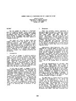
cava, a repeat MRI scan (1.5T) was performed. The MRI
results, in conjunction with our patient’shistory,were
considered by the radiologist to be indicative of epidural
varicosis. No further pathological abnormalities that
could cause the nerve root pain were found (Figure 1).
According to our vascular surgeons, no surgical cor-
rection for the hypoplasia of her inferior vena cava was
possible because it was a congenital defect. The optimal
therapy to manage t he progressive pain sympt oms of
our patient was then considered. Due to the increased
risk o f bleeding, the consideration of surgical interven-
tion was abandoned and she was treate d with perip heral
analgesics in combination with low-dos e pregabalin,
with satisfactory results. In addition, compression ther-
apy (class II) combined with Marcoumar (phenprocou-
mon) was carried out, which led to an acceptable
decrease in her symptoms (target international normal-
ized ratio; 2.0 to 3.0).
To date, our patient still complains of sciatic pain on
her right side, but is able to work while on interm ittent
pain medication.
Discussion
MRI is an important t ool in the diagnosis o f radicular
complaints. A review of the recent literature and the
case o f our patient shows that the presence of epidural
varicosis, without also b eing aware of a vascular
abnormality, can easily be misinterpreted as being her-
niated disc tissue [5]. Thrombosed veins appear hyperin-
tense on T1-weighted and T2-weighted images.
Depending on the degree of thrombosis, an epidural
vein on T2- weighted images may appear hypodense and
hyperintense. Therefore epidural varicosi s is oft en mis-
interpreted as herniated lumbar discs [6,7].
In the literature, several pathophysiological models for
the formation of venous epidural vascular anomalies are
discussed. Gümbel et al. postulated t he possibility of
primary epidural varicose veins without any underlying
or extra intraspinal pathology [8]. Wong et al. suggested
that varicose veins are due to the epidural mechanical
compression of the venous plexus by disc herniations,
spondylolisthesis or spinal steno sis [9]. Through veno us
stasis, an epidural vein thrombosis may occur over time
with subsequent irritation of nerve structures.
Epidural varicosis as a result of an obstruction of the
inferior vena cava has frequently been described in the
literature. When an obstruction and/or occlusion of the
inferior vena cava and vena iliaca communis is present,
there is increased blood flow into the azygos and hemia-
zygos veins. Expansion of the epidural venous plexus,
with potential compression of the neuronal structures,
also occurs. Trea tment with ant icoagulant medication in
combinati on with compression therapy, as in the case of
our patient, is usually sufficient [10].
In the literature, the alternative possibilities of throm-
bolysis and surgica l intervention have been described.
However, the results of thrombolysis are not convincing,
so it is rarely used [10]. Genevay et al. [1] consi der that
surg ical treatment of an epidural varix is obligatory, but
Figure 1 Epi dural varicosis (arrows).MRIscanofthelumbar
portion (transversal and sagittal).
Endres Journal of Medical Case Reports 2011, 5:537
/>Page 2 of 3
only if a neurological symptom is present. With respect
to the nature of the surgery, different approaches exist.
Reports on surgical thermocoagulation of the venous
plexus [2,9-12], interventional techniques [13] or surgi-
cal compression of the venous plexus with a resorbable
gelatin sponge [14] have been reported. In most cases,
this leads to a good surgical result with significant
reduction of the symptoms [9]. In cases of severe epi-
dural varicosis due to a faulty inferior vena cava and
dilation of all lumbar veins, the advice is against surgical
intervention. This is based on unsatisfactory surgical
results [14] and disproportional surgical risk [12].
Conclusions
Epidural varicosis with irritation o f nerve structures
observed on MRI should direct attention to the possibi-
lity of an inferior vena cava thrombosis or compression.
In such cases, an MRI scan of the region around the
inferior vena cava should be performed.
It is proposed that epidural varic osis due to inferior
vena cava pathology can cause radicular pain. Knowl-
edge of the existence of such a condition and its possi-
ble etiologies may assist in its recognition and improve
clinical management of affected patients.
In cases of epidural varicosis with irritation of neuro-
nal structures that develop due to hypoplasia o f the
inferior vena cava, surgical intervention gives unsatisfac-
tory results [9]. In contrast, interventions that resolve
the cause of the pathology in the inferior vena cava lead
to significantly better results [11].
This is not always possible where there is hypoplasia
and/or aplasia of the inferior vena cava, so, as in our
patient’s case, only sympt omatic therapy in combination
with anticoagulation and compression therapy is
possible.
Consent
Written informed consent was obtained from the patient
for publicatio n of this case report and any accompany-
ing images. A copy of the written consent is available
for review by the Editor-in-Chief of this journal.
Competing interests
The author declares that they have no competing interests.
Received: 16 February 2011 Accepted: 1 November 2011
Published: 1 November 2011
References
1. Genevay S, Palazzo E, Huten D, Fossati P, Meyer O: Lumboradiculopathy
due to epidural varices: two case reports and a review of the literature.
Joint Bone Spine 2002, 69:214-217.
2. Zimmerman GA, Weingarten K, Lavyne MH: Symptomatic lumbar epidural
varices. Report of two cases. J Neurosurg 1994, 80:914-918.
3. Pennekamp PH, Gemünd M, Kraft CN, von Engelhardt LV, Lüring C,
Schmitz A: Epidural varicosis as a rare cause of acute radiculopathy with
complete foot paresis: case report and literature review. Z Orthop Ihre
Grenzgeb 2007, 145:55-60.
4. Dudeck O, Zeile M, Poellinger A, Kluhs L, Ludwig WD, Hamm B: Epidural
venous enlargements presenting with intractable lower back pain and
sciatica in a patient with absence of the infrarenal inferior vena cava
and bilateral deep venous thrombosis. Spine (Phila Pa 1976) 2007, 32:
E688-E691.
5. Hanley EN, Howard BH, Brigham CD, Chapman TM, Guilford WB,
Coumas JM: Lumbar epidural varix as a cause of radiculopathy. Spine
(Phila Pa 1976) 1994, 19:2122-2126.
6. Yücesoy K, Acar M, Koyuncuoglu M: Acute foot drop caused by
thrombosed epidural vein. Acta Neurochir (Wien) 2001, 143:631-632.
7. Hammer A, Knight I, Agarwal A: Localized venous plexi in the spine
simulating prolapse of an intervertebral disc. A report of six cases. Spine
2003, 1:E5-E12.
8. Gümbel U, Pia HW, Vogelsang H: Lumbosacral vascular anomalies: cause
of sciatica. Acta Neurochir 1969, 20:131-151.
9. Wong CH, Thng PL, Thoo FL, Low CO: Symptomatic spinal epidural
varices presenting with nerve impingement: report of two cases and
review of the literature. Spine (Phila Pa 1976) 2003, 28:E347-E350.
10. Yun SS, Kim JI, Kim KH, Sung GY, Lee DS, Kim JS, Moon IS, Lim KW, Koh YB:
Deep venous thrombosis caused by congenital absence of inferior vena
cava, combined with hyperhomocysteinemia. Ann Vasc Surg 2004,
18:124-129.
11. Paksoy Y, Gormus N: Epidural venous plexus enlargements presenting
with radiculopathy and back pain in patients with inferior vena cava
obstruction or occlusion. Spine (Phila Pa 1976) 2004, 29:2419-2424.
12. Slin’ko EI, Al-Qashqish II: Surgical treatment of lumbar epidural varices. J
Neurosurg Spine 2006, 5:414-423.
13. Moonis G, Hurst RW, Simon SL, Zager EL: Intradural venous varix: a rare
cause of an intradural lumbar spine lesion. Spine (Phila Pa 1976) 2003, 28:
E430-E432.
14. Pekindil G, Yalniz E: Symptomatic lumbar foraminal epidural varix. Case
report and review of the literature.
Br J Neurosurg 1997, 11:159-160.
doi:10.1186/1752-1947-5-537
Cite this article as: Endres: Epidural varicosis as a possible cause of
radicular pain: a case report. Journal of Medical Case Reports 2011 5:537.
Submit your next manuscript to BioMed Central
and take full advantage of:
• Convenient online submission
• Thorough peer review
• No space constraints or color figure charges
• Immediate publication on acceptance
• Inclusion in PubMed, CAS, Scopus and Google Scholar
• Research which is freely available for redistribution
Submit your manuscript at
www.biomedcentral.com/submit
Endres Journal of Medical Case Reports 2011, 5:537
/>Page 3 of 3