báo cáo khoa học: "A non-healing corneal ulcer as the presenting feature of type 1 diabetes mellitus: a case report" pps
Bạn đang xem bản rút gọn của tài liệu. Xem và tải ngay bản đầy đủ của tài liệu tại đây (6.75 MB, 14 trang )
This Provisional PDF corresponds to the article as it appeared upon acceptance. Fully formatted
PDF and full text (HTML) versions will be made available soon.
A non-healing corneal ulcer as the presenting feature of type 1 diabetes mellitus:
a case report
Journal of Medical Case Reports 2011, 5:539 doi:10.1186/1752-1947-5-539
Alexander S Ioannidis ()
Sophia L Zagora ()
Alfred W Wechsler ()
ISSN 1752-1947
Article type Case report
Submission date 10 July 2011
Acceptance date 4 November 2011
Publication date 4 November 2011
Article URL />This peer-reviewed article was published immediately upon acceptance. It can be downloaded,
printed and distributed freely for any purposes (see copyright notice below).
Articles in Journal of Medical Case Reports are listed in PubMed and archived at PubMed Central.
For information about publishing your research in Journal of Medical Case Reports or any BioMed
Central journal, go to
/>For information about other BioMed Central publications go to
/>Journal of Medical Case
Reports
© 2011 Ioannidis et al. ; licensee BioMed Central Ltd.
This is an open access article distributed under the terms of the Creative Commons Attribution License ( />which permits unrestricted use, distribution, and reproduction in any medium, provided the original work is properly cited.
A non-healing corneal ulcer as the presenting feature of type 1 diabetes mellitus:
a case report
Alexander S Ioannidis*, Sofia L Zagora and Alfred W Wechsler
Address: Sydney Eye Hospital, 8 Macquarie Street, Sydney, NSW 2000, Australia.
*Corresponding author
ASI:
SLZ:
AWW:
Abstract
Introduction: Diabetic keratopathy is a rare complication of diabetes mellitus. This case
illustrates the importance of checking blood sugar levels of patients with non-healing
corneal ulcers to rule out the possibility of undiagnosed diabetes mellitus.
Case presentation: We report the unusual case of a 24-year-old southeast Asian
woman who presented with a sterile corneal ulcer to our hospital and later was found to
be diabetic after a prolonged hospital stay. Despite all efforts, the corneal ulcer had
failed to heal until treatment for previously undiagnosed diabetes was started. The
sterile corneal ulcer began to heal once blood sugar levels began to normalize.
Conclusions: Diabetic keratopathy is a rare complication of diabetes mellitus and
needs to be considered as a diagnosis in younger patients with non-healing sterile
corneal ulcers. Blood sugar levels should be checked in these cases for undiagnosed
diabetes mellitus.
Introduction
We report an unusual case of a 24-year-old southeast Asian woman who presented
with a sterile corneal ulcer to our hospital and later was found to be diabetic. Her
corneal ulcer had failed to heal until her blood sugar levels began to normalize. Diabetic
keratopathy is a rare complication of diabetes mellitus and needs to be considered as a
diagnosis in younger patients with non-healing sterile corneal ulcers. In a previous
publication, a 44-year-old man presented in a similar fashion, although in that instance
the condition was bilateral [1].
The case in our report highlights the importance of
investigating patients who present with unexplained corneal ulceration to exclude
undiagnosed diabetes mellitus.
Case presentation
A 24-year-old southeast Asian woman was admitted with a history of a white spot on the
right cornea and increasing discomfort. On examination, her vision was 6/36 on the right
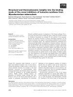
and 6/9 on the left. She had a corneal ulcer measuring 5.5 × 2mm on her right cornea. A
small localized area of scarring was present lateral to where the defect was present
(Figure 1). There was a +1 cell reaction in her right anterior chamber. She had a history
of bilateral anterior uveitis. Corneal sensation was normal in both eyes. There were
early bilateral posterior subcapsular cataracts.
In view of the findings, corneal scrapes were taken for microscopy, culture, and
sensitivity. Virology assays inclusive of herpes simplex virus and varicella-zoster virus
polymerase chain reaction were performed. Our patient had normal C-reactive protein,
rheumatoid factor, anti-nuclear antibody, extractable nuclear antigen, syphilis, and
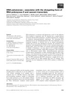
hepatitis B and C serology. She was started on topical g. cephalothin 5% and g.
gentamicin 0.9% hourly for 48 hours. She made a mild initial improvement and was
changed to topical g. chloramphenicol 1% four times each day and g. prednisolone
0.5% four times each day once her microbiology and virology results were negative. A
bandage contact lens was inserted to facilitate healing (Figure 2).
In the third week of admission, she complained of a headache and was found to be
mildly tachycardic. She was apyrexial with no reported malaise. A urinary dipstick
analysis was performed, and her urinary glucose level was 21mmol/L. Blood glucose
was urgently requested and was found to be 23mmol/L. A blood gas analysis showed a
pH of 7.38, a partial pressure of carbon dioxide (pCO
2
) of 44.7mmHg, and a partial
pressure of oxygen (pO
2
) of 89.5 mmHg.
She was transferred to the care of the medical team and a diagnosis of type 1 diabetes
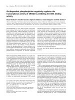
was made. She was started on treatment with insulin. Her corneal ulcer persisted and
punctal plugs were inserted to increase the tear film and facilitate healing. Autologous
serum drops were started every two hours during waking hours. There was a rapid
reduction of the epithelial defect as her blood glucose levels normalized (Figure 3).
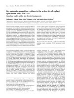
Four days after insulin treatment was started, her ulcer had healed and she was
discharged from the hospital and follow-up was conducted at her local diabetes clinic. At
a one-month review in the eye clinic, her ulcer remained healed, leaving a localized
area of subepithelial scarring (Figure 4).
Discussion
The ocular features of diabetes mellitus have been described in other reports. Impaired
glucose metabolism typically results in a localized microangiopathy that affects primarily
the retinal vasculature and that produces the classic lesions in the fundus with
microaneurysms, intraretinal hemorrhages, exudation, and new vessel formation [2]. A
combination of good glycemic control and regular visits to the eye clinic can often slow
or halt the progression of the disease.
Diabetic keratopathy is a rare complication of the condition. In this setting, impaired
epithelial healing is thought to be a consequence of an abnormal aldose reductase
pathway and secondary accumulation of polyol within the epithelial and endothelial cells
and thus result in cellular dysfunction [3,4]. This results in delayed healing responses
and loss of epithelial adhesion to the basement membrane, increasing the risk of
recurrent corneal erosions. Minor trauma and ocular manipulation with contact lenses
can also produce chronic non-healing defects [5]. Our patient had no history of trauma
or contact lens use. Other ocular features of diabetes mellitus include reduced corneal
sensation and tear production and basement membrane thickening [5-7].
It is important when considering the diagnosis of diabetic keratopathy to exclude other
treatable causes of non-healing defects, such as anterior basement membrane
dystrophy and recurrent erosion syndrome. Treatment options in cases of a persistent
ulceration include the use of frequent lubrication, bandage contact lenses, topical
autologous serum drops, and patching. If conservative measures fail, it may be
necessary to perform temporary tarsorrhaphy to facilitate healing.
Other conditions that delay epithelial healing need to be identified and treated
accordingly. Hence, coexisting neurotrophic keratopathy needs to be excluded by a
careful assessment of corneal sensation. Dry eye disease can also delay healing and
can be identified with rose-bengal staining of the cornea and conjunctiva and use of the
Schirmer test. Nocturnal lagophthalmos needs to be managed with appropriate
nocturnal padding and lubrication.
Animal studies have shown that the opioid antagonist naltrexone and insulin used
topically can facilitate healing in diabetic rats by enhancing DNA synthesis and re-
epithelialization through alterations in local opioid growth factors [8,9]. In the future,
these agents may be approved for use in the treatment of diabetic keratopathy.
Conclusions
Four days after insulin treatment was started, our patient’s ulcer had healed and
she was discharged from the hospital and follow-up was arranged at her local diabetes
clinic. The diagnosis of diabetes mellitus was thus made somewhat incidentally. Our
patient did not report weight loss, polyuria, or polydipsia to facilitate the diagnosis of
diabetes, and her only complaint was a brief history of headaches while in the hospital.
Her urinary glucose level was checked as part of an investigation for transient
tachycardia and elevated glucose levels were detected.
Using a combination of a bandage contact lens, temporary punctal plugs, and
autologous serum drops proved useful in facilitating ulcer healing. Although the corneal
ulcer showed initial signs of healing, it was after the blood sugar levels began to
normalize that the ulcer fully healed.
Consent
Written informed consent was obtained from the patient for publication of this case
report and any accompanying images. A copy of the written consent is available for
review by the Editor-in-Chief of this journal.
Competing interests
The authors declare that they have no competing interests.
Authors' contributions
ASI analyzed and interpreted the patient data regarding the clinical presentation. SLZ
and AWW were major contributors in writing the manuscript. All authors read and
approved the final manuscript.
References
1. Lockwood A, Hope-Ross M, Chell P: Neurotrophic keratopathy and diabetes
mellitus. Eye 2006, 20:837-839.
2. Ockrim Z, Yorston D: Managing diabetic retinopathy. BMJ 2010, 341:c5400.
3. Akagi Y, Yajima Y, Kador PF, Kuwabara T, Kinoshita JH: Localization of aldose
reductase in the human eye. Diabetes 1984, 33:562-566.
4. Kinoshita JH, Fukushi S, Kador P, Merola LO: Aldose reductase in diabetic
complications of the eye. Metabolism 1979, 28:462-469.
5. Kaji Y: Prevention of diabetic keratopathy. Br J Ophthalmol 2005, 89:254-255.
6. Azar DT, Spurr-Michaud SJ, Tisdale AS, Gipson IK: Decreased penetration of
anchoring fibrils into the diabetic stroma. A morphometric analysis. Arch
Ophthalmol 1989, 107:1520-1523.
7. Sakamoto A, Sasaki H, Kitagawa K: Successful treatment of diabetic
keratopathy with punctal occlusion. Acta Ophthalmol Scand 2004, 82:115-117.
8. Klocek MS, Sassani JW, McLaughlin PJ, Zagon IS: Naltrexone and insulin are
independently effective but not additive in accelerating corneal epithelial healing
in type I diabetic rats. Exp Eye Res 2009, 89:686-692.
9. Zagon IS, Jenkins JB, Sassani JW, Wylie JD, Ruth TB, Fry JL, Lang CM,
McLaughlin PJ: Naltrexone, an opioid antagonist, facilitates reepithelialization of
the cornea in diabetic rat. Diabetes 2002, 51:3055-3062.
Figure 1 Color photograph of the right eye shows the ulcer and an area of paracentral
intrastromal scarring.
Figure 2 Cobalt blue photograph of the right eye. Fluorescein dye was used to highlight
the large central ulcer (green stain).
Figure 3 Cobalt blue photograph of the right eye shows a smaller ulcer (green stain).
The ulcer began to heal rapidly once insulin treatment was initiated.
Figure 4 Color photograph of the right eye at one month. The defect has healed, leaving
a diffuse area of scarring.
Figure 1
Figure 2
Figure 3
Figure 4