báo cáo khoa học: "Isolation of specific and biologically active peptides that bind cells from patients with acute myeloid leukemia (AML)" doc
Bạn đang xem bản rút gọn của tài liệu. Xem và tải ngay bản đầy đủ của tài liệu tại đây (360.07 KB, 9 trang )
BioMed Central
Page 1 of 9
(page number not for citation purposes)
Journal of Hematology & Oncology
Open Access
Research
Isolation of specific and biologically active peptides that bind cells
from patients with acute myeloid leukemia (AML)
Naomi Galili*
†1
, Emmanuelle Devemy
†2
and Azra Raza
1
Address:
1
Saint Vincent's Comprehensive Cancer Center, New York, NY, USA and
2
McGill University, Montreal, Canada
Email: Naomi Galili* - ; Emmanuelle Devemy - ;
Azra Raza -
* Corresponding author †Equal contributors
Abstract
Purpose: In a departure from conventional strategies to improve treatment outcome for myeloid
malignancies, we report the isolation of leukemia-specific peptides using a phage display library
screened with freshly obtained human myeloid leukemia cells.
Results: A phage display library was screened by 5 rounds of biopanning with freshly isolated
human AML cells. Individual colonies were randomly picked and after purification, biologic activity
(growth and differentiation) on fresh AML cells was profiled. Ten peptides were synthesized for
further biological studies. Multiple peptides were found to selectively bind to acute myeloid
leukemia (AML) cells. The peptides bound to leukemia cells, were internalized and could induce
proliferation and/or differentiation in the target patient cells. Two of the peptides, HP-A2 and HP-
G7, appeared to have a novel mechanism of inducing differentiation since they did not cause G1
arrest in cycling cells even as the expression of the differentiation marker CD11b increased.
Conclusion: Peptide induced differentiation of leukemia cells offers a novel treatment strategy for
myeloid malignancies, whereas their ability to induce proliferation could be harnessed to make cells
more sensitive to chemotherapy. Conceptually, these leukemia specific peptides can also be used
to refine diagnosis, document minimal residual disease, and selectively deliver toxins to malignant
cells.
Background
We proposed to isolate leukemia specific peptides that
have the potential to target and deliver toxins to acute
myeloid leukemia cells (AML) or to modify the biological
behavior of the cells to which they bind using a phage dis-
play library. First described by George Smith in the mid
1980s [1], this technique allows repertoires of antibodies,
proteins or peptides displayed on the surface of phage par-
ticles to be screened by any chosen target. Single chain Fv
phage libraries have been used to isolate antibodies that
recognize cell surface antigens for clinical, diagnostic and
therapeutic applications [2-6] or for antigen epitope map-
ping [6,7]. An alternative approach was to use peptide
phage libraries to identify small molecules that can bind
either purified targets or cell surface receptors. Peptides
that are specific for surface expressed immunoglobulins
isolated from chronic lymphocytic leukemia (CLL) cells
[8] and multiple myeloma cells [9] have been identified
for potential patient specific targeted therapy. Our
approach was to use freshly obtained patient cells to iso-
late leukemia specific peptides from a phage library. In
this study we show that multiple myeloid leukemia spe-
Published: 10 July 2008
Journal of Hematology & Oncology 2008, 1:8 doi:10.1186/1756-8722-1-8
Received: 19 May 2008
Accepted: 10 July 2008
This article is available from: />© 2008 Galili et al; licensee BioMed Central Ltd.
This is an Open Access article distributed under the terms of the Creative Commons Attribution License ( />),
which permits unrestricted use, distribution, and reproduction in any medium, provided the original work is properly cited.
Journal of Hematology & Oncology 2008, 1:8 />Page 2 of 9
(page number not for citation purposes)
cific and non-specific peptides can be identified by this
method. In addition, we show that these peptides are
capable of altering the biological behavior of AML cells
while having no effect on normal marrow elements.
Methods
Patients and normal donors
Specimens were obtained from patients with chronic
myelogenous leukemia in blastic crisis (CML-BC), AML of
different FAB classifications, as well as from normal
donors. Informed consent was obtained from all patients
prior to study according to the regulations of the Institu-
tional Review Board of Rush Medical Center.
Cell preparation
The peripheral blood (PB) or bone marrow (BM) cells
were subjected to density cut centrifugation over Ficoll-
Hypaque. The mononuclear fractions were washed twice
and used directly. Granulocytes were obtained from the
high-density fraction. Immature CD34+ stem cells were
isolated by magnetic cell sorting and separation (Miltenyi
Biotec, California). The promyelocytic cell line HL60
(ATCC) was maintained in RPMI 1640/20% FBS. Cells
were induced to differentiate into the granulocytic lineage
by treatment with 1.5% (v/v) of DMSO during 7 days of
culture.
Isolation of phage binding to human malignant myeloid
cells
This work was performed using the Display PHAGE sys-
tem library, purchased from Display System Biotech
(Vista, California) with a diversity of approximately 3 ×
10
7
.
Initial selection of phage specific for myeloid leukemia
used cells from patients with CML-BC. CML-BC cells were
incubated with the library (10
12
cfu) in PBS/0.1% milk at
4°C for 90 minutes. Unbound phage was removed by 5
washes with buffer. Weakly bound phage was acid eluted
by glycine (0.1 M, pH2.2) followed by cell lysis (30 mM
Tris pH8.0, 1 mM EDTA) to elute the tightly bound phage.
This second fraction was amplified by re-infection and
growth in E. Coli. Amplified phage was purified by PEG
precipitation and used for another round of binding. The
"biopanning" step was repeated five times to obtain a
population that was highly enriched with phage express-
ing peptides that bind to leukemia cells. After growth on
agar plates using antibiotic selection, individual colonies
were randomly picked, amplified, and PEG purified.
Binding to leukemia cells was confirmed by ELISA assay.
ELISA Assay
Amplified phage from a single colony (10
9
cfu/ml) were
dispensed into a 96-well plate containing either leukemia
or normal peripheral or bone marrow mononuclear cells
(250,000 cells/well) previously fixed with glutaraldehyde
and blocked with PBS/2% milk. After 2 hours, wells were
washed and bound phage was detected by a monoclonal
anti-M13 antibody HRP conjugate (1:2,000 in PBS/1%
milk, Amersham Pharmacia Biotech, NJ). Following addi-
tion of HRP substrate, intensity of color was measured by
spectrophotometry. Each phage clone was tested in tripli-
cate with appropriate controls. Identical assays were per-
formed using synthesized peptides that had been
conjugated with biotin. The bound peptide was detected
by streptavidin-HRP diluted 1:1,000 in PBS/1% milk
(Amersham Pharmacia Biotech, NJ). A biotin control con-
firmed that binding to the cells was via the peptide moiety
itself and not via the biotin conjugate.
Peptide synthesis
Phage DNA was extracted using the QIAprep M13 kit
(Qiagen). The hypervariable oligonucleotide sequence
coding for the peptide was PCR amplified using the fol-
lowing primer set: Primer 1 = 5' GGG ATT TTG CTA AAC
AAC 3', Primer 2 = 5' GGA GGT CTA GAT AAC GAG 3'.
Each clone was amplified, purified and sequenced (Gene
Link, NY) in duplicate. The synthetic octapeptides with
terminal cysteine residues were commercially synthesized
(New England Peptide, Inc., Massachusetts) with more
than 95% purity. Peptides were not cyclized by oxidation;
therefore percentage of free sulfhydryl groups was evalu-
ated by Ellman's reaction using a cysteine standard (Pierce
Biotechnology). This reaction allowed us to determine the
percentage of reduced cysteines in the peptide solution
used for our experiments. The percentage ranged between
50% and 75% for most peptides except for HP-B6 (100%)
and HP-G2 (30%). A biotin molecule was conjugated to
the N-terminal of the peptide for cytochemistry studies.
Cytochemistry
Fresh cells, blocked with PBS/4% BSA for one hour at 37°C,
were incubated with biotin-conjugated peptide at 1 μM for
30 minutes unless otherwise stated. Free biotin was used to
confirm that binding of biotin-conjugated peptide was
mediated by the peptide moiety. After washing, cells were
fixed with 3.7% paraformaldehyde, permeabilized with
methanol and incubated with streptavidin-FITC (DAKO)
diluted 1:500 in PBS/4% BSA for 30 minutes at room tem-
perature and washed again. Alternatively, cytospin prepara-
tions of cells were fixed in 3.7% paraformaldehyde and kept
at -80°C for further study. The fluorescent signal was ana-
lyzed using AxioVision 2.05 software.
Liquid culture of HL60, AML and normal cells
To assess the biological effects of peptides on growth and
differentiation of HL60 cells, logarithmic growth phase
cells were seeded in 3 ml of RPMI 1640/10% FBS. Cul-
tures in the presence or absence of a single peptide (10
-6
M -10
-4
M) or DMSO (1.5%) were assessed for the level of
Journal of Hematology & Oncology 2008, 1:8 />Page 3 of 9
(page number not for citation purposes)
cell viability, cell number, and differentiation, after 7 days
without changing the medium.
AML or normal bone marrow cells were seeded at a con-
centration of 10
6
cells/ml in 3 ml of RPMI 1640/15% FBS.
Peptides were added at a single dose of 10
-4
M without
addition of fresh medium or peptide for the duration of
the cultures. Granulocyte-Macrophage Colony-Stimulat-
ing Factor (GM-CSF, 100 U/ml) was used as a positive
control. Viability was evaluated at day 14.
Analysis of cell cycle
After 7 days of culture, HL60 cells (1 × 10
6
) were washed
in PBS, fixed in 1% paraformaldehyde and permeabilized
with 0.5% Triton X100 in acid solution. After neutraliza-
tion, cells were incubated with RNase A and stained with
propidium iodide. The relative DNA content was analyzed
by flow cytometry.
CD11b analysis by flowcytometry
After 7 days of culture, HL60 cells (1 × 10
6
) were stained
with fluorescence conjugated monoclonal antibodies rec-
ognizing the myeloid maturation marker CD11b (Becton
Dickinson Immunocytometry system) for 20 minutes at
4°C. The percentage of cells with a fluorescence intensity
above the control was measured.
Colony assay
Mononuclear cells (10
4
cells/ml) were plated in 0.8%
methylcellulose (Methocult, StemCell Technologies Inc.,
Vancouver, Canada) in Iscoves modified Dulbecco
medium (IMDM, Gibco Laboratories, Grand Island, NY)
with the peptides at a single dose of 10
-4
M or GM-CSF at
100 U/ml without addition of fresh medium or peptide.
The solvent used for peptide preparation served as a con-
trol. Following 14 days of incubation, the number of col-
onies was assessed and cytospin preparations were made
for Giemsa morphology staining. Percentage of differenti-
ated cells was evaluated by counting 200 cells.
Statistical analysis
All analyses were performed using SYSTAT software ver-
sion 10.0. The relationship between AML patients' clinical
data and responses of AML cells to the peptides was ana-
lyzed by Fisher's exact test.
Results
Isolation of phage able to bind cells from leukemia
patients
Phage clones that bind to leukemia cells were isolated by
incubating a library of M13 phage bearing 8 mer pep-
tides with BM mononuclear cells from CML-BC patients.
After five rounds of biopanning, 450 individual phage
were picked randomly and tested by ELISA against cells
from CML-BC patients to confirm the phage binding.
Only clones with an O.D at least two times higher than
controls were considered positive for the specimen stud-
ied and only those positives for at least three different
CML-BC cell populations were considered leukemia pos-
itive. Among the 450 clones, 129 (28%) were positive for
3 different CML-BC patients. These clones were then pro-
filed with AML and normal cells. Sixty-eight (15%)
clones bound to CML-BC and AML cells, and were used
for further studies with PB and BM cells from normal
donors. Of these 68 clones, 18 did not bind normal PB
specimens (n = 3), and 17/18 did not bind normal BM
specimens (n = 3). The 68 phage clones were sequenced
and 51 gave a good sequence chromatogram. Twelve dif-
ferent sequences of 8 amino acids surrounded by two
cysteine residues were detected with an enrichment of
the 3 sequences: CVSEDIYDAC (22/51), CEFQQWSGKC
(8/51) and CNHVCSRLGC (7/51). Table 1 shows the
Table 1: Peptide sequences and binding profiles of phage tested on leukemia and normal cells in an ELISA assay.
Phage
Clones
Sequences AML
BM
CML
BM
Normal
PB
Normal
BM
Normal
CD 34+
HP
A
-A6 CNETTVREYC (4) + + ND
B
ND ND
HP-A2 CIEETARKGC (2) + + - - -
HP-B6 CNHVCSRLGC (7) + + - - -
HP-G7 CNELHMKQHC (2) + + - - -
HP-G2 CNNATFEDGC (1) + + + - ND
HP-C8 CNNATVEDEC (1) + + - ND ND
HP-F6 CEFLQWSGKC (1) + + + ND ND
HP-G5 CEFQQWSGKC (8) + + + - ND
HP-D4 CETGERIVLC (1) + + + ND ND
HP-B4 CDEKRGPNEC (1) + + - - -
HP-B11 CVSEDIYDAC (22) + + + ND ND
HP-D2 CHSWKPDKLC (1) + + + ND ND
A
HP; Harvey Preisler.
B
ND not done.
The number in bracket represents the number of clones expressing the same sequence.
Journal of Hematology & Oncology 2008, 1:8 />Page 4 of 9
(page number not for citation purposes)
sequences as well as the binding characteristics of the
phage tested on leukemia and normal cells. Two phage,
HP-G2 and HP-C8, differ by two amino acids and dis-
played different binding profiles. To confirm the specifi-
city of the clones for leukemia cells, 4 clones were tested
on isolated normal marrow CD34+ cells (under-repre-
sented in normal donors). No binding was detected. The
binding profile of the phage clones was nearly identical
for AML and CML-BC cells, implying that the binding of
the peptides was myeloid leukemia specific rather than
CML-BC specific. Since AML specimens were more read-
ily available, all further studies were done using AML
cells.
Peptide binding profiles
Ten biotin-conjugated peptides were synthesized and
tested for their ability to bind leukemia and normal BM
cells in a cytochemistry assay (Table 2). The HP-G2 pep-
tide differed from that of the corresponding phage in that
it could bind to normal bone marrow cells. Additionally,
3 peptides showed differential binding profiles on normal
bone marrow cells that differed from the phage binding of
normal peripheral blood cells. This may be due to cell lin-
eage differences in blood versus bone marrow popula-
tions. Four peptides bound to mononuclear cells from
normal BM donors, HP-C8 (41%), HP-B11 (95%) HP-G2
(18%) and HP-D4 (45%), but did not bind high-density
cells. This suggests that while these peptides are not spe-
cific for leukemia cells they appear to be specific for a sub-
set of cells included in the mononuclear population. To
investigate whether this subset could be CD34+ cells,
three peptides HP-A2, HP-G7 and HP-B11 were tested on
purified normal BM CD34+ cells. Only HP-B11 was found
to bind to a small population of cells (Table 2). Impor-
tantly, the 10 peptides did not bind to human skin fibrob-
last cells, Hs68, which express epitopes common to
multiple cell types. The 6 peptides that did not bind nor-
mal bone marrow cells together with the peptide HP-A2A
(alanine substituted for arginine of HP-A2) and HP-A6
(both used in further studies) were then profiled on
mononuclear cells from patients with AML (Table 3). The
percentage of cells recognized by the individual peptides
varied, as expected, due to the heterogeneous nature of the
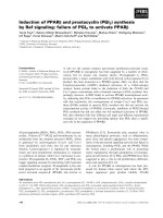
epitopes recognized by the peptide. Figure 1 illustrates the
staining for two peptides, HP-A2 and HP-C8, on one nor-
mal and one AML specimen. HP-C8 bound cells in both
specimens unlike HP-A2 that bound only AML cells. Spe-
cificity of peptide binding was further confirmed by a
competition assay on cells from two different AML
patients. Incubation with 100-fold concentration of non-
biotinylated HP-A2 resulted in blocking the binding of
the biotinylated HP-A2. Similarly, HP-G7 and HP-B6
binding was specifically blocked by the non-labeled
peptide.
Table 3: Percentage of BM cells from AML patients recognized by peptides not binding to normal BM cells in a cytochemistry assay.
Patient Number HP-A2 HP-A2A HP-G5 HP-G7 HP-D2 HP-B6 HP-B4 HP-A6
15492 29 21 18 29 7 10 38 14
15480 36 ND ND 31 ND 23 ND ND
17026 44 ND ND 0 0 34 0 ND
17648 32 40 314450521921
16313 55 40 ND 54 48 56 25 31
16352 76 60 63 0 77 85 59 62
16278 22 30 25 0 22 41 24 22
15727 80 72 517482872717
1340 32 21 34 45 46 90 31 27
Table 2: Binding profiles of synthesized peptides on leukemia and normal bone marrow cells.
Peptide name Peptide sequence AML bone
marrow cells
Normal bone
marrow cells
Normal
CD34+ cells
HP-A2 CIEETARKGC + - -
HP-B6 CNHVCSRLGC + - ND
HP-G7 CNELHMKQHC + - -
HP-B4 CDEKRGPNEC + - ND
HP-C8 CNNATVEDEC + + ND
HP-G2 CNNATFEDGC + + ND
HP-D4 CETGERIVLC + + ND
HP-G5 CEFQQWSGKC + - ND
HP-B11 CVSEDIYDAC + + +
HP-D2 CHSWKPDKLC + - ND
Journal of Hematology & Oncology 2008, 1:8 />Page 5 of 9
(page number not for citation purposes)
Using fixed AML cells, only a cytoplasmic fluorescent sig-
nal was observed. The same binding assay was therefore
repeated with fresh cells obtained from AML patients
using a time-lapse format (Figure 2). The fluorescence was
first observed on the surface membrane at 1 minute, fol-
lowed by appearance of some signal in the cytoplasm at 5
minutes, and clear cytoplasmic signal at 30 minutes. This
suggests internalization of the peptide following surface
binding.
HL60 cell differentiation induced by peptides
HL60 cells, a human promyeloid cell line, is often used as
a model to study myeloid cell differentiation. All the pep-
tides that bind AML cells were also found to bind to HL60
cells. Therefore, these cells were used to study the biolog-
ical effects triggered by peptide binding. Since peptides
HP-A2 and HP-G7 bind only AML cells and not normal or
CD34+ bone marrow cells, they were used for these stud-
ies. HL60 cells were cultured in RPMI1640/10% FBS for 7
days with peptides HP-A2 or HP-G7 at concentrations
ranging from 10
-6
M to 10
-4
M. DMSO (1.5%) was used as
a positive control for differentiation [10]. As shown in
Table 4, the cell proliferation was inhibited by DMSO but
not by the peptides. Cells remained viable under all con-
ditions. Differentiation was measured by following
appearance of the myeloid maturation marker CD11b.
Percentage of control cells expressing CD11b was of 40.6
± 1.3% indicating a low level of spontaneous differentia-
tion. After differentiation induced by DMSO, the CD11b
marker was found on 82.2 ± 1.6% of cells. Similarly, cells
incubated in the presence of 10
-4
M of either peptide
showed increased CD11b expression with peptide HP-A2
inducing levels equal to those found with DMSO (79.7 ±
3.1%). Lower concentrations of peptide did not induce
CD11b expression. Giemsa staining for morphology
showed that only 5.5% of the control cells were granulo-
cytes whereas cultures with DMSO contained 90.7% gran-
ulocytes. At 10
-4
M, both peptides induced a more limited
morphological effect. Since the biological activity of the
peptides on both marker expression and morphology was
observed at only 10
-4
M, we limited our further studies on
patient cells to this concentration. As differentiation is
usually associated with arrest in the cell cycle, we looked
at the correlation between CD11b expression and the cell
cycle. Cultures induced to differentiate with DMSO exhib-
ited cell cycle arrest as evidenced by an increase in G0/G1
(from 44.5 ± 9.1% to 58.0 ± 13.0%). In contrast, the pep-
tide cultures showed no change in the proportion of cells
in G0/G1 despite the marked increase in CD11b expres-
sion.
Peptides are not toxic to human cells
In order to show that these peptides were not directly toxic
to both malignant and normal cells, viability was evalu-
ated at 2 weeks for 2 AML and 3 normal BM specimens in
the presence of a single dose of 10
-4
M peptide. There was
no significant decrease in the viability of cells cultured in
the presence of any of the 7 peptides tested and used in
further studies (HP-A2, HP-A2A, HP-B4, HP-B6, HP-D2,
HP-G5, HP-G7 >90% viability).
Proliferation and Differentiation of freshly obtained
human leukemia cells are altered by peptides
Methylcellulose culture permits assessment of the effects
of peptides on the cloning efficiency of myeloid progeni-
tor cells. Cells were cultured with peptide for 14 days
using GM-CSF as a positive control. Due to the limited
number of cells available from each patient, not all pep-
HP-A2 is internalized after binding to AML cellsFigure 2
HP-A2 is internalized after binding to AML cells. AML
cells were incubated with biotinylated-peptide HP-A2 (1 μM)
at various time points. The bound peptides were initially
detected at the cell surface and subsequently internalized.
Peptides can bind to AML cellsFigure 1
Peptides can bind to AML cells. AML and normal BM
cells were incubated with biotinylated-peptides or non-con-
jugated biotin (1 μM), which were detected by streptavidin-
FITC. For this specimen, HP-A2 was detected in AML but
not normal cells while HP-C8 was detected in both popula-
tions.
Journal of Hematology & Oncology 2008, 1:8 />Page 6 of 9
(page number not for citation purposes)
tides could be tested on all patient samples. Results from
a representative patient in whom 3 different peptides were
tested are presented in Figure 3. Similar studies were per-
formed on multiple samples from different patients and
are summarized in the section below. Peptides HP-A2,
HP-B6 and HP-G2 induced an increase in the number of
colonies in the example shown (Figure 3). This increase
was equivalent to the one observed after GM-CSF stimula-
tion. In contrast, incubation of normal cells with the pep-
tide did not induce any change in colony number.
Cellular differentiation evaluated by morphological
changes was assessed after 14 days of in vitro cultures.
Using the AML specimen shown in Figure 3, the propor-
tion of differentiated monocytes/macrophages increased
after incubation with the peptides HP-A2 and HP-G2
(47% and 52% respectively). Spontaneous differentiation
in control cells was only 31% (Figure 4). Peptide HP-B6
failed to induce differentiation in this patient. The propor-
tion of mature monocytes/macrophages was significantly
higher in the presence of peptides than in the presence of
GM-CSF.
Summary of the effects of 7 different peptides on
proliferation and differentiation of AML progenitor cells
The biologic effects of 7 different peptides on the behavior
of AML progenitor cells from 20 patients were evaluated.
Five peptides do not bind to normal bone marrow cells
(HP-A2, HP-B4, HP-B6, HP-G5 and HP-G7) while 2 pep-
tides bind to both leukemia and normal cells (HP-C8 and
HP-G2). It was of interest to include both types of pep-
tides to see whether there was an obvious difference. Fig-
ure 5 is a summary of the biological activity of each
peptide. Table 5 is a summary of clinical characteristics
and biological activity induced by at least one peptide for
each individual patient. As seen in Figure 5 for example,
40% of 15 AML patients incubated with HP-A2 showed
induced proliferation. Eleven of these 15 patients were
evaluated for differentiation and 25% showed a response.
All peptides induced proliferation in at least several
patient samples. Four peptides however could also inhibit
colony formation in a percentage of the samples (18%
with peptide HP-B6, 10% with peptides HP-G2 and HP-
G7, 8% with peptide HP-G5). Peptides HP-C8 and HP-G2
were the least (10–20% of marrow specimens studied)
and peptides HP-B4 and HP-G7 were the most likely to
induce differentiation (45% and 60% respectively). Pep-
tide induced differentiation ranged between 9–75% and
spontaneous differentiation ranged between 5.5–29%.
As seen in Table 5, an increase in colony number (prolif-
eration) was induced in 10/19 specimens by at least one
peptide (column 8). Differentiation (column 12) was
seen in 9/15 for at least one peptide. Three specimens
showed both differentiation and proliferation after cul-
ture with a specific peptide. Interestingly, in only 2 AML
specimens, inhibition of proliferation by a specific pep-
tide was accompanied by cell differentiation (AML
#15857 and #14499).
Single amino acid mutation effects biological activity
Binding specificity should be dependent on peptide con-
formation. Mutation of one of the amino acids may alter
the binding properties and/or biological activity. An inter-
nal arginine residue (large and polar) of peptide HP-A2
(IEETARKG) was replaced by an alanine residue (small
and non-polar); HP-A2A. Two additional peptides con-
tained a polar amino acid at the same position HP-G7 and
HP-B6 (NELHMKQH, and NHVCSRLG). These three pep-
tides did not bind normal BM cells suggesting that this
amino acid may be important to peptide specificity. While
binding properties of the mutated peptide were not
changed (Table 3), the number of patients in whom pro-
liferation was stimulated was significantly lower (from 40
to 22%). In addition, this peptide actually inhibited pro-
liferation in a number of samples (16%) (Figure 5).
Discussion
Therapeutic progress in AML has been painfully slow
despite significant improvement in our understanding of
the underlying pathology. A major conundrum in design-
ing treatment strategies relates to the issue of targeting
only the malignant cells while sparing their normal coun-
Table 4: Effects of DMSO, HP-A2 and HP-G7 on proliferation, cell cycle and granulocytic differentiation in HL60 cells.
Control DMSO HP-A2 HP-G7
10
-6
M10
-5
M10
-4
M10
-6
M10
-5
M10
-4
M
Cell number
(% of the control)
100 38 ± 0.3
A
87 ± 3.7 86 ± 1.6 87 ± 1.4 95 ± 1.3 73 ± 4.7 89 ± 1.3
G0/G1 (%) 44.5 ± 9.1 58.0 ± 13.0 38.0 ± 2.3 34.0 ± 4.7 41.8 ± 6.1 43.6 ± 3.9 39.6 ± 6.0 42.5 ± 7.9
CD11b 40.6 ± 1.3 82.2 ± 1.6 38.1 ± 1.2 40.4 ± 2.7 79.7 ± 3.1 37.7 ± 2.9 34.1 ± 3.9 57.8 ± 3.1
Granulocytic
cells (%)
5.5 ± 1.7 90.7 ± 4.3 13.0 ± 5.2 10.3 ± 3.4 16.5 ± 4.2 7.0 ± 1.3 10.1 ± 2.2 15.0 ± 2.2
A
All results are mean ± S.D. (n = 3).
Journal of Hematology & Oncology 2008, 1:8 />Page 7 of 9
(page number not for citation purposes)
terparts. In this work, we have shown that using a phage
library, it is possible to isolate peptides that can bind and
induce biologic activity only in leukemia cells. Since cell
lines are often not representative of the physiological state
of malignant cells, we used freshly obtained patient spec-
imens for the initial screening, and in order to avoid iso-
lating patient specific peptides, the screening and
profiling were performed with cells from multiple
patients with different myeloid leukemia subtypes. It is of
interest that only 12 unique peptide sequences were
found and that one sequence (HP-B11) was found in 22
clones. The redundancy of the phage sequence may be
related to the copy number of each epitope expressed on
hematopoietic cells. Ten synthesized peptides were re-
profiled on AML and normal cells. As expected, all the
peptides were able to bind to the malignant cells, but four
peptides also were found to bind to normal BM cells. The
peptides were able to bind to all AML-FAB subtypes in our
small sample size, but the percentage of cells recognized
by the peptide was lower at relapse.
We have found at least 2 peptides, HP-A2 and HP-G7, that
bind only to AML cells and not to normal cells including
normal CD34+ cells. Cytochemistry studies showed that
after surface binding, these two peptides were internalized
within minutes. Intracellular localization is a particularly
attractive feature since it offers a novel method to deliver
drugs and may reduce toxicity by allowing lower concen-
trations to be administered. In addition, these studies
showed that the peptides do not always label all the leuke-
mia cells which is not surprising since AML blasts are not
a homogeneous population [11] and AML marrows fre-
quently contain normal mononuclear cells.
In order to develop clinically useful strategies, we were
looking for peptides that, upon binding, could induce
some biological change in the leukemia cell growth and
differentiation. The peptides that we have identified could
clearly affect both these parameters although the biologic
response elicited was variable. A given peptide could
induce proliferation, differentiation, both or none in the
cells of different AML patients. Of the two leukemia spe-
cific peptides, HP-A2 stimulated proliferation in 40% and
differentiation in 25% of the patients tested while HP-G7
stimulated proliferation in 35% and differentiation in
60% of the patients. This includes some patients in whom
the peptides induced proliferation followed by differenti-
ation. To be therapeutically useful in future, the biologic
effects of a given peptide would have to be defined for the
individual patient. While the peptides were screened to be
myeloid specific and not patient specific, the biologic
activity elicited by a peptide could depend on multiple
variables. The maturation state at which the AML clone is
arrested is defined by the FAB subtype and this may be a
factor that determines the specific response to the peptide.
We have not analyzed sufficient numbers of AML patients
to correlate activity with subtype. Additionally, activity
could be determined by the number of epitopes on the
cells of the AML clone.
Blast analysis of the sequence of the two leukemia specific
peptides revealed that 7 of 8 amino acids of HP-A2
matched mostly to proteins of lower organisms. For HP-
G7, the best match was 6 amino acids identical to two dif-
ferent proteins; the Toll-like receptor 2 and the neramini-
dase protein of the streptococcus pneumonia bacteria.
Further studies, however, will be needed to clarify the
molecular epitope that is recognized by these peptides in
order to understand the mechanism(s) responsible for
their biological activity.
The high concentration of peptides used for these studies
is non-physiological. However, the peptide solution,
HP-A2 and HP-G2 but not HP-B6 induced differentiation of cells from the AML patient shown in Fig.4Figure 4
HP-A2 and HP-G2 but not HP-B6 induced differenti-
ation of cells from the AML patient shown in Fig.4.
AML cells were incubated in methylcellulose culture in the
presence of peptides at 10
-4
M or GM-CSF (100 U/ml). After
14 days, cell morphology was evaluated. The percentage of
each cell population was determined by counting 200 cells.
Peptides are capable of increasing the cloning efficiency of cells from a patient with AML but not normal cellsFigure 3
Peptides are capable of increasing the cloning effi-
ciency of cells from a patient with AML but not nor-
mal cells. AML and normal cells were incubated in
methylcellulose culture in the presence of peptides at 10
-4
M. After 14 days, the number of colonies was assessed.
Journal of Hematology & Oncology 2008, 1:8 />Page 8 of 9
(page number not for citation purposes)
administered as a single dose at the start of cell culture,
was found to contain a mixture of various conformations
including linear, cyclic or concatamers. It is possible that
the structure exhibiting biological activity represented
only a fraction of the total and its stability in the culture
medium is unknown. This may explain the need for a high
initial concentration to elicit a biological effect. It will be
necessary in the future to identify the conformation
responsible for biological activity. In addition to confor-
mation, peptide sequence determines the nature of the
biological response. For example, HP-C8 and HP-G2,
identical for 6 amino acids, elicited different activities
despite binding similarities. Finally, a single amino acid
substitution in HP-A2 altered the ability of the peptide to
induce proliferation. These observations support a specif-
icity of the structure-function relationships of the peptide
and its target.
HP-A2 and HP-G7 were also found to bind HL60 cells
that have often been used as a model to study myeloid cell
growth and differentiation. We have used HL60 cells to
study the effect of these two peptides on cell cycle and dif-
ferentiation. While DMSO was found to increase CD11b
expression and arrest the cell cycle, we found that CD11b
expression was increased by the peptides without con-
comitant cell cycle arrest. Studies with other differentiat-
ing agents show accumulation of cells during
differentiation in G1 by day 3 or day 4 [12-14], we have
not found a G1 block or decrease in cell number in pep-
tide treated differentiating cells even after 7 days of cul-
ture. Thus the mechanism of differentiation by the
peptides appears to be novel and different than that of the
known conventional differentiating agents. It may be that
the binding of these peptides results in an uncoupling of
the expression of the proliferation and differentiation
markers. In the absence of growth arrest, it is conceivable
that the peptides can also be used concomitantly with
chemotherapeutic agents which depend upon cellular
synthesis to increase the therapeutic index.
The leukemia specific peptides are expected to be non-
toxic based on in vitro results, and theoretical in vivo clin-
ical applications are multiple. Coupled to toxins, the pep-
tides could specifically target and kill tumor cells as first
line therapy and/or as therapy targeting residual disease.
When coupled with an imaging reagent, the peptides
could be used to locate sites of tumor sanctuary. The pep-
tides could also be combined with peptides that selec-
tively target the tumor vasculature [15]. The challenge
now is to identify the molecular target on the surface of
leukemia cells that binds these peptides, demonstrate that
the in vitro effects are reproducible in vivo by conducting
similar experiments in animal models and finally translat-
ing these findings into clinical trials for human use; areas
Table 5: Clinical characteristics of AML patients at the time the bone marrow was obtained and biological response after 14 days of
culture in the presence of GM-CSF (100 U/ml) or one peptide (10
-4
M).
Patient
number
Age FAB Blast
Count
Karyotype
Abnormalities
Prior
MDS
Prior
Toxic
Exposure
Increase in
colony number
Decrease in
colony number
Differentiation
peptide GM-CSF peptide GM-CSF peptide GM-CSF
15857 76 M0 20 yes yes - - +
A
-++
1474258M088 nonoyes NDND
14924 76 M1 8 yes + + - - - -
16249 M177 +
14372 56 M1 49 no yes yes + + - - - -
14122 72 M1 63 yes yes yes + + - - ND ND
15492 45 M1 55 no no yes - + + - + -
15480 55 M1 88 no yes - ND - ND - ND
14373 66 M1 69 yes yes + + + - + +
1507166M127 noyes
15019 77 M2 34 no yes no + + - - + +
15891 58 M2 52 yes - + - - + +
16313 48 M2 38 no no no + - - - + +
1136933M220yesyesyes NDND
1499648M3 yesnono NDND
1635274M434 yesyesno+-+-+-
16278 25 M4 35 no no + + - - + -
14499 74 Unclas-sified 3 no - + + - + +
13459 59 Un-known 80 no yes + + - - - -
A
A modification of cell behavior in response to GM-CSF or to at least one peptide is noted +
Publish with Bio Med Central and every
scientist can read your work free of charge
"BioMed Central will be the most significant development for
disseminating the results of biomedical research in our lifetime."
Sir Paul Nurse, Cancer Research UK
Your research papers will be:
available free of charge to the entire biomedical community
peer reviewed and published immediately upon acceptance
cited in PubMed and archived on PubMed Central
yours — you keep the copyright
Submit your manuscript here:
/>BioMedcentral
Journal of Hematology & Oncology 2008, 1:8 />Page 9 of 9
(page number not for citation purposes)
that are currently being actively pursued in our laboratory
at present.
Competing interests
The authors declare that they have no competing interests.
Authors' contributions
NG participated in study design, conducted functional
studies, analysis and wrote the manuscript. ED partici-
pated in study design, isolated the peptides conducted
functional studies and analysis. AR participated in study
concept, design and coordination and wrote the manu-
script. All authors read and approved the manuscript.
Acknowledgements
The Coleman Foundation supported this work, which was conceived and
initiated by the late Harvey Preisler MD.
References
1. Smith GP: Filamentous fusion phage: novel expression vectors
that display cloned antigens on the virion surface. Science
1985, 228(4705):1315-1317.
2. Poul MA, Becerril B, Nielsen UB, Morisson P, Marks JD: Selection of
tumor-specific internalizing human antibodies from phage
libraries. J Mol Biol 2000, 301(5):1149-1161.
3. Nie YZ, He FT, Li ZK, Wu KC, Cao YX, Chen BJ, Fan DM: Identifi-
cation of tumor associated single-chain Fv by panning and
screening antibody phage library using tumor cells. World J
Gastroenterol 2002, 8(4):619-623.
4. Huls GA, Heijnen IA, Cuomo ME, Koningsberger JC, Wiegman L, Boel
E, Vuurst de Vries AR van der, Loyson SA, Helfrich W, van Berge
Henegouwen GP, van Meijer M, de Kruif J, Logtenberg T: A recom-
binant, fully human monoclonal antibody with antitumor
activity constructed from phage-displayed antibody frag-
ments. Nat Biotechnol 1999, 17(3):276-281.
5. Adams GP, Schier R: Generating improved single-chain Fv mol-
ecules for tumor targeting. J Immunol Methods 1999, 231(1–
2):249-260. Review
6. Wong C, Waibel R, Sheets M, Mach JP, Finnern R: Human scFv
antibody fragments specific for the epithelial tumour
marker MUC-1, selected by phage display on living cells. Can-
cer Immunol Immunother 2000, 50(2):93-101.
7. Kjaer S, Wind T, Ravn P, Ostergaard M, Clark BF, Nissim A: Gener-
ation and epitope mapping of high-affinity scFv to eukaryotic
elongation factor 1A by dual application of phage display. Eur
J Biochem 2001, 268(12):3407-3415.
8. Buhl L, Szecsi PB, Gisselo GG, Schafer-Nielsen C: Surface immu-
noglobulin on B lymphocytes as a potential target for specific
peptide ligands in chronic lymphocytic leukaemia. Br J Haema-
tol 2002, 116(3):549-554.
9. Szecsi PB, Riise E, Roslund LB, Engberg J, Turesson I, Buhl L, Schafer-
Nielsen C: Identification of patient-specific peptides for detec-
tion of M-proteins and myeloma cells. Br J Haematol 1999,
107(2):357-364.
10. Koeffer HP: Induction of differentiation of human acute mye-
logenous leukemia cells: therapeutic implications. Blood 1983,
62(4):709-721.
11. Sutherland HJ, Blair A, Zapf RW: Characterization of a hierarchy
in human acute myeloid leukemia progenitor cells. Blood
1996, 87(11):4754-4761.
12. Drayson MT, Michell RH, Durham J, Brown G: Cell proliferation
and CD11b expression are controlled independently during
HL60 cell differentiation initiated by 1,25 alpha-dihydroxyvi-
tamin D(3) or all-trans-retinoic acid. Exp Cell Res 2001,
266(1):126-134.
13. Rots NY, Iavarone A, Bromleigh V, Freedman LP: Induced differen-
tiation of U937 cells by 1,25-dihydroxyvitamin D3 involves
cell cycle arrest in G1 that is preceded by a transient prolif-
erative burst and an increase in cyclin expression. Blood 1999,
93(8):2721-2729.
14. Brown G, Choudhry MA, Durham J, Drayson MT, Michell RH: Mono-
cytically differentiating HL60 cells proliferate rapidly before
they mature. Exp Cell Res 1999, 253(2):511-518.
15. Pasqualini R, Ruoslahti E: Organ targeting in vivo using phage
display peptide libraries. Nature 1996, 380(6572):364-366.
The probability of a biologic response of AML patients to each peptideFigure 5
The probability of a biologic response of AML
patients to each peptide. Cells from different AML speci-
mens were incubated in the presence of peptides at 10
-4
M
for 14 days in methylcellulose culture. The numbers of colo-
nies and cell morphology were evaluated for each specimen.
The probability of the AML specimen to respond to the pep-
tide (increase of colony number, decrease of colony number
and cell differentiation) was assessed. The numbers on the
bars indicate the number of patients evaluated.