báo cáo khoa học: "Induction of endogenous γ-globin gene expression with decoy oligonucleotide targeting Oct-1 transcription factor consensus sequence" pps
Bạn đang xem bản rút gọn của tài liệu. Xem và tải ngay bản đầy đủ của tài liệu tại đây (688.39 KB, 11 trang )
BioMed Central
Page 1 of 11
(page number not for citation purposes)
Journal of Hematology & Oncology
Open Access
Research
Induction of endogenous γ-globin gene expression with decoy
oligonucleotide targeting Oct-1 transcription factor consensus
sequence
Xiaoxin S Xu*, Xin Hong and Gan Wang*
Address: Institute of Environmental Health Sciences, Wayne State University, 2727 Second Avenue, Detroit, MI 48201, USA
Email: Xiaoxin S Xu* - ; Xin Hong - ; Gan Wang* -
* Corresponding authors
Abstract
Human β-globin disorders are relatively common genetic diseases cause by mutations in the β-
globin gene. Increasing the expression of the
γ
-globin gene has great benefits in reducing
complications associated with these diseases. The Oct-1 transcription factor is involved in the
transcriptional regulation of the γ-globin gene. The human
γ
-globin genes (both A
γ
and G
γ
-globin
genes) carry three Oct-1 transcription factor consensus sequences within their promoter regions.
We have studied the possibility of inducing
γ
-globin gene expression using decoy oligonucleotides
that target the Oct-1 transcription factor consensus sequence. A double-stranded 22 bp decoy
oligonucleotide containing the Oct-1 consensus sequence was synthesized. The results obtained
from our in vitro binding assay revealed a strong competitive binding of the decoy oligonucleotide
for the Oct-1 transcription factor. When K562 human erythroleukemia cells were treated with the
Oct-1 decoy oligonucleotide, significant increases in the level of the
γ
-globin mRNA were observed.
The results of our western blots further demonstrated significant increases of the fetal hemoglobin
(HbF, α2γ2) in the Oct-1 decoy oligonucleotide-treated K562 cells. The results of our
immunoprecipitation (IP) studies revealed that the treatment of K562 cells with the Oct-1 decoy
oligonucleotide significantly reduced the level of the endogenous
γ
-globin gene promoter region
DNA co-precipitated with the Oct-1 transcription factor. These results suggest that the decoy
oligonucleotide designed for the Oct-1 transcription factor consensus sequence could induce
expression of the endogenous
γ
-globin gene through competitive binding of the Oct-1 transcription
factor, resulting in activation of the
γ
-globin genes. Therefore, disrupting the bindings of the Oct-1
transcriptional factors with the decoy oligonucleotide provides a novel approach for inducing
expression of the
γ
-globin genes. It also provides an innovative strategy for the treatment of many
disease conditions, including sickle cell anemia and β-thalassemia.
Introduction
Human β-hemoglobin disorders, such as sickle cell ane-
mia and β-thalassemia, are relatively common genetic dis-
eases that affect millions of people worldwide. The
diseases cause severe clinical symptoms including heart
disease, stroke, kidney failure, infection, and other com-
plications. It is well documented that these diseases are
caused by mutations in the β-globin gene: a T-to-A muta-
tion at the sixth amino acid codons of the β-globin gene
causes sickle cell anemia and various deletions that occur
Published: 27 March 2009
Journal of Hematology & Oncology 2009, 2:15 doi:10.1186/1756-8722-2-15
Received: 18 December 2008
Accepted: 27 March 2009
This article is available from: />© 2009 Xu et al; licensee BioMed Central Ltd.
This is an Open Access article distributed under the terms of the Creative Commons Attribution License ( />),
which permits unrestricted use, distribution, and reproduction in any medium, provided the original work is properly cited.
Journal of Hematology & Oncology 2009, 2:15 />Page 2 of 11
(page number not for citation purposes)
at the β-globin gene lead to β-thalassemia [1]. When
γ
-
globin genes are highly expressed, the presence of high
levels of fetal hemoglobin (HbF, α2γ2) in erythrocytes
(~20–30%) can compensate for the defective β-globin
product and significantly reduce disease symptoms [1].
Therefore, increased expression of the
γ
-globin genes has
important clinical relevance in the treatment of β-globin
disorders. However, the
γ
-globin genes are developmen-
tally regulated and normally expressed at high levels only
during the fetal stage of human development [2-5]. In
adults, the β-globin gene is predominantly expressed and
the adult hemoglobin (HbA, α2β2) consists of over 98%
of total hemoglobin whereas the
γ
-globin genes are
expressed at very low levels and the HbF consists of less
than 1% of the total hemoglobin. Several strategies have
been developed to induce expression of
γ
-globin genes for
the treatment of sickle cell anemia and β-thalassemia [6-
9]. However, new strategies still need to be developed so
that more effective treatments can be provided for these
patients.
Oct-1 is a member of the POU family of transcription fac-
tors that specifically interacts with the octamer motif
ATGCAAAT, a regulatory element that is important for tis-
sue- and cell-specific transcription as well as for the tran-
scription of a number of housekeeping genes [10]. Studies
reveal that the promoter region of each of the human
γ
-
globin genes carries three Oct-1 transcription factor con-
sensus sequences, which are located at the -280, -220, and
-175 regions, respectively [1]. Clinical studies reveal that
mutations occurring at the -175 Oct-1 consensus
sequence of the
γ
-globin gene lead to elevated levels of
γ
-
globin transcription and increased levels of HbF in indi-
viduals with a hereditary persistence of fetal hemoglobin
(HPFH) condition [11-13]. In our previous studies, muta-
tions generated at the -280 Oct-1 consensus sequence of
the
γ
-globin genes also resulted in increased transcription
of the genes [14]. All of these observations suggest that the
Oct-1 transcription factor negatively regulates transcrip-
tion of the
γ
-globin genes, and therefore, disrupting the
binding of the Oct-1 transcription factor at these consen-
sus sequences may lead to increased expressions of the
γ
-
globin genes.
Decoy oligonucleotides provide an attractive approach to
manipulating transcription factors and regulating the
expression of the desired target genes [15-19]. Theoreti-
cally, when a decoy oligonucleotide containing the con-
sensus sequence of a specific transcription factor is
introduced into the cells, the presence of high levels of the
decoy oligonucleotide will compete with the endogenous
gene targets for binding to the transcription factor, which
will lead to the removal of the transcription factor from
the endogenous gene targets and cause alteration in tran-
scription of the target genes. Most published studies uti-
lize the decoy oligonucleotides to down regulate
transcription of target genes [20-29]. If decoy oligonucle-
otides can also be used to up regulate expression of the
desired target genes, it will significantly extend its poten-
tial applications. Induced transcription of target genes
with decoy oligonucleotides will also have important clin-
ical implications in the treatments of many disease condi-
tions.
We have studied the possibility of using decoy oligonucle-
otides targeting an Oct-1 transcription factor consensus
sequence to induce expression of the endogenous
γ
-
globin gene. Using a double-stranded decoy oligonucle-
otide containing the Oct-1 consensus sequence, the
results obtained from our in vitro protein binding study
revealed a strong competitive binding of the decoy oligo-
nucleotide for the Oct-1 transcription factor. When K562
human erythroleukemia cells were treated with the decoy
oligonucleotide, significant increases in the level of
γ
-
globin mRNA and HbF protein were detected. The results
obtained from our immunoprecipitation (IP) study fur-
ther demonstrated that the treatment of the K562 cells
with the decoy oligonucleotide caused a significant
decrease in the binding of the Oct-1 transcription factor to
the endogenous
γ
-globin gene promoter region DNA. All
of these results suggest that the decoy oligonucleotide
designed to target the Oct-1 transcription factor consensus
sequence can effectively induce expression of the endog-
enous
γ
-globin genes by competing with the Oct-1 con-
sensus sequences of the endogenous
γ
-globin gene for the
Oct-1 binding, resulting in activation of the endogenous
γ
-globin gene and increased accumulation of the HbF pro-
tein.
Materials and methods
Cell line and oligonucleotides
The K562 Human erythroleukemia cells were purchased
from the American Type Culture Collection (ATCC, Rock-
ville, MD) and maintained in RPMI 1640 medium supple-
mented with 10% heat-inactivated fetal bovine serum.
All the oligonucleotides used in this study were synthe-
sized either by the Keck Oligo Synthesis Laboratory at the
Yale University School of Medicine or by Integrated DNA
Technologies, Inc (Coralville, IA) and are listed in Table 1.
A pair of phosphorothioate-modified complementary 24
mer oligonucleotides containing the Oct-1 consensus
sequence was synthesized and annealed to form a 24 bp
double-stranded DNA fragment, which was designated as
the Oct-1 decoy oligonucleotide. A pair of phospho-
rothioate-modified complementary 24 mer oligonucle-
otides containing a scrambled sequence was also
synthesized and annealed to form a 24 bp double-
stranded DNA fragment, which was designated as the
scrambled oligonucleotide.
Journal of Hematology & Oncology 2009, 2:15 />Page 3 of 11
(page number not for citation purposes)
In vitro Oct-1 binding assay
A pair of complementary 24-mer oligonucleotides con-
taining the Oct-1 consensus with a sequence of
5'
ACGTCAGTATGCAAATCGTATCAG
3'
was synthesized.
The complementary oligonucleotides were annealed to
form a 24 bp Oct-1 consensus DNA fragment. The DNA
fragment was radioactively labeled with [γ-
32
P]-ATP by T4
polynucleotide kinase. The radioactively labeled Oct-1
consensus DNA fragment (1 × 10
-11
mole or ~20,000 cpm
radioactively labeled Oct-1 DNA fragment) was incubated
with a HeLa nuclear extract (5 μg) in a volume of 20 μl
containing 1× Protein Binding buffer (20 mM HEPES, pH
7.9, 100 mM KCl, 0.2 mM EDTA, 0.5 mM DTT, and 20%
glycerol) at room temperature for one hour. To reduce
unspecific binding, denatured salmon sperm DNA (4
μ
g)
was also supplemented in each binding reaction. The
unlabeled 24 bp scrambled or Oct-1 decoy oligonucle-
otide was added into some reactions for competitive bind-
ing of the Oct-1 transcription factor. The reactants were
analyzed by polyacrylamide gel electrophoresis using a
6% gel and visualized by autoradiography.
The Oct-1 decoy oligonucleotide treatment and
preparation of total RNA
The K562 cells were seeded at a density of 4 × 10
4
cells/ml
and treated with the Oct-1 decoy oligonucleotide at vari-
ous concentrations by adding the 24 bp Oct-1 decoy oli-
gonucleotide directly into the cell growth medium. As
controls, some K562 cells were treated with either 10
μ
M
scrambled oligonucleotide or 75
μ
M hemin, an effective γ-
globin gene inducer, in parallel experiments. At various
time points, the cells were harvested and total RNA was
isolated using an RNeasy mini kit (Qiagen, Santa Clarita,
CA). Total RNA was also isolated from the untreated K562
cells at the same time points in parallel experiments.
Real time PCR assay
A two-step real time PCR assay was performed to measure
the level of the
γ
-globin mRNA. The reverse transcription
(RT) reaction was carried out in 2 μg of total RNA using
the Taqman Reverse Transcription Master Mix (Applied
Biosystems, Foster City, CA). A primer optimization step
was then tested to determine the optimal primer concen-
trations. Once the optimal primer concentrations were
determined, the cDNA sample (10 ng) was used for a
quantitative PCR reaction to determine the value of cycle
threshold (C
t
) of the
γ
-globin gene from each RNA sample
using a Sybr Green Master Mix with ABI 7500 Fast Real
Time PCR System (Applied Biosystems). The Ct value of
the GADPH gene was also determined for each RNA sam-
ple. The real time PCR data was then analyzed to deter-
mine the levels of the
γ
-globin mRNA in each RNA
sample. The level of the
γ
-globin mRNA in the untreated
K562 cells of each time point was counted as 100% and
the level of the
γ
-globin mRNA in the treated K562 cells of
the same time point was then calculated as a percentage in
comparison to that of the untreated K562 cells. The
GAPDH gene was used as an internal control for normali-
zation. Relative expression of the
γ
-globin mRNA was
expressed as 2
-
ΔΔ
Ct
where
Δ
C
t
was calculated by subtracting
the average normalization gene C
t
(GAPDH) from the
average target gene (
γ
-globin gene) C
t
value in the same
cell line and the
ΔΔ
C
t
was obtained by subtracting the
Δ
C
t
of the untreated cells from the
Δ
C
t
of the treated cells.
Table 1: Oligonucleotides used in the study.
Name of oligonucleotide Sequences of the oligonucleotide
1. Gel shifting Oct-1 oligos.
Oct-1 binding probe (sense strand)
5'
-CTGATACGATTTGCATACTGACGT-
3'
Oct-1 binding probe (anti-sense strand)
3'
-GACTATGCTAAACGTATGACTGCA-
5'
2. Decoy oligonucleotides
Oct-1 Decoy oligo (sense strand)
5'
-TGTCGAATGCAAATCACTAGAA-
3'
Oct-1 Decoy oligo (anti-sense strand)
3'
-ACAGCTTACGTTTAGTGATCTT-
5'
3. Control oligonucleotides.
Scrambled oligo (sense strand)
5'
-AGTCGTCACGTAAGTCGAGCAC-
3'
Scrambled oligo (anti-sense strand)
3'
-TCAGCAGTGCATTCAGCTCGTG-
5'
4. Real time PCR primers.
Forward primer
5'
-TGGTGACCGTTTTGGCAATC-
3'
Reverse primer
5'
-GAAAGCTCTGCATCATGGGC-
3'
Journal of Hematology & Oncology 2009, 2:15 />Page 4 of 11
(page number not for citation purposes)
Western blot hybridization assay
The mouse anti-human fetal hemoglobin (HbF) mono-
clonal antibody (MHFH00) was purchased from Caltag
Laboratories (Burlingame, CA). Both the untreated and
the treated K562 cells were harvested and lysed in RIPA
cell lysis buffer (1 × PBS, 1% NP-40 (v/v), 0.5% Deoxy-
cholic acid (w/v), 0.1% SDS (w/v)). The cell lysates (30
μ
g
total protein) were analyzed by PAGE using a 10% gel.
The proteins were transferred to a PVDF membrane and
the level of HbF protein was determined by western blots
using the mouse anti-human HbF antibody. The same
membrane was then stripped in a stripping solution (62.5
mM Tris, pH6.8, 2% SDS (v/v), 0.7% 2-mercaptoethanol
(v/v)) and hybridized with an actin antibody (Oncogene
Research Products, San Diego, CA) to determine the level
of actin in each sample. The level of the HbF was calcu-
lated as a level relative to that of the actin in each sample
to minimize the experimental variations.
Immunoprecipitation (IP) assay
The K562 cells were seeded at a density of 6 × 10
4
cells/ml.
The cells were treated with either the 24 bp Oct-1 decoy
oligonucleotide (2
μ
M and 10
μ
M) or the 24 bp scram-
bled oligonucleotide (10
μ
M) for four days and then har-
vested. Approximately 6 × 10
6
cells were harvested for
each study in the experiment. As a control, the same
number of cells was also harvested from the untreated
K562 cells. The cells were fixed in 1% formaldehyde for 20
minutes at room temperature and washed in 1 × PBS three
times. The cells were resuspended in the SDS cell lysis
buffer at a density of 1 × 10
6
cells/200
μ
l and incubated on
ice for 10 minutes. The cells were then sonicated (four
cycles of 10 second sonications with a 30 second pulse) to
lyse the cells. The cell lysates were centrifuged at 4°C for
10 minutes at 14,000 rpm to remove any insoluble cell
debris and the supernatants were used in the IP study. The
cell lysates (100
μ
l) were diluted with 900
μ
l of ChIP dilu-
tion buffer (0.01% SDS, 1.1% Triton X-100, 1.2 mM
EDTA, 16.7 mM Tris-HCl, pH8.1, 167 mM NaCl) and
then incubated with 20
μ
l of Oct-1 antibody (Santa Cruz
Biotechnology, Inc., Santa Cruz, CA) at 4°C overnight.
Protein A-conjugated agarose beads (40
μ
l beads) (Sigma
Inc., St. Louise, MO) were then added and the reactants
were incubated at 4°C for two hours. The beads were col-
lected from the reactants by centrifugation and washed
three times with low salt washing buffer (0.1%SDS, 1.1%
Triton X-100, 2 mM EDTA, 20 mM Tris-HCl, pH8.1, 150
mM NaCl) and three times with high salt washing buffer
(0.1% SDS, 1% Triton X-100, 0.5 mM EDTA, 20 mM Tris-
HCl, pH8.1, 500 mM NaCl) to remove any unspecific
binding proteins. Half of the beads were analyzed by west-
ern blot to determine the level of the Oct-1 protein precip-
itated with the beads in each reaction. The rest of the
beads were resuspended in 100
μ
l of 50 mM NaCl and
incubated at 65°C for 5 hours to reverse the DNA-protein
crosslinks. The proteins were removed by phenol chloro-
form extraction and the DNA was recovered by ethanol
precipitation. The DNA was dissolved in 10
μ
l of TE buffer
and a quantitative PCR assay was performed to determine
the amount of the
γ
-globin gene promoter region DNA
contained in each reaction using a pair of primers that
bind to the
γ
-globin gene at the positions of -350 to -330
and +50 to +30 respectively with the ABI 7500 Fast Real
Time PCR System. The Ct value was determined for each
reaction. The amount of
γ
-globin gene promoter DNA pre-
cipitated from the untreated K562 cells by the IP was cal-
culated as 100% and the amount of
γ
-globin gene
promoter DNA precipitated from the treated K562 cells by
the IP was calculated as a percentage in comparison to
that of the untreated K562 cells.
Statistical analysis
Results are expressed as the mean + S.D. Statistically sig-
nificant differences were determined using a one-factor
analysis of variance with p < 0.01. The quantification of
the γ-globin mRNA in these studies was obtained from at
least three independent experiments.
Results
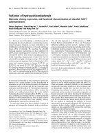
The Oct-1 decoy oligonucleotides effectively competed
with the Oct-1 consensus probe for an Oct-1 binding in
vitro
We first tested whether the decoy oligonucleotide
designed for the Oct-1 transcription factor can effectively
compete with the Oct-1 consensus probe in vitro. For this
study, a 24 bp DNA fragment containing the Oct-1 con-
sensus sequence was radioactively labeled with [γ-
32
P]-
ATP and used as a probe for the in vitro DNA-protein bind-
ing study. The DNA probe was incubated with HeLa
nuclear extracts at room temperature for one hour to
allow for the Oct-1 transcription factor to bind to the
DNA probe. For a competitive binding, the HeLa nuclear
extracts were incubated with both the radioactively
labeled Oct-1 DNA probe and the unlabeled Oct-1 decoy
oligonucleotide at room temperature for one hour. The
reactants were then analyzed by polyacrylamide gel elec-
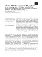
trophoresis using a 6% gel (Fig. 1). The binding of the
Oct-1 transcription factor caused a mobility shift of the
Oct-1 DNA probe in the gel (Fig. 1, lane 2 vs lane 1).
When the HeLa nuclear extracts were incubated with both
the radioactively labeled Oct-1 DNA probe and the unla-
beled Oct-1 decoy oligonucleotide, however, a dose-
dependent reduction in the level of the shifted Oct-1 DNA
probe was observed (Fig. 1, lanes 9–11). The Oct-1 tran-
scription factor binding-caused gel mobility shift of the
Oct-1 DNA probe was completely diminished when 1 ×
10
-6
M of the Oct-1 decoy oligonucleotide was supple-
mented in the binding reaction (Fig. 1, lane 11 vs lane 2).
As a control, no clear reduction in the level of the shifted
Oct-1 DNA probe was observed when similar concentra-
Journal of Hematology & Oncology 2009, 2:15 />Page 5 of 11
(page number not for citation purposes)
tions of the scrambled oligonucleotide were supple-
mented into the binding reactions (Fig. 1, lanes 6–8). This
result suggests that the Oct-1 decoy oligonucleotide effec-
tively competes with the Oct-1 consensus DNA probe for
the binding of the Oct-1 transcription factor.
The Oct-1 decoy oligonucleotide treatment did not affect
cell viability
To determine if the Oct-1 decoy oligonucleotide treat-
ment could result in reduced cell viability, we studied the
cell viability of the K562 human erythroleukemia cells
under the Oct-1 decoy oligonucleotide treatment. The
K562 cells were seeded at a density of 4 × 10
3
cells/ml and
treated with the Oct-1 decoy oligonucleotide at various
concentrations (0, 2, 5, and 10
μ
M). As controls, some
K562 cells were either untreated or treated with the scram-
bled oligonucleotide (10
μ
M). The cell density was deter-
mined at various time points (1, 2, 3, 4, and 5 days) and
the cell growth curve was determined for each treatment
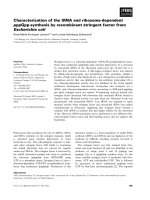
(Fig. 2). The cell growth was not significantly affected by
the Oct-1 decoy oligonucleotide treatment even with the
oligonucleotide at concentrations as high as 10
μ
M (Fig.
2).
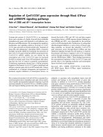
The Oct-1 decoy oligonucleotide treatment caused an
increase in expression of the γ-globin gene in K562 cells
The results obtained from our in vitro protein binding
assays revealed a strong competitive binding of the Oct-1
decoy oligonucleotide for the Oct-1 transcription factor.
To determine if the Oct-1 decoy oligonucleotide can
induce expression of endogenous
γ
-globin genes, the
K562 cells were treated with the Oct-1 decoy oligonucle-
otide and the effect of the decoy oligonucleotide treat-
ment on
γ
-globin expression was determined.
We first determined the Oct-1 decoy oligonucleotide treat-
ment-induced transcription of the
γ
-globin gene. The
K562 cells were either untreated or treated with the Oct-1
decoy oligonucleotide at various concentrations (0, 2, 5,
and 10
μ
M). As controls, some K562 cells were treated
with the scrambled oligonucleotide (10
μ
M) or hemin
(75
μ
M), an effective hemoglobin inducer [30-32]. At var-
ious time points, the cells were harvested and total RNA
was isolated. A real time PCR assay was then performed to
determine the level of the
γ
-globin mRNA in each RNA
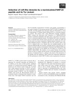
sample (Fig. 3A). When treated with the scrambled oligo-
nucleotide at 10
μ
M, no significant increase in the level of
γ
-globin mRNA was observed in the K562 cells (Fig. 3A).
When treated with the Oct-1 decoy oligonucleotide, how-
ever, significant increases in the level of the
γ
-globin
mRNA were detected in the K562 cells even with the Oct-
1 decoy oligonucleotide at concentrations as low as 2
μ
M.
For example, when the K562 cells were treated with 5 μ
MOct-1 decoy oligonucleotide for two, three, four and
five days, the level of the
γ
-globin mRNA increased 1.92,
2.93, 2.85, and 5.13 fold respectively. As a positive con-
trol, when the K562 cells were treated with 75
μ
M hemin
Determination of the competitive binding of the Oct-1 decoy oligonucleotide in vitroFigure 1
Determination of the competitive binding of the Oct-
1 decoy oligonucleotide in vitro. The radioactively
labeled Oct-1 consensus DNA probe was incubated with
HeLa nuclear extracts at room temperature for one hour
and then analyzed by polyacrylamide gel electrophoresis
using a 6% gel. Some reactions also contained the unlabeled
Oct-1 decoy oligonucleotide or the scrambled oligonucle-
otide for competitive binding. The Oct-1 binding-caused gel
mobility shift was confirmed by a western blot hybridization
assay using an Oct-1 antibody (data not shown).
The cell viability of K562 cells under the Oct-1 decoy oligo-nucleotide treatmentFigure 2
The cell viability of K562 cells under the Oct-1 decoy
oligonucleotide treatment. The K562 cells were seeded
at a density of 2 × 10
4
cells/ml and treated with either the
scrambled oligonucleotide or the Oct-1 decoy oligonucle-
otide at the concentrations indicated. The cell density was
determined at various time points (0, 1, 2, 3, 4, 5 and 6 days).
The cell growth curves represent the mean data of three
independent experiments.
Journal of Hematology & Oncology 2009, 2:15 />Page 6 of 11
(page number not for citation purposes)
for two, three, four, and five days, the levels of the
γ
-globin
mRNA increased 2.41, 4.47, 4.00, and 3.71 fold respec-
tively (Fig. 3A). This result suggests that the Oct-1 decoy
oligonucleotide treatment increases the transcription of
the
γ
-globin genes in the K562 cells as effectively as that of
hemin, a well-known γ-globin inducer.
Although the results obtained from our real time PCR
study demonstrated increases in transcription of the
γ
-
globin genes in the Oct-1 decoy oligonucleotide-treated
K562 cells, whether these increases in transcription led to
increases in the protein level of fetal hemoglobin (HbF,
α2γ2) was unknown. Therefore, we further performed an
immuno-blotting study to determine the level of HbF in
the Oct-1 decoy oligonucleotide-treated K562 cells (Fig.
3B). Indeed, the results obtained from our immuno-blot-
ting studies revealed that the treatment of the Oct-1 decoy
oligonucleotide (2, 5, or 10
μ
M) resulted in increases in
the level of HbF in the K562 cells (Fig. 3B). As a negative
control, the treatment of the K562 cells with 10
μ
M scram-
bled oligonucleotide did not cause a significant increase
in the level of HbF (Fig. 3B). As a positive control, the
treatment with 75
μ
M hemin also resulted in an increase
in the level of HbF in the K562 cells (Fig. 3B).
The Oct-1 decoy oligonucleotide-induced expression of the endogenous
γ
-globin genes in K562 cellsFigure 3
The Oct-1 decoy oligonucleotide-induced expression of the endogenous
γ
-globin genes in K562 cells. The K562
cells were treated with either the decoy or the scrambled oligonucleotides at indicated concentrations. As a positive control,
some K562 cells were treated with 75 μM hemin. Total RNA was isolated from both untreated and treated cells at various
time points following the treatment. The level of
γ
-globin mRNA was determined by a reverse transcription-based quantitative
PCR (real time PCR). In order to determine the level of HbF in the treated K562 cells, the cells were treated with the decoy
or the scrambled oligonucleotide for four days and then harvested and analyzed by western blots using an antibody that recog-
nized the HbF. (A) The Oct-1 decoy oligonucleotide treatment-induced
γ
-globin transcription as determined by real time PCR
assay. The level of the
γ
-globin mRNA in the untreated K562 cells was counted as 100% and the levels of the
γ
-globin mRNA in
the treated K562 cells were calculated as relative levels to that of the untreated K562 cells. The results are from at least three
independent experiments. (B) The Oct-1 decoy oligonucleotide treatment-induced accumulation of HbF in the K562 cells. The
results are from three individual experiments. * Statistical significance between the untreated and the treated K562 cells at the
same time point with p < 0.01.
Journal of Hematology & Oncology 2009, 2:15 />Page 7 of 11
(page number not for citation purposes)
These results suggest that the treatment of K562 human
erythroleukemia cells with the Oct-1 decoy oligonucle-
otide caused an increased expression of the endogenous
γ
-
globin genes in the K562 cells.
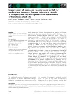
The Oct-1 decoy oligonucleotide treatment caused a
reduction in binding of the Oct-1 transcription factor to
the promoter region sequence of the endogenous γ-globin
gene
To determine whether the increased
γ
-globin gene tran-
scription in the Oct-1 decoy oligonucleotide-treated K562
cells was caused by the competitive binding of the Oct-1
decoy oligonucleotide to the Oct-1 transcription factor,
we further performed an immunoprecipitation (IP) study
[33,34] to determine the binding status of the Oct-1 bind-
ing sites within the promoter region of the endogenous γ-
globin gene under the Oct-1 decoy oligonucleotide treat-
ment. Both untreated and the Oct-1 decoy oligonucle-
otide-treated K562 cells were fixed in 1% formaldehyde
and sonicated to shear the chromosomal DNA into small
fragments. The Oct-1 transcription factor was then immu-
noprecipitated from both the untreated and the decoy oli-
gonucleotide-treated K562 cells by the IP protocol using
an Oct-1 antibody and Protein A-conjugated agarose
beads. As controls, the Oct-1 transcription factor was also
immunoprecipitated from the scrambled oligonucleotide
and hemin-treated K562 cells. Half of the beads were ana-
lyzed by western blot to determine the level of Oct-1 pro-
tein precipitated from each cell lysate (Fig. 4A) and rest of
the beads were analyzed by a quantitative PCR assay to
determine the level of the
γ
-globin gene promoter region
DNA co-precipitated with the Oct-1 transcription factor
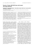
from each cell lysate (Fig. 4B). The results of our western
blots revealed that similar amounts of Oct-1 transcription
factor were precipitated from individual cell lysates, sug-
gesting a very successful IP of the study. The results of our
quantitative PCR studies indicated that the Oct-1 decoy
oligonucleotide treatment caused significant decreases in
the level of the endogenous
γ
-globin gene promoter
region DNA co-precipitated with the Oct-1 transcription
factor by the IP protocol. In comparison to the amount of
γ
-globin gene promoter region DNA co-precipitated with
the Oct-1 transcription factor in the untreated K562 cells,
only 44% and 17% of the
γ
-globin gene promoter region
DNA was co-precipitated with the Oct-1 transcription fac-
tor when the K562 cells were treated with 2
μ
M and 10
μ
M
Oct-1 decoy oligonucleotide, respectively. When the K562
cells were treated with 10 μM scrambled oligonucleotide,
however, more than 83% of the
γ
-globin gene promoter
region DNA was still co-precipitated with the Oct-1 tran-
scription factor. The treatment of K562 cells with 75
μ
M
hemin also resulted in a decrease in the level of the co-pre-
cipitated
γ
-globin gene promoter region DNA to 61% of
the untreated K562 cells. This result revealed that the
treatment of K562 cells with the Oct-1 decoy oligonucle-
otide caused a dose-dependent decrease in the binding of
Oct-1 transcription factor to the endogenous
γ
-globin
gene promoter region DNA sequence, suggesting that the
Oct-1 decoy oligonucleotide effectively competed with
the endogenous
γ
-globin gene promoter region Oct-1 con-
sensus sequences for binding with the Oct-1 transcription
factor, which resulted in activation of the endogenous
γ
-
globin gene and increases in the level of
γ
-globin mRNA
and HbF in the K562 cells.
Discussion
In this work, we have studied the possibility of using a
decoy oligonucleotide targeting the Oct-1 transcription
factor to induce expression of the endogenous
γ
-globin
gene. The results obtained from our in vitro protein bind-
ing study indicate that the Oct-1 decoy oligonucleotide
effectively competes with the Oct-1 consensus DNA probe
for binding to the Oct-1 transcription factor. The results
obtained from our Oct-1 decoy oligonucleotide treatment
studies demonstrate that the treatment of the K562 cells
with the Oct-1 decoy oligonucleotide results in an
increase in transcriptions of the
γ
-globin genes and an
increase in accumulation of the HbF in the K562 cells.
These results provide strong evidence to suggest that the
decoy oligonucleotide targeting the Oct-1 transcription
factor consensus sequence can be used to induce expres-
sion of the endogenous
γ
-globin gene. Since increased
expression of the
γ
-globin gene has already shown its clin-
ical benefit in the treatment of β-globin disorders such as
sickle cell anemia and β-thalassemia, this work provides a
novel approach for the treatment of these diseases. In
addition, the knowledge obtained from this study may
also lead to innovative strategies for the treatment of
many other disease conditions via manipulating expres-
sion of the desired target genes through the decoy oligo-
nucleotides.
The target of the decoy oligonucleotide used in this study
is the Oct-1 transcription factor consensus sequence. It is
known that the endogenous
γ
-globin genes promoter
region contains three Oct-1 transcription factor-binding
sites, the -280, -220 and -175 binding sites, respectively
[1]. The naturally occurring mutations at the -175 Oct-1
binding-site of the
γ
-globin gene lead to elevated levels of
HbF in individuals with hereditary persistence of fetal
hemoglobin (HPFH) [11-13]. Biochemistry studies also
reveal that these mutations diminish the binding of the
Oct-1 transcription factor to the site. The results obtained
from our previous studies demonstrated that mutations
generated at the -280 Oct-1 binding site of the
γ
-globin
gene cause increased expressions of the
γ
-globin gene [14].
The results obtained from this study further reveal that the
treatment of the K562 cells with the Oct-1 decoy oligonu-
cleotide leads to the activation of the endogenous
γ
-
globin genes. All of these results suggest that the Oct-1
Journal of Hematology & Oncology 2009, 2:15 />Page 8 of 11
(page number not for citation purposes)
transcription factor negatively regulates the transcription
of the endogenous
γ
-globin gene expression. Therefore,
these results provide important insight into the mecha-
nism of
γ
-globin gene regulation and a possible mecha-
nism regarding the
γ
-globin gene silencing.
Many studies have demonstrated the down-regulation of
the target genes' expression using decoy oligonucleotides
[15,16,20-29,35-38]. In comparison, much less work has
been conducted in the exploration of up-regulation of
gene expression for the desired target genes using the
decoy oligonucleotide strategy although the clinical rele-
vance of up-regulation of target genes for the treatment of
many disease conditions clearly exists. One of the obsta-
cles for this up-regulation is to identify the transcription
factors that negatively regulate transcription of the target
genes. It is also a great challenge to develop a methodol-
ogy that can sequence-specifically disrupt the interactions
The effect of Oct-1 decoy oligonucleotide treatment on binding of Oct-1 transcription factor to the endogenous γ-globin gene promoter region DNA in K562 cellsFigure 4
The effect of Oct-1 decoy oligonucleotide treatment on binding of Oct-1 transcription factor to the endog-
enous γ-globin gene promoter region DNA in K562 cells. The K562 cells were treated with either the decoy or the
scrambled oligonucleotides for four days. The cells were fixed in 1% formaldehyde and sonicated to lyse the cells. An immuno-
precipitation protocol was performed to pull down the Oct-1 transcription factor using an Oct-1 antibody and the Protein A-
conjugated agarose beads. Half of the beads were analyzed by western blots to determine the level of Oct-1 transcription fac-
tor precipitated by the IP and the rest of the beads were treated in 5 M NaCl at 65°C for four hours to reverse the protein-
DNA crosslinks and the recovered DNA was analyzed by a quantitative PCR (qPCR) protocol to determine the level of γ-
globin gene promoter region DNA co-precipitated with the Oct-1 transcription factor. A pair of primers that bind to the
γ
-
globin gene at the -350 to -330 and +50 to + 30 region sequences respectively was used in the qPCR study. (A) Detection of
the Oct-1 transcription factor precipitated by the IP protocol. (B) Quantification of the levels of γ-globin gene promoter region
DNA co-precipitated with the Oct-1 transcription factor in the IP assay. The results are from three independent experiments.
Journal of Hematology & Oncology 2009, 2:15 />Page 9 of 11
(page number not for citation purposes)
between the transcription factors and their consensus
sequences. The work described here takes advantage of the
well-studied human
γ
-globin gene by disrupting the bind-
ing of Oct-1 transcription factor to the endogenous
γ
-
globin gene promoter region Oct-1 consensus sequences
using a decoy oligonucleotide, and therefore, achieving
activation of the endogenous
γ
-globin genes in the treated
cells. A similar strategy can also be applied to other target
genes by targeting different transcription factor consensus
sequences. Therefore, the work described in this study has
broad implications in the treatment of many disease con-
ditions.
The results obtained from our K562 cell study revealed
that the transcription of the γ-globin genes was signifi-
cantly increased when the K562 cells were treated with the
Oct-1 decoy oligonucleotide at concentrations as low as 2
μ
M, which is the lowest concentration of the decoy oligo-
nucleotide used in the study. Therefore, it is likely that the
transcription of the
γ
-globin genes can be achieved at even
lower concentrations of the decoy oligonucleotide. This
result suggests that the concentration of the Oct-1 decoy
oligonucleotide required to induce transcription of the
γ
-
globin genes is much lower than the concentrations of
antisense oligonucleotides used in most studies [39-42].
One possible explanation for this difference is the target
choice in the strategies: when the antisense strategy is used
to regulate expression of target genes, high levels of anti-
sense oligonucleotides need to be maintained inside the
cells in order to effectively inhibit translation from the
mRNA, which is continuously transcribed throughout the
treatment; when the decoy oligonucleotide strategy is
used, however, the limited copy numbers of the target
genes result in the requirement of much fewer molecules
of decoy oligonucleotide for efficient regulation of tran-
scription of the target genes. Therefore, the decoy oligonu-
cleotides strategy may provide a better approach in
regulating expression of the target genes than that of the
antisense strategy.
The results obtained from our real time PCR analysis indi-
cated that the transcription of the
γ
-globin genes was
maintained at high levels for a relatively long period of
time following a single Oct-1 decoy oligonucleotide treat-
ment. The mechanism that leads to this long duration of
transcription is unknown. One possibility is that the dou-
ble-stranded phosphorothioate-modified decoy oligonu-
cleotide is more stable than that of the single-stranded
phosphorothioate-modified oligonucleotides inside cells
and the requirement of fewer molecules of the decoy oli-
gonucleotide leads to this relatively long period of tran-
scription for the
γ
-globin gene. It is also possible that the
disruption of the Oct-1 transcription factor binding to its
endogenous gene targets due to the decoy oligonucleotide
has a prolonged effect on transcription of the genes,
which can also lead to the long duration of transcription
of the
γ
-globin gene following the Oct-1 decoy oligonucle-
otide treatment. Further studies are needed in order to
determine the mechanism that causes this extended tran-
scription duration of the
γ
-globin gene following the Oct-
1 decoy oligonucleotide treatment.
The results obtained from our IP studies revealed that the
Oct-1 decoy oligonucleotide treatment led to reduced
Oct-1 transcription factor binding at the
γ
-globin pro-
moter. This provides strong evidence to suggest that the
Oct-1 decoy oligonucleotide induces expression of the
γ
-
globin genes through its competitive binding with the
Oct-1 transcription factor, resulting in activation of the
γ
-
globin genes. The hemin treatment also led to reduced
Oct-1 binding at the
γ
-globin promoter. Although the
mechanism underlying this effect is not known, it is pos-
sible that the induced transcription of the
γ
-globin genes
by the hemin reduces the binding of the Oct-1 to the
γ
-
globin promoter.
The decoy oligonucleotides used in this study are phos-
phorothioate-modified oligonucleotides. The results
obtained from our cell viability study indicate that the
cells are relatively tolerant towards the decoy oligonucle-
otide treatment even with the decoy oligonucleotide at
concentrations as high as 10
μ
M. The in vitro protein-DNA
binding results also suggest that the Oct-1 transcription
factor has a strong binding affinity towards the decoy oli-
gonucleotide. The gene expression studies also demon-
strate the cell permeability for the decoy oligonucleotide.
Therefore, this modified decoy oligonucleotide is effective
in inducing expression of the endogenous
γ
-globin genes.
However, other modifications (e.g. peptide nucleic acids
(PNA)) are also available in the oligonucleotide synthesis.
Although some studies have already used these modifica-
tions in the decoy oligonucleotide synthesis [15,27,37],
whether or not these modifications can be used to induce
expression of endogenous gene targets is still largely
unclear. Further studies are needed to evaluate the effect
of these modifications on decoy oligonucleotide-induced
endogenous gene expression.
Competing interests
The authors declare that they have no competing interests.
Authors' contributions
XSX is involved in the overall study design, performed
data analysis, and drafted the manuscript. XH carried out
the gel mobility study, cell treatment, and real time PCR
study. GW is involved in the overall study design, carried
out the chromatin-immunoprecipitation (ChIP) experi-
ments, and involved in the manuscript preparation.
Journal of Hematology & Oncology 2009, 2:15 />Page 10 of 11
(page number not for citation purposes)
Acknowledgements
We thank L. Chuang, X. Zhang, and S. Lomonaco for their experimental
assistance. We also thank Dr. T. Kocarek and L. Wang for their critical
readings of this manuscript. Performance of this work was facilitated by the
Cell Culture Facility Core and the Imaging and Cytometry Facility Core of
the Environmental Health Sciences Center in Molecular and Cellular Toxi-
cology with Human Applications at Wayne State University (P30 ES06639).
This work is supported by the grant R01HL62551 from the National Heart,
Lung, and Blood Institute (NHLBI), NIH to G. W. and Wayne State Univer-
sity new faculty start-up fund to X.S.X.
References
1. Stamatoyannopoulos G, Nienhuis A, Majerus X, Varmus X: The
molecular Basis of Blood diseases. Second edition. W.B. Saun-
ders Company, Philadelphia, PA; 1994.
2. Stamatoyannopoulos G: Control of globin gene expression dur-
ing development and erythroid differentiation. Exp Hematol
2005, 33:259-271.
3. Chakalova L, Carter D, Debrand E, Goyenechea B, Horton A, Miles J,
Osborne C, Fraser P: Developmental regulation of the beta-
globin gene locus. Prog Mol Subcell Biol 2006, 38:183-206.
4. Pace BS, Zein S: Understanding mechanisms of gamma-globin
gene regulation to develop strategies for pharmacological
fetal hemoglobin induction. Dev Dyn 2006, 235:1727-1737.
5. Mahajan MC, Karmakar S, Weissman SM: Control of beta globin
genes. J Cell Biochem 2007, 102:801-810.
6. Yang YM, Pace B: Pharmacologic induction of fetal hemoglobin
synthesis: cellular and molecular mechanisms. Pediatr Pathol
Mol Med 2001, 20:87-106.
7. Wang G, Xu X, Pace B, Glazer PM, Chan P, Goodman SR, Shokolenko
I: Induction of human gamma-globin gene expression via
peptide nucleic acids (PNAs). Nucleic Acids Res 1999,
27:2806-2813.
8. Quek L, Thein SL: Molecular therapies in beta-thalassaemia. Br
J Haematol 2007, 136:353-365.
9. Lisowski L, Sadelain M: Current status of globin gene therapy
for the treatment of beta-thalassaemia. Br J Haematol 2008,
141:335-345.
10. Phillips K, Luisi B: The virtuoso of versatility: POU proteins
that flex to fit. J Mol Biol 2000, 302:1023-1039.
11. Surrey S, Delgrosso K, Malladi P, Schwartz E: A single base change
at position -175 in the 5'-flanking region of the Gγ-globin
gene from a black with Gγβ+-HPFH. Blood 1988, 71:
807-810.
12. Ottolenghi S, Nicolis S, Taramelli R, Malgaretti N, Mantovani R, Comi
P, Giglioni B, Longinotti M, Dore F, Oggiano LEA: Sardinian G
gamma-HPFH: a T-C substitution in a conserved "octamer"
sequence in the G gamma-globin promoter. Blood 1988,
71:815-817.
13. Stoming TA, Stoming GS, Lanclos KD, Fei YI, Altay C, Kutlar F, Huis-
man THJ: An Aγ type of nondeletional hereditary persistence
of fetal hemoglobin with a T->C mutation at position -175 to
the cap site of the Aγ globin gene. Blood 1989, 73:329-333.
14. Xu X, Glazer PM, Wang G: Activation of human γ-globin gene
expression via triplex-forming oligonucleotide (TFO)-
directed mutations in the γ-globin gene 5' flanking region.
Gene 2000, 242:219-228.
15. Gambari R: New trends in the development of transcription
factor decoy (TFD) pharmacotherapy. Curr Drug Targets 2004,
5:419-430.
16. Cutroneo KR, White SL, Chiu JF, Ehrlich HP: Tissue fibrosis and
carcinogenesis: divergent or successive pathways dictate
multiple molecular therapeutic targets for oligo decoy ther-
apies. J Cell Biochem 2006, 97:1161-1174.
17. Isomura I, Morita A: Regulation of NF-kappaB signaling by
decoy oligodeoxynucleotides. Microbiol Immunol 2006,
50:559-563.
18. Tomita N, Kashihara N, Morishita R: Transcription factor decoy
oligonucleotide-based therapeutic strategy for renal disease.
Clin Exp Nephrol 2007, 11:7-17.
19. Edwards MR, Bartlett NW, Clarke D, Birrell M, Belvisi M, Johnston
SL: (2009) Targeting the NF-kappaB pathway in asthma and
chronic obstructive pulmonary disease. Pharmacol Ther 2009,
121:1-13.
20. Eller MS, Yaar M, Ostrom K, Harkness DD, Gilchrest BA: A role for
interleukin-1 in epidermal differentiation: regulation by
expression of functional versus decoy receptors. J Cell Sci
1995, 108:
2741-2746.
21. Sharma HW, Perez JR, Higgins-Sochaski K, Hsiao R, Narayanan R:
Transcription factor decoy approach to decipher the role of
NF-kappa B in oncogenesis. Anticancer Res 1996, 16:61-69.
22. Tomita N, Horiuchi M, Tomita S, Gibbons GH, Kim JY, Baran D, Dzau
VJ: An oligonucleotide decoy for transcription factor E2F
inhibits mesangial cell proliferation in vitro. Am J Physiol 1998,
275:278-284.
23. Shiratsuchi T, Ishibashi H, Shirasuna K: Inhibition of epidermal
growth factor-induced invasion by dexamethasone and AP-1
decoy in human squamous cell carcinoma cell lines. J Cell Phys-
iol 2002, 193:340-348.
24. Wang LH, Yang XY, Zhang X, Mihalic K, Xiao W, Farrar WL: The cis
decoy against the estrogen response element suppresses
breast cancer cells via target disrupting c-fos not mitogen-
activated protein kinase activity. Cancer Res 2003,
63:2046-2051.
25. Novak EM, Metzger M, Chammas R, da Costa M, Dantas K, Manabe
C, Pires J, de Oliveira AC, Bydlowski SP: Downregulation of TNF-
alpha and VEGF expression by Sp1 decoy oligodeoxynucle-
otides in mouse melanoma tumor. Gene Ther 2003,
10:1992-1997.
26. Penolazzi L, Borgatti M, Lambertini E, Mischiati C, Finotti A, Romanelli
A, Saviano M, Pedone C, Piva R, Gambari R: Peptide nucleic acid-
DNA decoy chimeras targeting NF-kappaB transcription
factors: Induction of apoptosis in human primary osteo-
clasts. Int J Mol Med 2004, 14:145-152.
27. Borgatti M, Boyd DD, Lampronti I, Bianchi N, Fabbri E, Saviano M,
Romanelli A, Pedone C, Gambari R: Decoy molecules based on
PNA-DNA chimeras and targeting Sp1 transcription factors
inhibit the activity of urokinase-type plasminogen activator
receptor (uPAR) promoter. Oncol Res 2005, 15:373-383.
28. Fabian RH, Perez-Polo JR, Kent TA: A decoy oligonucleotide
inhibiting nuclear factor-kappaB binding to the IgGkappaB
consensus site reduces cerebral injury and apoptosis in neo-
natal hypoxic-ischemic encephalopathy. J Neurosci Res 2007,
85:1420-1426.
29. Osako MK, Tomita N, Nakagami H, Kunugiza Y, Yoshino M, Yuyama
K, Tomita T, Yoshikawa H, Ogihara T, Morishita R: Increase in
nuclease resistance and incorporation of NF-kappaB decoy
oligodeoxynucleotides by modification of the 3'-terminus.
J
Gene Med 2007, 9:812-819.
30. Tsiftsoglou AS, Wong W, Robinson SH, Hensold J: Hemin increase
production of beta-like globin RNA transcripts in human
erythroleukemia K-562 cells. Dev Genet 1989, 10:311-317.
31. Fibach E, Kollia P, Schechter AN, Noguchi CT, Rodgers GP: Hemin-
induced acceleration of hemoglobin production in immature
cultured erythroid cells: preferential enhancement of fetal
hemoglobin. Blood 1995, 85:2967-2974.
32. Tahara T, Sun J, Nakanishi K, Yamamoto M, Mori H, Saito T, Fujita H,
Igarashi K, Taketani S: Heme positively regulates the expression
of beta-globin at the locus control region via the transcrip-
tional factor Bach1 in erythroid cells. J Biol Chem 2004,
279:5480-5487.
33. Bigler J, Eisenman RN: Isolation of a thyroid hormone-respon-
sive gene by immunoprecipitation of thyroid hormone
receptor-DNA complexes. Mol Cell Biol 1994, 14:7621-7632.
34. Weinmann AS, Farnhamm PJ: Identification of unknown target
genes of human transcription factors using chromatin
immunoprecipitation. Methods 2002, 26:37-47.
35. Morishita R, Gibbons GH, Horiuchi M, Kaneda Y, Ogihara T, Dzau VJ:
Role of AP-1 complex in angiotensin II-mediated transform-
ing growth factor-beta expression and growth of smooth
muscle cells: using decoy approach against AP-1 binding site.
Biochem Biophys Res Commun 1998, 243:361-367.
36. Lee YN, Park YG, Choi YH, Cho YS, Cho-Chung YS: CRE-tran-
scription factor decoy oligonucleotide inhibition of MCF-7
breast cancer cells: cross-talk with p53 signaling pathway.
Biochemistry 2000, 39:4863-4868.
37. Borgatti M, Finotti A, Romanelli A, Saviano M, Bianchi N, Lampronti I,
Lambertini E, Penolazzi L, Nastruzzi C, Mischiati C, Piva R, Pedone C,
Gambari R: Peptide nucleic acids (PNA)-DNA chimeras tar-
Publish with BioMed Central and every
scientist can read your work free of charge
"BioMed Central will be the most significant development for
disseminating the results of biomedical researc h in our lifetime."
Sir Paul Nurse, Cancer Research UK
Your research papers will be:
available free of charge to the entire biomedical community
peer reviewed and published immediately upon acceptance
cited in PubMed and archived on PubMed Central
yours — you keep the copyright
Submit your manuscript here:
/>BioMedcentral
Journal of Hematology & Oncology 2009, 2:15 />Page 11 of 11
(page number not for citation purposes)
geting transcription factors as a tool to modify gene expres-
sion. Curr Drug Targets 2004, 5:735-744.
38. Cho JW, Kim JY, Kim CW, Lee KS: Down-regulation of TGF-
beta1-induced type I collagen synthesis by AP-1 transcrip-
tion factor decoy in scleroderma fibroblasts. J Dermatol Sci
2006, 43:207-209.
39. Bitko V, Barik S: Intranasal antisense therapy: preclinical mod-
els with a clinical future? Curr Opin Mol Ther 2007, 9:119-125.
40. Eckstein F: The versatility of oligonucleotides as potential
therapeutics. Expert Opin Biol Ther 2007, 7:1021-1034.
41. Spurgers KB, Sharkey CM, Warfield KL, Bavari S: Oligonucleotide
antiviral therapeutics: antisense and RNA interference for
highly pathogenic RNA viruses. Antiviral Res 2008, 78:26-36.
42. Rayburn ER, Zhang R: Antisense, RNAi, and gene silencing
strategies for therapy: mission possible or impossible? Drug
Discov Today 2008, 13:513-521.