báo cáo khoa học: "The transplant iron score as a predictor of stem cell transplant survival" pptx
Bạn đang xem bản rút gọn của tài liệu. Xem và tải ngay bản đầy đủ của tài liệu tại đây (433.25 KB, 9 trang )
BioMed Central
Page 1 of 9
(page number not for citation purposes)
Journal of Hematology & Oncology
Open Access
Research
The transplant iron score as a predictor of stem cell transplant
survival
Jonathan A Storey
1,7
, Rebecca F Connor
1,7
, Zachary T Lewis
2
, David Hurd
1,7
,
Gregory Pomper
2
, Yi K Keung
1,7
, Manisha Grover
3
, James Lovato
4,6
,
Suzy V Torti
5,7
, Frank M Torti*
1,6,7
and István Molnár
1,7
Address:
1
Department of Internal Medicine, Section on Hematology and Oncology, Wake Forest University School of Medicine, Winston-Salem,
NC, USA,
2
Department of Pathology, Wake Forest University School of Medicine, Winston-Salem, NC, USA,
3
Department of Medicine, New York
University Downtown Hospital, New York, NY, USA,
4
Department of Public Health Sciences, Wake Forest University School of Medicine, Winston-
Salem, NC, USA,
5
Department of Biochemistry, Wake Forest University School of Medicine, Winston-Salem, NC, USA,
6
Department of Cancer
Biology, Wake Forest University School of Medicine, Winston-Salem, NC, USA and
7
Comprehensive Cancer Center of Wake Forest University,
Wake Forest University School of Medicine, Winston-Salem, NC, USA
Email: Jonathan A Storey - ; Rebecca F Connor - ; Zachary T Lewis - ;
David Hurd - ; Gregory Pomper - ; Yi K Keung - ;
Manisha Grover - ; James Lovato - ; Suzy V Torti - ;
Frank M Torti* - ; István Molnár -
* Corresponding author
Abstract
Recent studies have suggested that the presence of iron overload prior to stem cell transplantation
is associated with decreased survival. Within these studies, the criteria used to define iron overload
have varied considerably. Given the lack of consensus regarding the definition of iron overload in
the transplant setting, we sought to methodically examine iron status among transplant patients.
We studied 78 consecutive patients at risk for transfusion-related iron overload (diagnoses
included AML, ALL, MDS, and aplastic anemia) who received either autologous or allogeneic stem
cell transplant. Multiple measures of iron status were collected prior to transplantation and
examined for their association with survival. Using this data, three potentially prognostic iron
measures were identified and incorporated into a rational and unified scoring system. The resulting
Transplant Iron Score assigns a point for each of the following variables: (1) greater than 25 red cell
units transfused prior to transplantation; (2) serum ferritin > 1000 ng/ml; and (3) a semi-
quantitative bone marrow iron stain of 6+. In our cohort, the score (range 0 to 3) was more closely
associated with survival than any available single iron parameter. In multivariate analysis, we
observed an independent effect of iron overload on transplant survival (p = 0.01) primarily
attributable to an increase in early treatment-related deaths (p = 0.02) and lethal infections. In
subgroup analysis, the predictive power of the iron score was most pronounced among allogeneic
transplant patients, where a high score (≥ 2) was associated with a 50% absolute decrease in
survival at one year. In summary, our results lend further credence to the notion that iron overload
prior to transplant is detrimental and suggest iron overload may predispose to a higher rate of
lethal infections.
Published: 24 October 2009
Journal of Hematology & Oncology 2009, 2:44 doi:10.1186/1756-8722-2-44
Received: 17 July 2009
Accepted: 24 October 2009
This article is available from: />© 2009 Storey et al; licensee BioMed Central Ltd.
This is an Open Access article distributed under the terms of the Creative Commons Attribution License ( />),
which permits unrestricted use, distribution, and reproduction in any medium, provided the original work is properly cited.
Journal of Hematology & Oncology 2009, 2:44 />Page 2 of 9
(page number not for citation purposes)
Introduction
Long-standing iron overload can lead to heart and liver
failure, resulting in premature death [1]. As our ability to
treat iron overload improves, it is increasingly important
to identify patients at risk for developing complications
secondary to iron overload. Stem cell transplant patients
are at risk for excess accumulation of iron resulting from
repeated blood transfusions both before and during trans-
plantation [2]. Because of this risk, it is recommended that
transplant survivors with good long-term prognoses be
assessed for iron overload [3]. Because iron overload has
been perceived to be of primarily long term detriment, the
measurement of iron status prior to transplant has not rou-
tinely been performed. However, recent evidence suggests
that the determination of iron status before transplant has
important prognostic implications [4-6].
Iron overload prior to transplantation was initially identi-
fied as a marker of poor prognosis in pediatric β-tha-
lassemia patients [7]. Among those allogeneic transplant
recipients, the presence of iron-induced portal fibrosis or
hepatomegaly was associated with decreased survival. A
later study by Altes et al. suggested that iron overload also
adversely impacted those with hematologic malignancies
[4]. In that study, very high levels of serum ferritin and
transferrin saturation greater than 100% were used as sur-
rogates for iron overload. Meanwhile, a larger study by
Armand et al. defined iron overload based solely on
serum ferritin, using the highest quartile for each disease
type [6]. Using that definition of iron overload, a signifi-
cant association with transplant survival was seen in
patients with myelodysplastic syndrome (MDS) and acute
myeloid leukemia (AML).
While each of these retrospective studies suggests that iron
overload adversely affects transplant outcome, the clinical
definition of iron overload varied considerably between
studies. We set out to examine multiple measures of pre-
transplant iron status with the goal of determining which
marker(s) were most closely associated with clinical out-
come following transplant. We chose to study patients at
risk for transfusion related iron overload (diagnoses
included acute leukemia, MDS, and aplastic anemia)
undergoing either autologous or allogeneic transplant.
Three measures related to transfusional iron overload
were closely associated with transplant survival: (1)
number of blood unit transfusions, (2) serum ferritin, and
(3) bone marrow iron stores. These readily available
measures were combined into a clinical scoring system
termed the Transplant Iron Score.
The Transplant Iron Score showed a strong independent
association with overall survival. Our findings further val-
idate the detrimental impact of iron overload in the set-
ting of stem cell transplantation and identify a potential
mechanism of action.
Methods
We evaluated 78 consecutive adult patients admitted to
the Wake Forest transplant unit with a diagnosis of AML,
MDS, acute lymphoblastic leukemia (ALL), or aplastic
anemia. The included patients were all undergoing their
first hematopoietic stem cell transplant between Septem-
ber 9, 1999 and March 19, 2004. The patient demograph-
ics and characteristics are summarized in Table 1. This
study was approved by both the Protocol Review Com-
mittee of the Comprehensive Cancer Center of Wake For-
est University and the Institutional Review Board of Wake
Forest University School of Medicine.
All serum samples were obtained upon admission to our
bone marrow transplant unit, prior to the initiation of the
preparative chemotherapy. Samples were continuously
stored at -20°C, until measurements of iron parameters
were performed. Serum ferritin levels were measured
using a two-site chemiluminometric sandwich immu-
noassay (ADVIA Centaur
®
Ferritin assay, Bayer Diagnos-
tics, Tarrytown, NY). Transferrin saturation was calculated
using the method by Huebers and Finch [8]. Serum levels
of transferrin receptor (sTfR) were measured using a com-
mercially available sandwich enzyme immunoassay (EIA)
(Ramco Laboratories, Inc. Stafford, TX). C-reactive protein
was measured using an enzyme-linked immunosorbent
assay (high sensitivity) kit from American Laboratory
Products Company, Windham, NH. The kit shows no
cross-reactivity against albumin, lysozyme, alpha-1 antit-
rypsin and other acute phase proteins. Values for aspartate
transaminase (AST), alanine transaminase (ALT), total
bilirubin (TB), and the international normalized ratio
(INR) for blood clotting time were obtained by review of
the medical record. All recorded values were within 1
month of the admission date to our unit.
Bone marrow samples obtained within two months of
transplantation were reviewed by two of the authors for
specimen adequacy (I.M. and Z.L.), and representative
sections of the patient's samples were stained with the
Gomori's iron stain method [9]. Iron content of the bone
marrow was graded by one of the authors (Z.L.) who was
blinded to patients' laboratory and clinical parameters.
Grading of marrow iron stores was scored according to
previously published methods using a 0 to 6+ classifica-
tion scheme described in detail by Gale et al [10]. Higher
grades were associated with increased visible iron with a
score of 6+ having large visible iron clumps that obscure
cellular details. The number of packed red cell blood cell
(pRBC) transfusions prior to transplantation was deter-
mined by blood bank records. The ejection fraction (EF)
for each patient was based on the most recent pre-trans-
Journal of Hematology & Oncology 2009, 2:44 />Page 3 of 9
(page number not for citation purposes)
plant echocardiogram or MUGA scan. All measures of EF
were performed within 3 months of stem cell transplant.
The highest quartile of total transaminases, TB, and INR
among our study group was used as a surrogate for early
liver dysfunction, while the lowest quartile of EFs was
used to identify early pre-existing heart dysfunction.
Based on univariate quartile analysis, the three iron
parameters with the strongest survival association were
identified. Cutoff values for each parameter were deter-
mined independently using comparative statistics. Using
this method, multiple pre-defined cutoffs were examined
and compared for their association with survival. In order
to maintain an adequate sample size on both sides of the
cutoff, only values within the second and third quartile
were considered. The selected cutoff values demonstrated
the highest ability to discriminate survival based on a
comparative analysis of hazard ratios.
Ultimately, these three iron parameters were incorporated
into a unified scoring system. For each patient, a score was
calculated by assigning a single point for each of the fol-
lowing: (1) serum ferritin ≥ 1,000 ng/mL, (2) greater than
25 transfused units of red cells, and (3) marrow iron stain
of 6+. The sum of points, ranging between 0 and 3, was
defined as the Transplant Iron Score. For missing data, no
points were assigned. In the single patient where less than
two parameters were available for scoring, the iron score
was deemed indeterminate and was excluded from addi-
tional statistical analysis. The remaining 77 patients (99
percent) were included for analysis in our study. To allow
further analysis within our study, a score of two or greater
was deemed "high" and those patients were considered to
have transfusion related iron overload. Meanwhile, a
score of zero or one was classified as a "low" score. For
purposes of comparing the Transplant Iron Score to other
individual or combinations of iron parameters, each iron
parameter was scaled be scored on a 0 to 3 scale. Using
this approach, the individual iron parameters were
divided into one of four quartile groups, similar to the
four possible score groups defined by the Transplant Iron
Score. Based on these groupings, a univariate relative risk
of death was calculated for each iron parameter using haz-
ard regression analysis.
Survival time was measured from the date of transplant to
the date of death or last known follow-up. All data was
censored as of July 1
st
2007. The following clinical and
demographic parameters were collected for statistical
analysis: age at the time of transplant, gender, diagnosis,
Table 1: Patient characteristics
Patient Characteristics All Patients Number High Iron Score number (percent) Low Iron Score number (percent)
Number 77 27 50
Median age 46 49 44
Sex
Male 38 15 (56) 23 (46)
Female 39 12 (44) 27 (54)
Diagnosis
AML 55 18 (67) 37 (74)
ALL 9 5 (19) 4 (8)
MDS 8 3 (11) 5 (10)
Aplastic anemia 5 1 (4) 4 (8)
Cytogenetics
Favorable 3 2 (7) 1 (2)
Average 29 9 (33) 20 (40)
Poor 20 7 (26) 13 (26)
Disease state
Non-proliferative 14 4 (15) 10 (20)
First remission 35 6 (22) 29 (58)
Second remission 17 9 (33) 8 (16)
No remission 11 8 (30) 3 (6)
Transplant type
Autologous 31 8 (30) 23 (46)
Allogeneic 46 19 (70) 27 (54)
Matched related 27 9 (33) 18 (36)
Unrelated 19 10 (37) 9 (18)
Non-ablative 9 4 (15) 5 (10)
Values indicate the number of patients unless otherwise indicated. Percentages (%) may not add up to 100 due to rounding. A high iron score refers
to a Transplant Iron Score of 2 or 3, while a low score represents a 0 or 1. AML indicates acute myeloid leukemia; ALL, acute lymphoblastic
leukemia; MDS myelodysplastic syndrome; AA, aplastic anemia.
Journal of Hematology & Oncology 2009, 2:44 />Page 4 of 9
(page number not for citation purposes)
disease status (no remission, first remission, second
remission), transplant type (autologous, related or unre-
lated allogeneic stem cell transplantation), and cytoge-
netic data for acute myeloid leukemia patients at the time
of diagnosis. Cytogenetic information was grouped into
poor, average and favorable categories based on the study
by Byrd et al [11]. The specific cause of death for each
patient was determined by chart review and categorized
into disease related mortality, treatment related mortality,
or not determined. All deaths following documented dis-
ease relapse were categorized as disease related mortality.
Deaths attributable to treatment related mortality were
further subdivided into deaths resulting from infection,
graft-versus-host disease (GVHD), or veno-occlusive dis-
ease (VOD) of the liver. Documented infectious deaths
were defined by the presence of a positive culture. Cases
of suspected lethal infection met strict criteria for sepsis
including radiologic imaging consistent with infection
[12]. Kaplan-Meier curves were used to estimate median
survival and overall survival differences. Cox proportional
hazards regression was used to perform multivariate anal-
ysis. Results were considered significant when p-values
were less than 0.05.
Results
Multiple measures related to iron homeostasis were col-
lected prior to stem cell transplant and are listed in Table
2. The individual iron parameter most closely associated
with overall survival was the transfusion total, defined as
the number of red cell units received prior to transplant.
For each increase in quartile (e.g. 50
th
to 75
th
quartile) of
transfused blood, the risk of death following transplant
increased by a factor of 1.4. Serum ferritin was also signif-
icantly associated with transplant survival (p = 0.02),
while the marrow iron stain showed a strong trend
towards statistical significance (p = 0.08). Though not sig-
nificant by quartile analysis, a bone marrow iron stain
score of +6 was significantly associated with increased
mortality (p = 0.04). The number of patients above the
cutoff for transfusion number, ferritin, and iron stain were
30, 41, and 7, respectively. The Transplant Iron Score,
when compared with individual and a combination of
iron parameters, was most closely associated with survival
(p = 0.0006). The risk of death nearly doubled with each
point increase of the Transplant Iron Score.
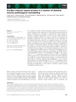
Trend analysis further supported that higher Transplant
Iron Scores were associated with decreased overall survival
(Figure 1A, p = 0.0003 by log-rank trend). The median
survival for patients with no evidence of iron overload
(score of 0) was estimated at over 6 years. Patients with
higher scores had lower median survival times: 2.4 years
for a score of 1; 6.5 months for a score of 2; and 8 days in
those patients with a score of 3. The 27 patients (35%)
with a high score had a substantially lower median sur-
vival of 5.0 months compared to 29.3 months in those
with a low score (Figure 1B). The unadjusted hazard ratio
associated with a high score was 2.60 (95% CI of 1.47 to
4.61). Our sample size did not allow us to perform rigor-
ous subgroup analyses by disease type, however we did
note that ALL (p = 0.02), AML (p = 0.06), and MDS (p =
0.004) patients with a high iron score exhibited a signifi-
cant decrease in survival. Iron overload also resulted in
decreased survival among aplastic anemia patients, how-
ever this did not reach statistical significance (p = 0.35).
The increase in mortality associated with iron overload
resulted primarily from early deaths. In the first six
months following transplant, 56% of those with a high
iron score had died as compared to 22% among those
with a low score (Figure 1B). This equates to a 34% abso-
lute risk associated with transfusion related iron overload
Table 2: Association of iron parameters on transplant survival
Iron Parameter(s) Median Relative Risk p-value Included in iron score
Blood Transfusions (units) 22 1.40 0.007 YES
Serum Ferritin (ng/mL) 1103 1.36 0.02 YES
Marrow Iron Stain Grade
10
4+ 1.34 0.08 YES
Transferrin 193 0.77 0.11 No
Transferrin Receptor 6.1 0.80 0.12 No
Serum Iron (mcg/dL) 90 0.91 0.50 No
Transferrin Saturation (%) 30 1.08 0.54 No
Ferritin + Transfusions* 1.43 0.002
Ferritin + Iron Stain* 1.49 0.010
Transfusion + Iron Stain* 1.58 0.003
Transplant Iron Score* 1.77 0.0006
The relative risk represents the relative risk of death associated with each incremental increase in quartile (e.g. 50
th
to 75
th
quartile) among the iron
parameters. The following cutoff values were used to assign patients to the various groupings: (1) serum ferritin ≥ 1,000 ng/mL, (2) greater than 25
transfused units of red cells, and (3) bone marrow iron stain of 6+. The Transplant Iron Score is calculated by assigning patients one point for each
of the values above the cutoff.
*The calculated relative risks were scaled (i.e. scored on a 0-3 scale) to allow comparisons to the individual iron quartiles.
Journal of Hematology & Oncology 2009, 2:44 />Page 5 of 9
(page number not for citation purposes)
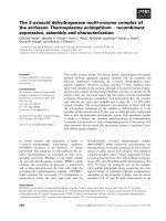
Overall survival stratified by the Transplant Iron ScoreFigure 1
Overall survival stratified by the Transplant Iron Score. Patients were stratified based on the calculated Transplant
Iron Score. (A) Score of 0 to 3 as defined by the scoring system. (B) High score (≥ 2) versus a low score (0 or 1). A number at
risk table is included for each score group.
Journal of Hematology & Oncology 2009, 2:44 />Page 6 of 9
(page number not for citation purposes)
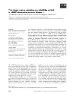
within the first six months. Subsequently, mortality rates
were nearly identical between groups after the six-month
time point. The difference in early survival was primarily
due to an increased number of treatment related deaths (p
= 0.018) (Figure 2B). Meanwhile, iron overload was not
associated with a significant increase in relapse rate (p =
0.84) or disease related mortality (p = 0.40). The effect of
iron overload was most pronounced among the patients
undergoing allogeneic transplantation (p < 0.001) with a
50% absolute mortality difference at one year. Addition-
ally, of the 19 allogeneic transplant patients with a high
iron score, only two patients survived more than three
years after transplant.
We further examined the cause of death among the 20
patients that died as a result of treatment related compli-
cations. The majority of these patients (55%) had either
documented (5 patients) or suspected (6 patients) lethal
infection. Furthermore, the rate of infection related mor-
tality was disproportionately high among those with a
high iron score (26%) as compared to those with a low
score (8%) (p = 0.04 by Fisher's exact test). Clinical infor-
mation regarding the 7 patients with a high iron score
with infection-related mortality is detailed in Table 3.
Rates of graft-versus-host disease (GVHD) and veno-
occlusive disease (VOD) of the liver were statistically sim-
ilar between groups (p = 0.9 and p = 0.3 by Fisher's exact
test, respectively).
Multivariate analysis was used to establish whether the
Transplant Iron Score was independently associated with
transplant survival. Covariables included established pre-
dictors of transplant outcome, such as age, gender, donor-
type, and remission status. In addition to standard trans-
Transplant outcomes stratified by the Transplant Iron ScoreFigure 2
Transplant outcomes stratified by the Transplant Iron Score. (A) Disease related mortality for all patients. (B) Treat-
ment related mortality for all patients. (C) Overall survival of autologous transplant patients. (D) Overall survival of allogeneic
transplant patients.
Journal of Hematology & Oncology 2009, 2:44 />Page 7 of 9
(page number not for citation purposes)
plant risk factors, an inflammatory marker (C-reactive
protein) was included along with measures of end-organ
damage (transaminase levels, INR, TB, and EF). Because
frank organ failure is not likely to be present in eligible
transplant patients, quartiles were used in the evaluation
of end-organ damage in an attempt to identify early organ
damage (i.e. mild transaminitis or a low-normal ejection
fraction). The Transplant Iron Score had a significant
independent effect on overall survival (p = 0.01) (Table
4). Among the subgroup of patients with AML, cytoge-
netic data (a marker for aggressive disease) did not influ-
ence the significance of this finding when added to the
multivariate model. Furthermore, the iron score was inde-
pendently associated with treatment related mortality (p =
0.04), infection related mortality (p = 0.02), and alloge-
neic transplant mortality (p = 0.03). When analyzing the
subgroup of allogeneic transplant patients, a statistically
significant difference in treatment related mortality (p =
0.01) and infection related mortality (p = 0.03) was main-
tained.
Discussion
Estimating systemic iron stores in stem cell transplant
patients is challenging. Serum ferritin has frequently been
used as an estimate of systemic iron stores, but is prone to
false elevation in the setting of inflammation and malig-
nancy [13]. Other blood markers, such as transferrin satu-
ration and soluble transferrin receptor, have proven to be
even less successful in establishing the diagnosis of iron
overload [14,15]. Non-invasive imaging techniques, such
as T2* MRI, show promise for determining tissue iron
stores but have not been extensively studied in transplant
patients [16]. The current "gold standard" for assessing
systemic iron overload remains dependent on liver biopsy
[17]. However, invasive procedures are often not practical
in patients awaiting stem cell transplant, as thrombocyto-
penia and neutropenia are common. Because of these dif-
ficulties, there is no consensus on how to best determine
iron status in the transplant setting [18].
We identified three clinical markers of iron overload that
were associated with decreased survival: (1) transfusion
burden, (2) serum ferritin, and (3) bone marrow iron
Table 3: Clinical characteristics of the patients with a high iron score and treatment-related death AML indicates acute myeloid
leukemia
Demographics Disease Donor Conditioning Iron Score (pRBCs) Survival Cause of Death
53 y/o male ALL (CR2) MRD Cytoxan/TBI 2 (45) 16 days Clostridial sepsis
24 y/o female AML (CR1) MUD Busulfan/Cytoxan 2 (42) 26 days Septic shock (culture negative)
48 y/o female AML (CR2) MRD Cytoxan/TBI 3 (38) 8 days Pneumonia/ARDS
62 y/o female Refractory AML MRD Non-ablative 2 (43) 50 days CMV pneumonia
50 y/o female MDS MUD Cytoxan/TBI 3 (35) 6 days Pneumonia/ARDS
32 y/o male AML (CR1) MRD Cytoxan/TBI 2 (58) 13 days Septic shock (culture negative)
49 y/o female Aplastic anemia MUD Cytoxan/TBI 2 (30) 27 days Gram-negative sepsis
ALL, acute lymphoblastic leukemia; MDS myelodysplastic syndrome; MRD matched related donor; MUD matched unrelated donor; TBI total body
irradiation; ARDS acute respiratory distress syndrome.
Table 4: Multivariate analysis of prognostic factors for stem cell transplant survival
Potential Risk Factors Covariables Hazard Ratio 95% CI p-value
Iron Overload Iron Score (0-3) 1.8 1.1 to 2.7 0.01
BMT Risk Factors
Age <40, 40s, 50s, 60s 1.5 0.9 to 2.3 0.08
Gender Male 0.8 0.4 to 1.6 0.54
Donor-Type Auto, Sibling, MURD 1.6 0.9 to 2.7 0.06
Remission Status CR1, CR2, No remission 1.1 0.7 to 1.7 0.70
End-Organ Damage
Heart Damage Ejection Fraction 1.4 1.0 to 1.9 0.03
Liver synthetic function INR 1.3 0.9 to 1.9 0.10
Hepatocellular damage AST + ALT 0.8 0.6 to 1.2 0.32
Liver obstruction Total Bilirubin 0.8 0.6 to 1.1 0.25
Inflammation C-reactive protein 1.0 0.7 to 1.4 0.97
Potential risk factors were divided into ordered categorical variables when appropriate. The ejection fraction, INR, total transaminases, bilirubin,
and C-reactive protein were categorized based on quartiles.
Journal of Hematology & Oncology 2009, 2:44 />Page 8 of 9
(page number not for citation purposes)
stores. Intuitively, each of these is a marker of transfusion
related iron overload, and all have been used separately to
estimate iron overload in transplant and non-transplant
studies [5,6,19]. Each marker we identified also has the
advantage of being readily available in the clinical setting,
as exemplified by the high availability within our study.
In comparison to individual iron parameters, the Trans-
plant Iron Score was more closely associated with trans-
plant outcomes. Specifically, the iron score was more
closely associated with survival than ferritin quartiles,
which have previously been used to estimate pre-trans-
plant iron overload [6]. Additionally, the iron score iden-
tified 35 percent of our study patients as having a "high"
score, whereas quartiles by definition only identify the
highest 25 percent. This suggests the Transplant Iron Score
may be simultaneously more accurate and more inclusive
than other proposed markers of iron overload in the
transplant setting. Further evidence of the potential power
of the iron score was seen in multivariate analysis, where
the iron score maintained significance while controlling
for other risk factors.
Using the Transplant Iron Score, we investigated the
mechanism by which iron overload influences transplant
survival. Classically, excess iron accumulates over decades
resulting in progressive heart and liver dysfunction and
eventually leading to premature death [1]. In contrast, our
results demonstrate that iron overload at the time of trans-
plant results in early mortality, and suggest that this proc-
ess is not dependent on end-organ damage. Also differing
from "classic" iron overload, our data suggests that a rela-
tively low systemic iron burden is sufficient to substan-
tially alter transplant survival. In adults, transfusion with
more than 100 units of blood is generally required prior
to clinical evidence of iron overload [20,21]. Meanwhile,
even our patients with a high iron score had only 46 units
of pRBCs on average. Taken together, our results suggest
that iron overload in the transplant setting influences
mortality by an alternate mechanism of action, differing
from the classic model of chronic free-radical induced
organ damage. Interestingly, the degree of iron overload
necessary to impact transplant survival appears to be sub-
clinical, underscoring the need for a more sensitive clini-
cal marker of iron such as the Transplant Iron Score.
To further explore the mechanism by which iron overload
influences survival, we closely examined the cause of
death for each of the transplant recipients. We observed
that treatment related mortality occurred more frequently
in those patients with a high iron score. The majority of
these deaths resulted from infection, thereby suggesting
that lethal infection is the dominant mechanism by which
iron overload influences transplant survival. While it has
been suggested that iron overload predisposes to infection
[4,5,22], to our knowledge, this is the first report showing
an independent association between iron overload and
infection related mortality.
In addition to adding insight into the mechanism of
action of transplant iron overload, our study also helps to
define its clinical applicability. Specifically, our study
simultaneously compares the impact of iron overload in
both the autologous and allogeneic transplant setting.
Using the Transplant Iron Score to define iron overload in
both groups, we found allogeneic transplant patients to
be at a disproportionately high risk of death associated
with iron overload. Our data suggests that iron overload
as a prognostic marker may be limited to, or at least more
pronounced in, patients undergoing allogeneic stem cell
transplant.
We acknowledge the limitations inherent in our small sin-
gle-institution study and believe that validation of the
Transplant Iron Score is necessary prior to its incorpora-
tion into clinical practice. Nevertheless, our results
strongly support the notion that iron overload prior to
transplant is detrimental and provide rationale to study
chelation therapy within the transplant setting. Typically,
there is only a small window of opportunity between
when a patient is identified as needing transplantation
and when the transplant is undertaken. If patients with
iron overload are detected early in this process, it is con-
ceivable that iron chelation could minimize the negative
impact of iron overload on transplant survival. This excit-
ing possibility merits study in a prospective randomized
fashion.
Competing interests
The authors declare that they have no competing interests.
Authors' contributions
JAS co-authored the manuscript, participated in the study
design, collected clinical data, and performed the statisti-
cal analysis. RFC co-authored the manuscript, collected
clinical data, and performed the laboratory testing. ZL
performed the iron stain grading and provided pathology
expertise. DH provided the blood samples and partici-
pated in the design of the study. GP provided data and
expertise from our blood bank. YKK participated in the
design of the study. MG assisted with data collection. JL
participated in the statistical design. SVT participated in
design of the study and assisted with proofreading. FMT
participated in the design and coordination of the study.
IM conceived of the study, and participated in its design
and coordination.
Acknowledgements
This research was supported, in part, by the Doug Coley Fund for Leukemia
Research (I.M.), the Leukemia Research Fund of Wake Forest University
Publish with BioMed Central and every
scientist can read your work free of charge
"BioMed Central will be the most significant development for
disseminating the results of biomedical research in our lifetime."
Sir Paul Nurse, Cancer Research UK
Your research papers will be:
available free of charge to the entire biomedical community
peer reviewed and published immediately upon acceptance
cited in PubMed and archived on PubMed Central
yours — you keep the copyright
Submit your manuscript here:
/>BioMedcentral
Journal of Hematology & Oncology 2009, 2:44 />Page 9 of 9
(page number not for citation purposes)
School of Medicine (I.M.), the Bob MacKay Memorial Fund and a grant from
the National Institute of Health (R37 DK42412).
References
1. Knovich MA, Storey JA, Coffman LG, Torti SV, Torti FM: Ferritin for
the clinician. Blood Rev 2009, 23(3):95-104.
2. Butt NM, Clark RE: Autografting as a risk factor for persisting
iron overload in long-term survivors of acute myeloid leu-
kaemia. Bone Marrow Transplant 2003, 32(9):909-13.
3. Rizzo JD, Wingard JR, Tichelli A, Lee SJ, Van Lint MT, Burns LJ, et al.:
Recommended screening and preventive practices for long-
term survivors after hematopoietic cell transplantation:
joint recommendations of the European Group for Blood
and Marrow Transplantation, Center for International Blood
and Marrow Transplant Research, and the American Society
for Blood and Marrow Transplantation (EBMT/CIBMTR/
ASBMT). Bone Marrow Transplant 2006, 37(3):249-61.
4. Altes A, Remacha AF, Sureda A, Martino R, Briones J, Canals C, et al.:
Iron overload might increase transplant-related mortality in
haematopoietic stem cell transplantation. Bone Marrow Trans-
plant 2002, 29(12):987-9.
5. Miceli MH, Dong L, Grazziutti ML, Fassas A, Thertulien R, Van Rhee
F, et al.: Iron overload is a major risk factor for severe infec-
tion after autologous stem cell transplantation: a study of
367 myeloma patients. Bone Marrow Transplant 2006,
37(9):857-64.
6. Armand P, Kim HT, Cutler CS, Ho VT, Koreth J, Alyea EP, et al.:
Prognostic impact of elevated pretransplantation serum fer-
ritin in patients undergoing myeloablative stem cell trans-
plantation. Blood 2007, 109(10):4586-8.
7. Lucarelli G, Galimberti M, Polchi P, Angelucci E, Baronciani D, Giar-
dini C, et al.: Bone marrow transplantation in patients with
thalassemia. N Engl J Med 1990, 322(7):417-21.
8. Huebers HA, Finch CA: The physiology of transferrin and trans-
ferrin receptors. Physiol Rev 1987, 67(2):520-82.
9. Mathis C: Gomori's Iron Stain. Theory and Practice of Histotechnol-
ogy: Mosby Co 1980:1076-82.
10. Gale E, Torrance J, Bothwell T: The quantitative estimation of
total iron stores in human bone marrow.
J Clin Invest 1963,
42:1076-82.
11. Byrd JC, Mrozek K, Dodge RK, Carroll AJ, Edwards CG, Arthur DC,
et al.: Pretreatment cytogenetic abnormalities are predictive
of induction success, cumulative incidence of relapse, and
overall survival in adult patients with de novo acute myeloid
leukemia: results from Cancer and Leukemia Group B
(CALGB 8461). Blood 2002, 100(13):4325-36.
12. Levy MM, Fink MP, Marshall JC, Abraham E, Angus D, Cook D, et al.:
2001 SCCM/ESICM/ACCP/ATS/SIS International Sepsis
Definitions Conference. Crit Care Med 2003, 31(4):1250-6.
13. Pippard MJ: Detection of iron overload. Lancet 1997,
349(9045):73-4.
14. Fernandez-Rodriguez AM, Guindeo-Casasus MC, Molero-Labarta T,
Dominguez-Cabrera C, Hortal-Casc n L, Perez-Borges P, et al.: Diag-
nosis of iron deficiency in chronic renal failure. Am J Kidney Dis
1999, 34(3):508-13.
15. Wish JB: Assessing iron status: beyond serum ferritin and
transferrin saturation. Clin J Am Soc Nephrol 2006, 1(Suppl
1):S4-8.
16. Kornreich L, Horev G, Yaniv I, Stein J, Grunebaum M, Zaizov R: Iron
overload following bone marrow transplantation in children:
MR findings. Pediatr Radiol 1997, 27(11):869-72.
17. Kowdley KV, Trainer TD, Saltzman JR, Pedrosa M, Krawitt EL, Knox
TA, et al.: Utility of hepatic iron index in American patients
with hereditary hemochromatosis: a multicenter study. Gas-
troenterology 1997, 113(4):1270-7.
18. Kamble R, Mims M: Iron-overload in long-term survivors of
hematopoietic transplantation. Bone Marrow Transplant 2006,
37(8):805-6.
19. Davies S, Henthorn JS, Win AA, Brozovic M: Effect of blood trans-
fusion on iron status in sickle cell anaemia. Clin Lab Haematol
1984, 6(1):17-22.
20. Brittenham GM, Griffith PM, Nienhuis AW, McLaren CE, Young NS,
Tucker EE,
et al.: Efficacy of deferoxamine in preventing com-
plications of iron overload in patients with thalassemia
major. N Engl J Med 1994, 331(9):567-73.
21. Marcus RE, Huehns ER: Transfusional iron overload. Clin Lab Hae-
matol 1985, 7(3):195-212.
22. Altes A, Remacha AF, Sarda P, Sancho FJ, Sureda A, Martino R, et al.:
Frequent severe liver iron overload after stem cell trans-
plantation and its possible association with invasive aspergil-
losis. Bone Marrow Transplant 2004, 34(6):505-9.