báo cáo khoa học: "Multiple Myeloma Involving Skin and Pulmonary Parenchyma after Autologous Stem Cell Transplantation" potx
Bạn đang xem bản rút gọn của tài liệu. Xem và tải ngay bản đầy đủ của tài liệu tại đây (425.2 KB, 2 trang )
BioMed Central
Page 1 of 2
(page number not for citation purposes)
Journal of Hematology & Oncology
Open Access
Letter to the Editor
Multiple Myeloma Involving Skin and Pulmonary Parenchyma after
Autologous Stem Cell Transplantation
Yuan Yuan
1,3,4
, Rosemary Wieczorek
2
, David L Green
1,3
, Perry Cook
3
,
Harold Ballard
1
and David J Araten*
1,3
Address:
1
Division of Hematology, New York Veteran's Hospital, New York, USA,
2
Department of Pathology, New York Veteran's Hospital, New
York, USA,
3
Division of Hematology, New York University School of Medicine and the NYU Langone Clinical Cancer Center, New York, USA and
4
Current Address: Division of Oncology/Hematology, Loma Linda University Cancer Center, 11234 Anderson Street, A600 Loma Linda, CA
92354, USA
Email: Yuan Yuan - ; Rosemary Wieczorek - ; David L Green - ;
Perry Cook - ; Harold Ballard - ; David J Araten* -
* Corresponding author
Abstract
Pulmonary involvement and skin involvement are rare complications of plasma cell neoplasms.
Here we describe what may be the first reported case of a patient with relapse in both of these
sites following autologous peripheral blood stem cell transplantation.
Text
We report a 58-year-old man with diffuse pulmonary
parenchymal plasma cell infiltration and skin nodules.
Five years previously he had been treated successfully with
VAD (vincristine, adriamycin and dexamethasone) chem-
otherapy and an autologous peripheral blood stem cell
transplant for multiple myeloma. Four years later he
relapsed with anemia, hypercalcemia, and an IgG of 6170
mg/dl. Skeletal survey showed no lytic lesions, the mar-
row was 30% infiltrated by plasma cells with a
t(11;14)(q13;q32) and a del 13q14.3 abnormality. The
paraprotein level transiently responded to thalidomide-
dexamethasone, but he in turn developed hyperammone-
mia, which responded to the addition of bortezomib and
liposomal pegylated doxorubicin to the regimen.
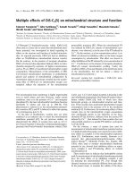
During the 3
rd
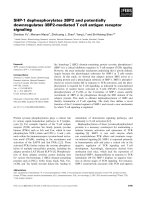
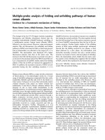
cycle of this regimen, subcutaneous nod-
ules appeared on all four extremities, and a needle aspi-
rate revealed plasma cells (figure 1A, B). The WBC rose to
> 40,000/cmm with 50% circulating plasma cells. He
developed dyspnea and cough, and CT revealed peri-
bronchial thickening and patchy opacities diffusely (fig-
ure 1C). Bronchoalveolar lavage (BAL) demonstrated
kappa restricted plasma cells (Figure 1D, E). He soon after
expired, concurrent with a rise in the ammonia level.
Discussion
In plasma cell neoplasms, pulmonary complications are
common, but infection is the most common cause. In a
series of 958 patients with myeloma, 10% had pulmonary
findings, but in only 1 case was plasma cell involvement
demonstrated histologically, and the clinical course was
suggestive of plasma cell involvement in only 3 other
cases [1]. In the literature, there are additional cases of
pulmonary involvement: some of these had advanced or
treatment-refractory disease, like our patient [2-4],
whereas in several cases, the lung was the initial site of
presentation [5-8]. Likewise, skin involvement is rare, but
this too has been reported [9,10]. We believe that this is
the first case patient reported to have both sites of involve-
Published: 13 November 2009
Journal of Hematology & Oncology 2009, 2:48 doi:10.1186/1756-8722-2-48
Received: 5 July 2009
Accepted: 13 November 2009
This article is available from: />© 2009 Yuan et al; licensee BioMed Central Ltd.
This is an Open Access article distributed under the terms of the Creative Commons Attribution License ( />),
which permits unrestricted use, distribution, and reproduction in any medium, provided the original work is properly cited.
Journal of Hematology & Oncology 2009, 2:48 />Page 2 of 2
(page number not for citation purposes)
ment as a complication of relapsed disease after autolo-
gous stem cell transplantation. The presence of circulating
plasma cells and hyperammonemia are both known to be
indicators of advanced disease. Plasma cell neoplasm
should be kept in the differential diagnosis of any pulmo-
nary infiltrate not responding to antibiotics or as a possi-
ble cause of skin nodules in a patient with a history of
myeloma, plasmacytoma, or MGUS.
Competing interests
The authors declare that they have no competing interests.
Authors' contributions
All authors were involved in the provision of clinical care
of the patient and the collection of data and the review of
the manuscript. RW performed immunohistochemical
analysis. YY drafted the manuscript and reviewed the liter-
ature, as did DA, who performed the final edits.
Consent
The Institutional IRB approved the submission of this
manuscript, which was considered by the editors to be a
proxy for the patient's informed consent for the publica-
tion of the manuscript and accompanying images, in light
of the fact that the patient had expired. A copy of the writ-
ten documentation of this is available for review by the
Editor-in-Chief of this journal.
References
1. Kintzer J, Rosenow E, Kyle R: Thoracic and Pulmonary Abnor-
malities in Multiple Myeloma. Archives of Internal Medicine 1978,
138:727-730.
2. Chejfec G, Natarelli J, Gould V: Myeloma lung: a previously unre-
ported complication of multiple myeloma. Human Pathology
1983, 14:558-561.
3. Schelle M, Schreiber C, Florschutz A, Kachel R, Schreiber J: Pulmo-
nary and pleural manifestations of multiple myeloma. Pneu-
mologie 2006, 60:743-748.
4. Yokote T, Akioka T, Miyamoto H, Oka S, Hara S, Yamano T, Takasu
T, Tsuji M, Hanafusa T: Pulmonary parenchymal infiltrates in a
patient with CD20-positive multiple myeloma. European Jour-
nal of Haematology 74:61-65.
5. Freeman Z: Myelomatosis with extensive pulmonary involve-
ment. Thorax 1961, 16:378-381.
6. Gabriel S: Multiple myeloma presenting as pulmonary infiltra-
tion. Dis Chest 1965, 47:123-136.
7. Gozdziuk K, Kedra M, Ryojad P, Sagam D: A rare case of solitary
plasmacytoma mimicking a primary lung tumor. Annals Tho-
racic Surg 2009, 87:25-26.
8. Kamble R, Rosenzweig T: Diffuse pulmonary parenchymal
involvement in multiple myeloma: antemortem diagnosis.
International Journal of Hematology 2006, 83:259-261.
9. Gambichler T, Othlinghaus N, Stucker M, Altmeyer P, AKCaE : Der-
matology Cutaneous giant plasmacytoma associated with
monoclonal gammopathy of unknown significance. Clinical
and Experimental Dermatology 2009, 34:417-418.
10. Walzer R, Shapiro L: Multiple Myeloma with Cutaneous
Involvement. Dermatologica 1967, 134:449-454.
(A) Skin nodules; (B) Wright Giemsa stain, aspirate of skin nodules; (C) Chest CT showing diffuse infiltrates; (D) H&E of BAL; (E) Kappa staining of BALFigure 1
(A) Skin nodules; (B) Wright Giemsa stain, aspirate of skin nodules; (C) Chest CT showing diffuse infiltrates;
(D) H&E of BAL; (E) Kappa staining of BAL.
C
D E
A B