báo cáo khoa học: "Acute dyspnoea and single tracheal localisation of mantle cell lymphoma" ppsx
Bạn đang xem bản rút gọn của tài liệu. Xem và tải ngay bản đầy đủ của tài liệu tại đây (426.08 KB, 2 trang )
LET T E R TO THE EDITOR Open Access
Acute dyspnoea and single tracheal localisation
of mantle cell lymphoma
Jean-Christophe Ianotto
1*
, Adrian Tempescul
1
, Jean-Richard Eveillard
1
, Norbert André
2
, Frederic Morel
3
,
Isabelle Quintin-Roué
4
, Christian Berthou
1
Abstract
Background: Mantle cell lymphoma is a lymphoid entity characterized by adenopathy, blood and bone marrow
involment which only recurrent mucosal localisation is the lymphomatoid polyposis. Few other mucosal infiltrations
have been already reported.
Results: We report here the first case of a unique tracheal localisation of mantle cell lymph oma at presentation of
the disease. The presence of classical t(11;14)(q13;q32) confirmed the diagnosis of mantle cell lymphoma by
eliminating MALT or cancer localisation.
Conclusion: This case illustrates the necessity to ensure the diagnosis of mucosal lymphoma versus MCL since
these diseases need different treatment regimens and prognoses.
To the Editor,
We report here an unusual case of a tracheal localisa-
tion of mantle cell lymphoma (MCL). The patient was
75-years-old and hospitalized for dyspnoea, dysphonia
and stridor, evolving from 3 months. No superficial
tumoural syndrome was observed and the patient did
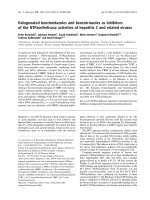
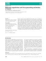
not express B-symp toms. The CT-scan showed the pre-
sence of an endotracheal tumour of two centimetres
under the glottis and two mediastinal centimetric lymph
nodes. No other localisations were found . The bronchial
endoscopy showed an obstructive vascularised tumour
(Figure 1), and the stomach endoscopy was negative.
The pneumologist took multiple biopsies and used both
laser and endotracheal prothesis to t reat the dyspnoea.
The anatomo-pathologist identified a massive prolifera-
tion of medium to large cells with abundant and clear
cytoplasm, round or o val nuclei. Mitosis were observed.
Those cells were CD20+/bcl-2+ lymphoid cells with no
lymphoepithelial lesions. Cells expressed the CD5+.
Many lymphoid cells expressed Cyclin D1 (Monoclonal
anti-mouse, clone SP4, Lab Vision). Some CD23+ den-
dritic cells were observed. CD138 and ALC stains were
negative, excluding plasmacytoma and solid tumour.
Fluorescence in situ hybridisation of t racheal tumour
revealed the presence of a t(11;14)(q13;q32) transloca-
tion. We made the diagnosis of MCL. Furthermore,
blood and bone marrow exams did not show any abnor-
mal lymphoid B cells with cytological and molecular
exams. The patient was treated with four courses of
Vincristine-Adriamycin-Dexamethasone-Chloramino-
phene followed by four injections of Rituximab. We
obtained a complete haematological, cytogenetical and
isotopic remission. The patient is still alive and in com-
plete remission, 4 years after the diagnosis.
Mantle cell lymphoma is a lymphoid entity defined by
clinical, cytological, immunological, biochemical and
cytogenetic criteria [1]. One particular entity of MCL,
lymphomatoid polyposis, is characterised by the involve-
ment of the gastrointestinal tract (30%), distinct from a
mucosal associated lymphoid tissue (MALT) localisation
[2,3]. The frequency of MALT in the trachea is very
low; however, nasopharynx and Waldeyer’s ring localisa-
tions of MCL mimicking MALT have been reported
[4,5]. Dyspnoea was previously described in mediastinal
involvement of MCL compressing the trachea [6]. Two
cases have been alread y reported but there were relapse
site or one of the multiple localisation of the MCL [7,8].
This case is different because of its unique localisation
and the fact that it is the first evolution of the disease.
Because tracheal involment is most seen in cancer and
* Correspondence:
1
Institut de Cancéro-hématologie, Département d’Hématologie, Hôpital
Morvan, CHRU Brest, France
Full list of author information is available at the end of the article
Ianotto et al. Journal of Hematology & Oncology 2010, 3:34
/>JOURNAL OF HEMATOLOGY
& ONCOLOGY
© 2010 Ianotto et al; licensee BioMed Central Ltd. T his is an Open Acc ess articl e distribut ed under the terms of the Creative Commons
Attribution Licens e ( nses/by/2.0), which pe rmits unrestricted use, distribution, and reproduct ion in
any medium, provided the original work is properly cited.
MALT lymphoma with different therapy and evolution,
it is important to maximise the chance of an accurate
diagnosis by correlating anatomo-pathologist and cyto-
genetic exams s o as not to underestimate the incidence
of atypical MCL in cancer/MALT localisation. This case
illustrates the necessity to ensure the diagnosis of muco-
sal lymphoma versus MCL since these diseases have dif-
ferent treatment regimens and prognoses.
Abbreviations
MALT: Mucosal Associated Lymphoid Tissue; MCL: Mantle Cell Lymphoma.
Author details
1
Institut de Cancéro-hématologie, Département d’Hématologie, Hôpital
Morvan, CHRU Brest, France.
2
Departement de Pneumologie, Hopital Cavale
Blanche, Brest, France.
3
Laboratoire de Cytogénétique, Faculté de Médecine,
Université de Bretagne Occidentale, Brest, France.
4
Laboratoire
d’Anatomopathologie, Hôpital Morvan, CHRU Brest, France.
Authors’ contributions
JCI wrote the paper; AT JRE and CB collected the data and reviewed the
paper; NA performed the endoscopic exam; FM did the cytogenetic exam
and IQR performed the anatomopathologic exam. All authors read and
approved the final manuscript.
Competing interests
The authors declare that they have no competing interests.
Received: 26 July 2010 Accepted: 28 September 2010
Published: 28 September 2010
References
1. Campo E, Raffeld M, Jaffe ES: Mantle-cell lymphoma. Seminars in
Hematology 1999, 36:115-127.
2. Rao DS, Said JW: Small lymphoid proliferations in extranodal locations.
Archives of Pathology and Laboratory Medicine 2007, 131:383-396.
3. Okubo K, Miyamoto N, Komaki C: Primary mucosa-associated lymphoid
tissue (MALT) lymphoma of the trachea: a case of surgical resection and
long term survival. Thorax 2005, 60:82-83.
4. Zinzani PL, Magagnoli M, Galieni P, Martelli M, Poletti V, Zaja F, Molica S,
Zaccaria A, Cantonetti AM, Gentilini P, Guardigni L, Gherlinzoni F,
Ribersani M, Bendandi M, Albertini P, Tura S: Nongastrointestinal low-
grade mucosa-associated lymphoid tissue lymphoma: analysis of 75
patients. Journal of Clinical Oncology 1999, 17:1254.
5. Jaffe ES: Lymphoid lesions of the head and neck: a model of lymphocyte
homing and lymphomagenesis. Modern Pathology 2002, 15:255-263.
6. Amemiya M, Takise A, Kaira K, Endou K, Horie T, Inazawa M: Obstructive
sleep apnea syndrome in a patient with superior vena cava syndrome
caused by malignant lymphoma. Nihon Kokyuki Gakkai Zasshi 2006,
44:197-201.
7. Kuppusamy Gounder S, Sikder M, Srinivas S, Chang VT, Kasimis B:
Asymptomatic mantle cell lymphoma in the trachea. Leukemia
Lymphoma 2009, 50:651-652.
8. Verde F, McGeehan A: Endotracheal involvement as an unusual
extranodal site of recurrence from mantle cell lymphoma. Radiolology
Case Report 2008, 3:194, Online.
doi:10.1186/1756-8722-3-34
Cite this article as: Ianotto et al.: Acute dyspnoea and single tracheal
localisation of mantle cell lymphoma. Journal of Hematology & Oncology
2010 3:34.
Submit your next manuscript to BioMed Central
and take full advantage of:
• Convenient online submission
• Thorough peer review
• No space constraints or color figure charges
• Immediate publication on acceptance
• Inclusion in PubMed, CAS, Scopus and Google Scholar
• Research which is freely available for redistribution
Submit your manuscript at
www.biomedcentral.com/submit
Figure 1 The sagittal CT-scan showed the presence of an endotracheal tumour under the glottis (left panel). Presence of a vascularised
tumor of the trachea visible by bronchial endoscopy (right panel).
Ianotto et al. Journal of Hematology & Oncology 2010, 3:34
/>Page 2 of 2