báo cáo khoa học: "Prolapsed sigmoid intussusception per anus in an elderly man: a case report" docx
Bạn đang xem bản rút gọn của tài liệu. Xem và tải ngay bản đầy đủ của tài liệu tại đây (2.35 MB, 4 trang )
CAS E REP O R T Open Access
Prolapsed sigmoid intussusception per anus in an
elderly man: a case report
Penn S Teyha
1
, Alphonce Chandika
1
and Vihar R Kotecha
2*
Abstract
Background: Intussusception in pediatrics is widely documented and well described. On the basis of the literature,
however, adult intussusception is a rare entity with a prevalence of from 1% to 5%. The majority of adult patients
with intussusception have an underlying pathology that needs to be identified by performing a proper physical
examination and a wide array of investigations.
Case presentation: We present a case of a 66-year-old African man who presented to our emergency department
with a mass protruding per anus with obstipation. During laparotomy, we found that the sigmoid colon had
intussuscepted into the rectum and out from the anus. Other abdominal viscera were normal and without any
obvious mesenteric lymphadenopathy. Sigmoid colectomy and spectacle colostomy were performed. Grossly, the
excised bowel looked normal, but the histologic results showed features of necrosis and chronic inflammation.
Conclusion: While 70% to 90% of cases of adult intussusception have an identifiable cause or lesion, most
pediatric intussusceptions are idiopathic. The presentation in an adult described herein was of an uncommon
idiopathic type with no identifiable cause found on the basis of the history, physical examination, or histological
findings.
Background
“Intussusception” refers to the telescoping of the proximal
bowel into the distal bowel or vice versa (retrograde intus-
susception). The telescoped part is referred t o as the
“ intussusceptum,” and the receiving part is called the
“intussuscipiens.” This condition is commonly described
in infants and rarely in adults. Adult intussusception
represents 5% of all cases of intussusception and accounts
for only 1% to 5% of intestinal obstructions in adults [1,2].
Almost 90% of cases of intussusception in adults have an
underlying bowel pathology such as carcinoma, polyps,
Meckel’s diverticulum, colonic diverticulum, strictures, or
benign neoplasms, which are usually discovered intra-
operatively [3]. In this report, we present a case of an
elderly man with a prolapsed intussusception.
Case presentation
A 66-year-old African man from a rural area presented
to our emergency department complaining of persistent,
dull lower-back pain of three months’ duration and a
mass protruding per anus for the previous four days.
His lower-back pain was radiating to both t highs and
was aggravated by farming activities. His pain was
relieved by resting, and the pain was not accompanied
by numbness of the limbs. He had no history of diffi-
culty in micturition. The mass protruded per anus spon-
taneously while he defecated and was associated with
severe pain followed by obstipation. There was no
obvious bleeding per rectum upon his presentation to
our emergency department. He had no history of consti-
pation prior to the protrusion of the mass. He had no
history of on-and-off mass protrusion per anus even
when lifting loads. In addition, he had no history of
changes in bowel habits or blood-stained stools, no his-
tory of tenesmus, and no history of incomplete bowel
emptying post-defecation. He had no history of feeling
any abdominal swelling and no history of significant
weight loss. An attempt to reduce the mass was made at
the district hospital, but the procedure failed; hence the
patient was referred to Bugando Medical Centre. His
medical history was uneventful.
* Correspondence:
2
Department of Surgery, Weill Bugando University College of Health
Sciences, P.O.Box 1464, Mwanza, Tanzania
Full list of author information is available at the end of the article
Teyha et al. Journal of Medical Case Reports 2011, 5:389
/>JOURNAL OF MEDICAL
CASE REPORTS
© 2011 Teyha et al; l icensee BioMed Central Ltd. This is an Open Access article distributed under the terms of the C reative Commons
Attribution License (http://c reativecommons.org/licenses/by/2.0), which permi ts unrestricted use, distribution, and reproduction in
any medium, provided the original work is properly cited.
His physical examination revealed that he was in pain,
alert, afebrile (body temperature 37°C), and dehydrated,
but he was not pale. His blood pressure was stable at
100/70 mmHg, and his radial pulse w as 90 beats/min-
ute, weak, regular, and syn chronous with other pulses.
He had no cervical lymphadenopathy, and no Virchow’s
node was palpable. He had a scaphoid abdomen that
moved with respiration, but no visible peristalsis was
observed, his abdomen was not tender, and no obvious
mass was palpable. His liver, spleen, and kidneys were
not palpable, and a tympanic percussion note was heard
throughout the abdomen. His bowel sounds were
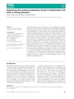
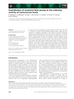
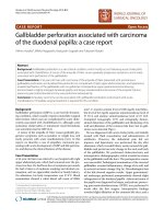
increased on auscultation. The digital rectal examination
revealed a large, foul-smelling, gangrenous sigmoid-
shaped mass protruding per anus (Figures 1 and 2). The
rest of his systemic examination was non-contributory.
A provisional diagnosis of sigmoid intussusception
with a differential diagnosis of rectal prolapse was made.
Blood samples were taken to measure his hemoglobin
level a nd blood grouping. His hemoglobin level was 10
g/dL. Pre-operatively the patient received 3 L of Ringer’s
lactate solution over the course of one hour, intravenous
ceftriaxone 1 g, and metronidazole 500 mg, and he was
prepared for emergency exploratory laparotomy. The
abdomen was approached through an extended mid-line
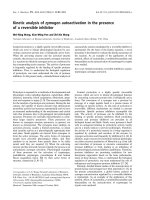
incision. T he intra-operative findings were that the sig-
moid colon had telescoped into the rectum and out per
anus (Figure 3). The large bowel was not dilated. No
othermassorpathologywasidentified intra-abdomin-
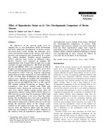
ally, nor were mesenteric lymph nodes palpable. Milking
of the intussusceptum was done followed by resection of
the gangrenous sigmoid colon ( Figure 4), and the two
bowel ends were exteriorized to form a spectacle colost-
omy. Macroscopically, normal rugae with no visible
lesions or hemorrhagic serosa were visualized. The
microscopic findings revealed features of chronic inflam-
mation and necrosis.
Discussion
Intussusception in adults is an uncommon condition,
representing 1% of ca ses of adult bowel obstruction and
less than 1% of hospital admissions [4]. It is a common
causeofbowelobstructionininfants,inwhomitpre-
sents with a classic triad of symptoms and signs: crampy
abdominal pain, a palpable sausage-shaped mass mainly
in the right upper quadrant, and currant jelly stools [5].
In one case series, it was noted that adult
Figure 1 The prolapsed bowel portion from the anus.
Figure 2 Closer view of the prolapsed bowel with foci of
hemorrhage.
Figure 3 Finger indicating the intussusception site.
Teyha et al. Journal of Medical Case Reports 2011, 5:389
/>Page 2 of 4
intussusception was slight ly more predominant among
men, with a male:female ratio of 1.8:1 [1]. Adult intus-
susception may present with acute, sub-acute, or
chronic non-specific features, which makes its diagnosis
difficult. In our patient, sub-acute non-specific com-
plaints of backache during strenuous activity culminated
in acute prolapsed intussusception of the sigmoid. In
one series, c omputed tomography, with an accuracy of
58% to 100%, was the most efficient tool in diagnosing
intussusception, followed by abdominal ultrasound [1, 2].
In our patient, the mass was obviously prot ruding from
the anus and did not warrant any complex investiga-
tions. Our case could possibly have been confused with
complete rectal prolapse. A retrospective study by
Rashid and Basson [6] showed that patients with rectal
prolapse exhibited a 4.2-fold relative risk for colorectal
cancer compared with the comparative group. In our
patient, the diagnosis of colorectal cancer was at the top
of the list as the underlying cause of intussusception,
mainly because of his age at presentation. With regard
to the management of adult sigmoid intussusception,
several schools of thought exist. However, there is a
common consensus that the treatment of choice is
resection of the affected portion of the sigmoid colon, as
the results reported in several series have revealed that
90% of cases have an underlying pathology. Lynn and
Agrez [7] reported the case of a patient with sigmoid
colon intussusception in whom the rectum was opened
circumferentially by using diathermy at the point of the
intussusception and the intussuscepted sigmoid colon
was removed from the rectum through the anus. How-
ever, this procedure could cause contamination of the
abdominal cavity. In our case, given that the intussus-
ceptum was edematous and gangrenous, a longitudinal
incision was made on the prolapsed bowel to facilitate
reduction with a milking motion. A decision to perform
reduction w as made after assessing the abdominal vis-
cera and the prese nce of me senteric lymph nodes for
any macroscopic evidence of a large bowel tumor. We
did a spectacle colostomy after resection of the gangre-
nous bowel, as the viability of the sigmoid colon could
not be guaranteed for primary end-to-end anastomosis.
While 70% to 90% of adult intussusceptions have an
identifiable cause or lesion, most pediatric intussuscep-
tions ar e idiopathic [ 1,2]. The case of an el derly patient
presented here was of the uncommon idiopathic type
with no identifiable cause found in the history, physical
examination, or histological findings.
Conclusion
Intussusception is a well-described condition that has
been documented mostly in pediatric patients. Adult
intussusception is a rare entity. Prolapsed intussuscep-
tion per anus has rectal prolapse as its most likely differ-
ential diagnosis; hence sigmoid prolapse has to be kept
in mind, since adult intussusception usually has an
underlying cause. All stigmata for t he cause should be
ruled out on the basis of the patient’s history and physi-
cal examination as well as during laparotomy.
Consent
Written informed consent was obtained from the patient
for publicatio n of this case report and any accompany-
ing images. A copy of the written consent is avail able
for review by the Editor-in-Chief of this journal.
Author details
1
Department of Surgery, Bugando Medical Centre, P.O.Box 1370, Mwanza,
Tanzania.
2
Department of Surgery, Weill Bugando University College of
Health Sciences, P.O.Box 1464, Mwanza, Tanzania.
Authors’ contributions
CA, PS, and VK operated on the patient. VK, PS, and CA wrote the
manuscript. All authors read and approved the final manuscript.
Competing interests
The authors declare that they have no competing interests.
Received: 31 January 2011 Accepted: 17 August 2011
Published: 17 August 2011
References
1. Azar T, Berger DL: Adult intussusception. Ann Surg 1997, 226:134-138.
Figure 4 The gangrenous sigmoid colon that was removed
during surgery.
Teyha et al. Journal of Medical Case Reports 2011, 5:389
/>Page 3 of 4
2. Marinis A, Yiallourou A, Samanides L, Dafnios N, Anastasopoulos G,
Vassiliou I, Theodosopoulos T: Intussusception of the bowel in adults: a
review. World J Gastroenterol 2009, 15:407-411.
3. Agha FP: Intussusception in adults. AJR Am J Roentgenol 1986,
146:527-531.
4. Ochiai H, Ohishi T, Seki S, Tokuyama J, Osumi K, Urakami H, Shimada A,
Matsui A, Isobe Y, Murata Y, Endo T, Ishii Y, Hasegawa H, Matsumoto S,
Kitagawa Y: Prolapse of intussusception through the anus as a result of
sigmoid colon cancer. Case Rep Gastroenterol 2010, 4:346-350.
5. Hackam DJ, Grikscheit TC, Wang KS, Newman KD, Ford HR: Pediatric
surgery. In Schwartz’s Principles of Surgery 9 edition. Edited by: Brunicardi
FC, Andersen DK, Billiar TR, Dunn DL, Hunter JG, Matthews JB, Pollock RE.
New York: McGraw-Hill; 2010:1433.
6. Rashid Z, Basson MD: Association of rectal prolapse with colorectal
cancer. Surgery 1996, 119:51-55.
7. Lynn M, Agrez M: Management of sigmoid colon intussusception
presenting through the anus. Aust N Z J Surg 1998, 68:683-685.
doi:10.1186/1752-1947-5-389
Cite this article as: Teyha et al.: Prolapsed sigmoid intussusception per
anus in an elderly man: a case report. Journal of Medical Case Reports
2011 5:389.
Submit your next manuscript to BioMed Central
and take full advantage of:
• Convenient online submission
• Thorough peer review
• No space constraints or color figure charges
• Immediate publication on acceptance
• Inclusion in PubMed, CAS, Scopus and Google Scholar
• Research which is freely available for redistribution
Submit your manuscript at
www.biomedcentral.com/submit
Teyha et al. Journal of Medical Case Reports 2011, 5:389
/>Page 4 of 4