báo cáo khoa học: "Isolated complete avulsion of the gallbladder (near traumatic cholecystectomy): a case report and review of the literature" potx
Bạn đang xem bản rút gọn của tài liệu. Xem và tải ngay bản đầy đủ của tài liệu tại đây (698.19 KB, 4 trang )
CASE REP O R T Open Access
Isolated complete avulsion of the gallbladder
(near traumatic cholecystectomy): a case report
and review of the literature
Theodoros E Pavlidis, Miltiadis A Lalountas
*
, Kyriakos Psarras, Nikolaos G Symeonidis, Anastasios Tsitlakidis,
Efstathios T Pavlidis, Konstantinos Ballas, Nikolaos Flaris, Georgios N Marakis and Athanassios K Sakantamis
Abstract
Introduction: Injury of the gallbladder after blunt abdominal trauma is an unusual finding; the reported incidence
is less than 2%. Three groups of injuries are describe d: simple contusion, laceration, and avulsion, the last of which
can be partial, complete, or total traumatic cholecystectomy.
Case presentation: A case of isolated complete avulsion of the gallbladder (near traumatic cholecystectomy) from
its hepatic bed in a 46-year-old Caucasian man without any other sign of injury is presented. The avulsion was due
to blunt abdominal trauma after a car accident. The rarity of this injury and the stable condition of our patient at
the initial presentation warrant a description. The diagnosis was made incidentally after a computed tomography
scan, and our patient was treated successfully with ligation of the cystic duct and artery, removal of the
gallbladder, coagulation of the bleeding points, and placement of a drain.
Conclusions: Early diagnosis of such injuries is quite difficult because abdominal signs are poor, non-specific, or
even absent. Therefore, a computed tomography scan should be performed when the mechanism of injury is
indicated.
Introduction
The first specimen of a lacerated gallbladder from a
blunt trauma was found in Guy’ sMuseuminLondon
and dates from 1388 [1]. The first known case of some-
one surviving a gallbladder traumatic rupture was in
1898 [1]. Penn [2] reported the incidence of gallbladder
trauma to be 1.9% in a collected review of 5670 cases of
blunt and penetrating trauma. Complete detachment of
the gallbladder from its hepatic bed, one of the rarest
consequences of blunt abdominal trauma, is r arer than
gallbladder contusion, perforation, and partial contusion.
The few reports in the literature are not clearly enumer-
ated [3-9], because of a lack of appropriate description
before the advanced classification of Losanoff and Kjos-
sev [4].
The gallbladder is a well-protected organ, being par-
tially embedded in the relatively massive liver substance,
cushioned on the surrounding omentum and intestines,
and covered by the bony cartilaginous rib cage. As a
result, gallbladder trauma due to a blunt injury is rare
and usually is associated with additional external or
visceral injuries [2,5,6,8]. Isolated complete avulsion of
the gallbladder after non-penetrating abdominal trauma
in a stable patient without any other sign of injury is
even rarer and is prone to delayed diagnosis and treat-
ment [5,9]. A computed tomography (CT) s can should
be performed when the mechanism of injury is indi-
cated, and an early explorative laparotomy is recom-
mended to reduce the high morbidity associated with
this condition [10-12].
Case presentation
A 46-year-old Caucasian man was involved in a car acci-
dent. He was a pedestrian when a car hit him. He fell
down on the road and one of the car’ s rear wheels
rolled over his lower chest. Two hours later, he pre-
sented in our emergency dep artment. On admission, he
was complaining of bilateral hypochondrial pain
* Correspondence:
Second Surgical Propedeutical Department, Medical School, Aristotle
University of Thessaloniki, Hippocration Hospital, Konstanti noupoleos 49, 546
42 Thessaloniki, Greece
Pavlidis et al . Journal of Medical Case Reports 2011, 5:392
/>JOURNAL OF MEDICAL
CASE REPORTS
© 2011 Pavlidis et al; licensee BioMed Central Ltd. This is an Open Access ar ticle distributed under the terms of the Creative Commons
Attribution License ( which permi ts unrestricted use, distribution, and reproduction in
any medium, provided the original work is properly cited.
radiating to his right shoulder; he was hemodynamically
stable after repeated blood tests and had a blood pres-
sure of 130/100 mm Hg and a pulse rate of 90 beats per
minute. An examination revealed no chest or abdominal
wall contusions. A chest X-ray was normal and there
were no rib fractures. The results of an ultrasound (US)
examination of the abdomen were normal, but the gall-
bladder could not be visualized.
. The results of all laboratory tests were normal except
for a leucocytosis level of 12.2 × 10
3
/mm
3
. Because of
the s uspicion of possible intra-abdominal injury due to
the severe mechanism of the accident, a CT scan was
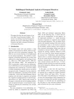
performed. The scan revealed pericholecystic fluid and
the possibility of an avulsed gallbladder (Figure 1). Mag-
netic resonance imaging (MRI) would have been another
option, but our patient had a contraindication because
of the presence of a pacemaker. An exploratory laparot-
omy was performed fiv e hours after admission, although
our patient remained hemodynamically stable.
During the laparotomy, a moderate amount of fresh
blood was identified in the right subhepatic space. The
gallbladder was lying freely avulsed, detached from its
liver bed, but there was no extrahepatic bile duct
injury. The gallbladder’s attachments to the cystic duct
and the cystic artery were intact and both of these
structures were subsequently ligated. The removed
gallbladder contained no stone. The abdomen had no
other pathology and was washed, drained, and closed
in layers. The postoperative course was une ventful,
and our patient was discharged on the fifth postopera-
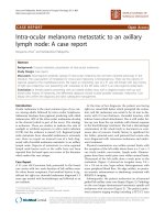
tive day. A pathology report confirmed gallbladder
injury with hemorrhage and chronic cholecystitis (Fig-
ure 2a, b).
Discussion
Blunt gallbladder injuries are classified as contusion,
perforation, or avulsion [4-6,8]. Contusion, defined as an
intramural hematoma, is most often diagnosed at the
time of laparotomy and i s probably underreported. Per-
foration, also known as “rupture” or “laceration”,isthe
most commonly reported injury. Avulsion has three sub-
types: partial avulsion, in which the gallbladder is par-
tially detached from the liver bed; complete avulsion, in
which the gallbladder is completely detached from the
liver bed but the cystic duct and artery are intact; and
total avulsion, in which the gall bladder lies fr ee in the
abdomen, torn from all attachments. To the best of our
knowle dge, only eight cases of total avu lsion (also called
“traumatic ch olecystectomy”) have been report ed. Trau-
matic cholecystitis is caused by a cystic duct obstruction
by blood clots from a liver or gallbladder injury. Losan-
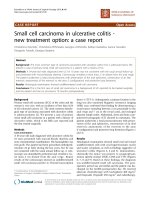
off and Kjossev [4] describe a more detailed classifica-
tion of blunt gallbladder injuries; according to their
classification, our patient belongs to type 3B (isolated
complete avulsion of the gallbladder or near traumatic
cholecystectomy; Figure 3 and Table 1[4]).
Earlier reports indicate that the most common etiolo-
gic factors in blunt trauma were falls, kicks, o r blows.
Figure 1 Computed tomography (CT) scan reveals
pericholecystic fluid (arrow) and indicates the potential for
some kind of injury of the gallbladder.
Figure 2 Photographs of fixed gallbladder prepared with
formaldehyde. (a) Successive sections of the gallbladder show
traumatic hemorrhagic filtering. (b) Inverted gallbladder with the
same findings.
Pavlidis et al . Journal of Medical Case Reports 2011, 5:392
/>Page 2 of 4
At present, motor vehicle crashes are the predominant
cause of blunt gallbladder trauma [2,4,5,8]. Factors pre-
disposing people to blunt gallbladder injuries are a th in-
walled normal gallbladder, a distended gallbladder, and
alcohol ingestion, the last of which increases the tone of
sphincter of Oddi and the biliary tract pressure. Our
patient had a history of chronic alcohol consumption.
Associated intra-abdominal injuries are common in
patients with a blunt gallbladder injury, averaging 2.7 to
3.3 associated injuries per patient. Liver injury is espe-
cially likely; the reported incidence is 83% to 91%.
Duodenum and spleen injuries occur in up to 54% of
patients with a blunt gallbladder injury [4,8]. Our
patient had no other injuries. We used ultrasonography
initially because of its low cost and the ability to per-
formthetestatthebedsideintheemergency
department.
Non-visualization of the gallbladder at ultrasonogra-
phy should raise the suspicion of a traumatic gallbladder
avulsion or rupture [7-12]. CT findings of gallbladder
injury are largely non-specific. Pericholecystic fluid is
most common but is least specific. Other signs of gall-
bladder injury are an ill-defined contour of the gallblad-
der wall, a mass effect on the duodenum, high-
attenuation intraluminal material (blood), a thickened
gallbladder wall, and a collapsed gal lbladder in a fasting
patient. Also, major liver injury often dominates the CT
picture and overshadows subtle abnormalities of the
gallbladder. It is not surprising that unsuspected gall-
bladder injury is often discovered during a laparotomy
for coexisting intra-abdominal injuries. Gallbladder inju-
ries, though infrequent, can be difficult to diagnose. CT
is the most reliable technique to diagnose a gallbladder
injury. However, benign entities can mimic gallbladder
injury. Delayed images through the gallbladder can be
useful in differentiating between a true gallbladder injury
and a relatively benign process [13]. In our case, the
possibility of an avulsed gallbladder was revealed from
an abdominal CT scan, which was performed bec ause of
Figure 3 Schematic drawing of all known types of gallbladder injury according to the classification by Losanoff and Kjossev [4]. Our
case is highlighted.
Table 1 Types of gallbladder injury according to the
classification by Losanoff and Kjossev [4] (Figure 3)
Type Injury of the gallbladder
1A Contusion with intramural hematoma
1B Contusion with perforation
2 Rupture
3A Avulsion with partial detachment
3B Avulsion with complete detachment from the liver but with
attachment to the structures of the hepatoduodenal ligament
(so-called “near traumatic cholecystectomy”)
3C Torn only from the hepatoduodenal ligament
3D Completely torn from all attachments (so-called “traumatic
cholecystectomy”)
4A Traumatic cholecystitis, secondary to hemobilia
4B Acute acalculus cholecystitis
5 Mucosal tear with leakage of bile
Pavlidis et al . Journal of Medical Case Reports 2011, 5:392
/>Page 3 of 4
the severe mechanism of the accident. An abdominal
CT scan, rather than US or MRI, is considered the “gold
standard” method to diagnose this kind of injury
[10-13]. In such cases, we recomme nd that a CT scan
be performed, even in the absence of other signs of
injury in a hemodynamically stable patient.
The choice of treatment depends on the severity of
the gallbladder injury and the general condition of the
patient. Patients with mild injuries such as contusion or
isolated partial avulsion may be observed, although late
necrosis and perforation have been reported [9,14,15].
Severe injuries generally require a cholecystectomy [16].
When the patient is hemodynamically stable, a di agnos-
tic l aparoscopy could play a role. Laparoscopic surgical
techniques may be safely used when the likelihood of
associated injuries is low and definitive treatment can be
rendered without increas ing patient morbidity and mor-
tality [17,18].
Conclusions
Early diagnosis of gallbladder injuries, such as near trau-
matic cholecystectomy, is quite difficult because abdom-
inal signs are poor, non-specific, or even absent.
Therefore, a CT scan should be performed when the
mechanism of injury is indicated. Such injuries have a
good prognosis if they are diagnosed early and there is
no serious associated trauma. T rauma surgeons should
always be aware of the existence of these injuries.
Consent
Written informed consent was obtained from the patient
for publicatio n of this case report and any accompany-
ing images. A copy of the written consent is available
for review by the Editor-in-Chief of this journal.
Abbreviations
CT: computed tomography; MRI: magnetic resonance imaging.
Authors’ contributions
TEP performed the procedure. MAL obtained the patient’s written informed
consent to publish the report, conducted the follow-up examinations,
analyzed and interpreted the patient data, and wrote part of the manuscript.
KP, NGS, AT, and ETP edited and wrote part of the manuscript. KB and GNM
were major contributors to the review and editing of the manuscript. NF
was the main pathologist and revised the manuscript. AKS made the
strategic plan and gave the final approval. All authors read and approved
the final manuscript.
Competing interests
The authors declare that they have no competing interests.
Received: 15 January 2011 Accepted: 18 August 2011
Published: 18 August 2011
References
1. Ricketts BM: Rupture of the gallbladder: with and without operation. A
historical review of 273 cases. St Louis Med Rev 1905, 51:108.
2. Penn I: Injuries of the gallbladder. Br J Surg 1962, 49:636.
3. Brown PJ: Traumatic cholecystectomy. Ann Surg 1932, 95:952-953.
4. Losanoff JE, Kjossev KT: Complete traumatic avulsion of the gallbladder.
Int J Care Injured 1999, 30:365-368.
5. Schechter DC: Solitary wounding of the gallbladder from blunt
abdominal trauma. NY State J Med 1969, 69:2895-2901.
6. Soderstrom CA, Maekawa K, DuPriest RW Jr, Cowley RA: Gallbladder
injuries resulting from blunt abdominal trauma: an experience and
review. Ann Surg 1981, 193:60-66.
7. Erb RE, Mirvis SE, Shanmuganathan K: Gallbladder injury secondary to
blunt trauma: CT findings. J Comp Ass Tomogr 1994, 18:778-784.
8. Sharma O: Blunt gallbladder injuries: presentation of twenty-two cases
with review of the literature. J Trauma 1995, 39:576-580.
9. Laffey DA, Hay DJ: Isolated perforation of the gallbladder following blunt
abdominal trauma. Postgrad Med J 1979, 55:212.
10. Jeffery RB Jr, Federle MP, Laing FC, Wing VW: Computed tomography of
blunt trauma to the gallbladder. J Comput Assist Tomogr 1986, 10:756.
11. Baumgartner FJ, Barnett MJ, Velez M, Chiu LC: Traumatic disruption of the
gallbladder evaluated by computerized tomography and magnetic
resonance imaging. Br J Surg 1988, 75:386.
12. Kambayashi M, Yong W, Watanabe K, Alam S: Hemobilia due to
gallbladder contusion following blunt trauma-sonography and CT
scanning for early detection: case report. J Trauma 1993, 34:440.
13. Wittenberg A, Minotti AJ: CT diagnosis of traumatic gallbladder injury. AJR
2005, 185:1573-1574.
14. Zantut LF, Machado MA, Volpe P, Poggetti RS, Birolini D: Gallbladder injury
in abdominal trauma: analysis of 32 cases. Rev Hosp Clin Fac Med Sao
Paulo 1993, 48:283-288.
15. Carrillo EH, Lottenberg L, Saridakis A: Blunt traumatic injury of the
gallbladder. J Trauma
2004, 57:408-409.
16. Bade PG, Thomson SR, Hirshberg A, Robbs JV: Surgical options in
traumatic injury to the extrahepatic biliary tract. Br J Surg 1989,
76:256-258.
17. Liess BD, Awad ZT, Eubanks WS: Laparoscopic cholecystectomy for
isolated traumatic rupture of the gallbladder following blunt abdominal
injury. J Laparoendosc Adv Surg Tech A 2006, 16:623-625.
18. Shope TR, Bass TL, Haluck RS: Laparoscopic management of traumatic
hemorrhagic cholecystitis. JSLS 2004, 8:93-95.
doi:10.1186/1752-1947-5-392
Cite this article as: Pavlidis et al.: Isolated complete avulsion of the
gallbladder (near traumatic cholecystectomy): a case report and review
of the literature. Journal of Medical Case Reports 2011 5:392.
Submit your next manuscript to BioMed Central
and take full advantage of:
• Convenient online submission
• Thorough peer review
• No space constraints or color figure charges
• Immediate publication on acceptance
• Inclusion in PubMed, CAS, Scopus and Google Scholar
• Research which is freely available for redistribution
Submit your manuscript at
www.biomedcentral.com/submit
Pavlidis et al . Journal of Medical Case Reports 2011, 5:392
/>Page 4 of 4