báo cáo khoa học: "Effect of growth hormone replacement therapy in a boy with Dent’s disease: a case report" ppt
Bạn đang xem bản rút gọn của tài liệu. Xem và tải ngay bản đầy đủ của tài liệu tại đây (1.24 MB, 6 trang )
CASE REP O R T Open Access
Effect of growth hormone replacement therapy
in a boy with Dent’s disease: a case report
Mira Samardzic
1*
, Snezana Pavicevic
1
, Michael Ludwig
2
and Radovan Bogdanovic
3
Abstract
Introduction: Dent’s disease is an X-linked recessive proximal tubulopathy characterized by low molecular weight
proteinuria, hypercalciuria, nephrocalcinosis, nephrolithiasis and progressive renal failure. To the best of our
knowledge, this is only the third report on the use of growth hormone therapy in a child with poor growth
associated with Dent’s disease.
Case presentation: We report on a 7-year-old Montenegrin boy with proteinuria, hypercalciuria , nephrocalcinosis,
rickets and short stature with unimpaired growth hormone secretion. A molecular genetic analysis showed S244L
substitution on the CLCN5 gene. After two years of conventional treatment with hydrochlorothiazide, laboratory
tests revealed more prominent proteinuria, mild hypophosphatemia, increased values of alkaline phosphatase and
features of rickets. Phosphate salts, calcitriol, potassium citrate and growth hormone were included in the therapy.
After three years of therapy, his adjusted parental stature was 1.53 standard deviations higher than at the initiation
of growth hormone therapy. His global kidney functions and levels of proteinuria and calciuria remained relatively
stable. In spite of the growth hormone therapy, his tubular reabsorption of phosphate deteriorated.
Conclusion: Treatment with recombinant human growth hormone may have a positive effect on final height in
poorly growing children with Dent’s disease and hypophosphatemic rickets. However, it is not possible to reach
definite conclusions due to the small sample within the literature and the brief duration of the therapy.
Introduction
Dent’s disease is an X-linked recessive p roximal tubulo-
pathy characterized by low molecular weight proteinuria,
hypercalciuria, nephrocalcinosis, nephrolithiasis, and
slowly progressive renal f ailure in affected males. Renal
acidification abnormalities are only rarely seen in Dent’s
disease, whereas the hypokalemic metabolic alkalosis
associated with hyperreninemic hyperaldosteronism
(Bartter-like syndr ome) has been reported in a few
patients [1]. Clinical characteristics of Dent’ s disease
include familial tubular syndromes such as X-linked
recessive nephrolithiasis, X-linked recessive hypopho-
sphatemic rickets and low-molecular weight proteinuria
with hypercalciuria and nephrocalcinosis in Japanese
children [2]. Dent’ s disease is caused by mutations in
the CLCN5 gene, which is located on the short arm of
the × chromosome (Xp11.22). The CLCN5 gene
encodes for th e 746 amino-acid CLC-5 chloride channel
that belongs to the voltage-gated chloride channel family
(CLC -7, CLC-Ka and CLC-Kb) implicated in membrane
excitability, transepithelial transport and possibly the
regulation of cell volume [3]. The mechanism b y which
CLC-5 dysfunction results in hypercalciuria and the
other features of Dent’s disease remains unclear.
The different cli nical features of Dent’s disease makes
treatment complex, and it requires simultaneous moni-
toring of the effects of therapy. There is still no clear
strategy for the management of patients with this condi-
tion. Thiazide diuretics have been used to reduce urin-
ary calcium excretion, and to prevent the recurrence of
nephrolithiasis [4]. A high citrate diet preserved the
renal function and delayed the progression of renal dis-
ease in CLC-5 knockout mice [5]. Rickets was a promi-
nent feature in about one third of patients reported with
Dent’s disease. The recommended treatment is based on
oral phosphate salts and calcitriol [6].
* Correspondence:
1
Institute for Sick Children, Department of Endocrinology and Nephrology,
Ljubljanska bb, 20 000 Podgorica, Montenegro
Full list of author information is available at the end of the article
Samardzic et al. Journal of Medical Case Reports 2011, 5:400
/>JOURNAL OF MEDICAL
CASE REPORTS
© 2011 Samardzic et al; li censee BioMed Central Ltd. This is an Open Access article distributed unde r the terms of the Creative
Commons Attribution Licen se ( which permits unrestricted use, distribution, and
reproduction in any medium, provided the original work is properly cited.
Case presentation
A 7-year-old Montenegrin boy was initially referred to
the pediatric endocrinology ward because of short sta-
ture. His height was 2.93 standard deviations (SD) below
the mean. His mid-parental height was 181.6 cm (+1
SD).Ourpatientwasbornattermfollowingan
uneventful pregnancy with a birth weight of 3.95 kg and
a birth length of 57 cm. His family history was negative
for short stature, delayed puberty and renal disease.
Except for dental caries, high palate and slight genus
valgus, he had no other abnormalities. His blood pres-
surewasnormalandhisboneagewas5years.A
laboratory test was positive for proteinuria. He had an
elevated urinary calcium level and b2-microglobulin
excretion. A renal ultrasound showed early medullary
nephrocalcinosis. His levels of serum calcium, phos-
phorus and alkaline phosphatase were normal. His tubu-
lar reabsorption of phosphate (TRP) was decreased. His
creatinine clearance test [7] was normal (92.8 ml/min/
1.73 m
2
). No other electrolyte or metabolic abnormal-
ities were observed. His overnight growth hormone
(GH) profiles were normal (> 3 peaks of > 10 μg/L). An
audiometry test was also unremarkable. In order to con-
firm a diagnosis of Dent’ s disease, molecular genetic
analysis was performed one year later and showed a
mutation in the CLCN5 gene, leading to S244L amino
acid substitution. The mutation carrier, the patient’ s
mother, was asymptomatic with slight hypercalciuria.
After two years of convent ional treatment with hydro-
chlorothiazide, our patient was ref erred for a new endo-
crine evaluation because of a failure to catch-up growth.
Laboratory tests showed more prominent urinary pro-
tein excretion, whereas the level of calciuria remained
unchanged. Clinical, radiological and laboratory signs of
hypophosphatemic ricke ts became noticeable. We found
hypophosphatemia (0.74 mmol/L), elevated serum alka-
line phosphatase activity (926 U/L), and a nearly normal
level of parathyroid hormone. X-rays showed enlarge-
ment of his wrists and knees and fraying of the meta-
physes of his distal ulna and radius. Our patient’ s
growth velocity was 4.7 cm/yr (-1 SD). His parental
adjusted height at that time was - 3.11 SD; his pubertal
status was Tanner stage one. A height prediction based
on his recent growth was approximately 160.7 cm (Sta-
tistical program: SAS v9.13). At the age of nine years
and three months we initiated recombinant human
growth hormone (rhGH) therapy. The indication for
GH therapy was markedly short stature and chronic
renal disease stage one [8]. The average do se of rhGH
was 0.04-0.05 mg/kg per day. Because of the overt hypo-
phosphatemic rickets and hypercalciuria, in addition to
hydrochlorothiazide we started him on calcitriol 20-40
ng/kg/day in two divided doses, phosphorus 20-40 mg/
kg/day, maximum 2.5 g/day in 3-5 divided doses and
potassium citrate. A follow-up was performed at three-
month intervals. We followed his growth velocity, serum
phosphate, serum creatinine, creatinine clearance, TRP,
protein and calcium excretion, insulin -like growth factor
1(IGF-1), insulin-like growth factor-binding protein 3
(IGFBP-3) and other laboratory tests in relation to
growth hormone therapy (Table 1, Table 2). In the first
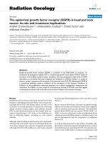
two years, our patient grew at an average rate of 9 cm
per year (> 95c), and in the third year he grew 6 cm
(50c) (Figure 1). His bone age remain ed retarded. Dur-
ing rhGH treatment and other therapies, there were no
relevant changes in his creatinine clearance or the
degree of nephrocalci nosis on renal ultrasonograms. His
cystatin C level was also normal at -0.85 mg/L (normal
range 0.53-0.95 mg/L). His level of proteinuria remained
stable whilst urinary calcium excretion was reduced.
Despite continued phosphaturia, his serum phosphate
level increased gradually, and his serum alkaline phos-
phatase returned to normal. No acceleration in bone age
or increase in glucose intolerance was noted. After sus-
pen ding GH t herapy for two months, GH secretion was
re-evaluated: IGF-1 level was under the normal range
and a clonidine stimulation test showed a peak serum
GH concentration of 16.5 μg/l, again confirming the
absence of a GH deficiency.
Discussion
Dent’ s disease is an inherited tubulopathy caused by
CLCN5 gene mutations. To date, more than 80 distinct
CLCN5 mutations have been reported [9]. S244, is the
most common mutation in CLCN5 thus far described.
In our patient’s family we also identified this mutation.
Tosetto et al. found that approximately 48% of patients
with Dent’s disease had rickets, which correlates with
only one mutation, S244L [10]. It is not completely
clear how the loss of function in the endosomal chloride
channel leads to a decrease in brush-border sodium/
inorganic phosphate co-transport. H owever, not all
patients with Dent’ s disease show a decrease in phos-
phate reabsorption [9]. Hoopes et al.,foundthathypo-
phosphatemic patients were not always affected by
rickets. Also, some patients with Dent’ sdiseasehave
been observed to have extrarenal manifestations such as
mild intellectual impairment, hypotonia and cataracts,
and such patients have been reported to share a muta-
tion in OCRL1 with the oculocerebrorenal syndrome of
Lowe. The occurrence of these extrarenal manifestations
with mutations relating to Lowe syndrome is referred as
Dent’s disease type 2 [11].
The most striking physical sign in the first described
patient with Dent’s disease and hypophosphatemic rick-
ets was the shortness of stature [12]. At the time of set-
ting the differential diagnosis our patient also had
growth failure (-2.89 SD), although he had normal
Samardzic et al. Journal of Medical Case Reports 2011, 5:400
/>Page 2 of 6
global kidney functions. Sheffer-Babila et al. [13] studied
the case of two brothers, 10 and 13 and a half years old,
suffering from Dent’s disease and GH deficiency, with-
out symptoms of rickets. At the time of setting the diag-
nosis their growth retardation was -2.2 and -1.2 SD
respectively. One brother had a diminished estimated
glomerular filtration rate (GFR) (creatinine clearance:
68-83 ml/min/1.73 m
2
); the other had normal estimated
GFR (creatinine clearance: 101-143 ml/min/1.73 m
2
).
These patients were treated with enalapril, hydrochlor-
othiazide, calcitriol, phosphate supplements, vitamin E,
vitamin C, po tassium citrate and growth hormo ne. Two
years after initiating GH therapy their growth velocity
was 8 and 10 cm/yr respectively. In cases of short sta-
ture of various origins but without GH deficiency, such
as Turner syndrome or short children born small for
gestational age (SGA), the treatment used is rhGH. Not
all patients with Dent’s disease have GH deficiency, but
reasons for treatment in our case were the presence of
short stature and chronic renal disease [8]. In the first
two years after starting our patient on GH and other
therapies, the boy grew 9 cm/yr, and in the third year
he grew 5.5 cm. His IGF-1 levels were below normal
range before treatment and increased to normal levels
after treatment. The acceleration in growth velocity
could be attributed to the increased concentration of
circulatin g IGF-1, the increase in efficiency of food utili-
zation with rhGH, and the conventional therapy for
hypophosphatemic rickets. Pharmacologic treatment of
X-linked hypophosphatemia rickets leads to an improve-
ment in the rickets, but effects on longitudinal growth
Table 1 Laboratory investigation before and after three years of combined conventional and GH replacement therapy
24mo pre-GH Baseline 24 mo post-GH 36mo post-GH
Hydrochlorothiazide Hydrochlorothiazide +
phosphate
+ K-citrate
+calcitriol+rhGH
Hydrochlorothiazide +
phosphate
+K-citrate +calcitriol
+rhGH
Hydrochlorothiazide +
phosphate
+K-citrate
+calcitriol+rhGH
> 2 GH peaks at night
(nl > 20 mU/L)
22.0-28.0 nd nd nd
GH peaks with provocative stimuli (nl
> 20 mU/L)
nd nd nd 33
(2 mo without GH therapy)
IGF-1
(9 y: nl 123-275 ng/ml)
(11 y: 139-395 ng/ml)
(12 y: 143-693 ng/ml)
nd 122
(before GH therapy)
300.5 - 496
(with GH therapy)
90-125
(2 mo without GH therapy)
Calcium (s)
(nl 2.1-2.5 mmol/L)
2.4 2.4 2.4 2.3
Phosphate (s)
(nl 0.8-1.5 mmol/L)
1.0 0.74 0.96 1-1.25
iPTH
(nl 0.95-5.7 pmol/l)
n.d 6.1 n.d 2.68
ALP (5-10 yr: 110-341 U/L)
(Puberty: < 500)
224.70 926.0 638 160-174
TRP (nl 85-98%) 65-77 75 57 50-60
Creatinine clearance
(nl 89-165 ml/min/1.73 m
2
)
92.8 99.8 107 112.7
b2-microglobulin
(nl < 0.03-0.37 mg/24 h)
81.6 nd nd nd
Protein excretion
(nl < 0.150 g/24 h)
1.86 3.21 2.1 2.0-3.0
Calcium excretion
(nl < 4 mg/kg/24 h)
10.1 10.9 8.9 6.7-7.2
ALP: alkaline phosphatase; iPTH intact parathyroid hormone; nd: not determined; nl: normal level
Table 2 Anthropometric characteristics of rhGH-treated
child with Dent’s disease
24mo
pre-GH
Baseline 24 mo
post-GH
36 mo
post-GH
Age (y) 7.3 9. 3 11. 3 12. 3
Bone age (y) 4 4/12 6 9 11
Height (cm) 110.5 120.0 138 143.5
Height (SDS) -2.89 -2.93 -1.83 -1.4
Sitting height (cm) 59 64 74 78
Sitting height/Leg length 1.16 1.14 1.19 1.18
BMI (SDS) +1,26 +1.02 +1.16 +1.14
Parental adjusted height (SDS) -3.08 -3.11 -2.02 -1.58
BMI: body mass index; SDS: standard deviation score
Samardzic et al. Journal of Medical Case Reports 2011, 5:400
/>Page 3 of 6
and ren al phosphate reabsorption are often disappoint-
ing [14].
Unlike in the case of the two brothers reported by
Shaffer-Babila et al.[13], we did not observe an effect on
renal phosphate reabsorption by rhGH treatment. It is
well k nown that GH, at least where mediated by IGF-1
(locally produced in the kidney), stimulates proximal
tubular sodium/inorganic phosphate co-transport [15].
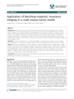
Figure 1 Growth chart of child with Dent’s disease before and during additional therapy with rhGH. G&P: assessment of bone age by
Greulich-Pyle method.
Samardzic et al. Journal of Medical Case Reports 2011, 5:400
/>Page 4 of 6
The growth of our patient was improved and propor-
tional to his pubertal state. Since the therapy was started
before the patient reached puberty it is not possible to
estimate how the GH therapy will continue to affect our
patient’s final height. However after two yea rs of ther-
apy, growth started to slow down.
Although a correlation was found between renal func-
tion and growth impairment, significant short stature
was seen at all levels of renal function. The etiology of
growth delay in children w ith chronic kidney disease is
multifactorial, inc luding rickets, GH resistance, reduced
GH secretion rate or greater loss of GH, functional IGF
deficiency and increased IGFBP -1,-2,-4 and -6 [16].
TheaimoftherapyinDent’ s disease and hypopho-
sphatemic rickets is to normalize serum alkaline phos-
phatase and achieve longitudinal growth. C onventional
treatment with oral phosphate and calcitriol can heal
rickets, but it does not always raise serum phosphate
concentrations significantly, nor does it always normal-
ize linear growth [12].
Both endogenous and exogenous GH result in an
increase in GFR. It is likely that the increased GFR is
mediated by IGF-1 [17]. It is of concern that long-term
rhGH treatment could produce hyperfiltration with
resultant glomerulosclerosis and an accelerated decline
in renal function [18]. In our patient’s case, GFR, mea-
sured as creatinine clearance at the beginning and at the
end of monitoring, remained normal, but it increased
from 92.8 to 112.7 ml/min/1.73 m
2
.Wewerealsocon-
cerned that rhGH might induce hypercalciuria during
calcitriol treatment, however calcium excretion did not
change significantly.
Conclusion
Effects of GH therapy in children with Dent’ sdisease
and short stature are positi ve, but it is difficult to reach
conclusions because of the small sample size in the lit-
erature, the short duration of the therapy and the lack
of a control group. Thus, further studies are needed to
deter minate the pathophysiological mechanism of rhGH
action in Dent’s disease.
Consent
Written informed consent was obtained from the
patient’s mother for publication of this case report and
any accompanying images. A copy of the written con-
sent is available for review by the Editor-in- Chief of this
journal.
Abbreviations
GH: gGrowth hormone; GRF: glomerular filtration rate; IGF-1: insulin-like
growth factor 1; IGFBP-3: insulin-like growth factor binding protein-3; SD:
standard deviation; TRP: tubular reabsorption of phosphate; rhGH:
recombinant human growth hormone.
Acknowledgements
This study was assessed and approved by the institutional review board and
the letter of approval is available for examination.
Author details
1
Institute for Sick Children, Department of Endocrinology and Nephrology,
Ljubljanska bb, 20 000 Podgorica, Montenegro.
2
Department of Clinical
Chemistry and Clinical Pharmacology, University of Bonn, Bonn, Germany.
3
Institute for Mother and Child Health Care of Serbia, Department of
Nephrology, Radoja Dakica 10, 11 000 Belgrade, Serbia.
Authors’ contributions
MS analyzed and interpreted the patient data regarding the
endocrinological follow- up and was a major contributor in writing the
manuscript. SP and RB performed nephrology management and consulted
in the case. ML performed the molecular genetic analysis. All authors read
and approved the final manuscript.
Competing interests
The authors declare that they have no competing interests.
Received: 28 December 2010 Accepted: 22 August 2011
Published: 22 August 2011
References
1. Bogdanovic R, Draaken M, Toromanovic A, Dordevic M, Stajic N, Ludwig M:
A novel CLCN5 mutation in a boy with Bartter-like syndrome and partial
growth hormone deficiency. Pediatr Nephrol 2010, 25(11):2363-2368.
2. Wrong OM, Norden AGW, Feest TG: Dent’s Disease; a familial proximal
renal tubular syndrome with low molecular weight proteinuria,
hypercalciuria, nephrocalcinosis, metabolic bone disease, progressive
renal failure and a marked male predominance. QJM 1994, 87(8):473-493.
3. Lloyd SE, Gunther W, Pearce SH, Thomson A, Bianchi ML, Bosio M, Craig IW,
Fisher SE, Scheinman SJ, Wrong O, Jentsch TJ, Thakker RV: Characterisation
of renal chloride channel, CLCN5, mutations in hypercalciuric
nephrolithiasis (kidney stones) disorders. Hum Mol Genet 1997,
6(8):1233-1239.
4. Raja KA, Schurman S, D’mello RG, Blowey D, Goodyer P, Van Why S, Ploutz-
Snyder RJ, Asplin J, Scheinman SJ: Responsiveness of hypercalciuria to
thiazide in Dent’s disease. J Am Soc Nephrol 2002, 13:2938-2944.
5. Cebotaru V, Kaul S, Devuyst O , Cai H, Recausen L, Guggino WB,
Guggino SE: High citrate diet delays progression of renal insufficiency
in the C1C-5 knockout mouse model of Dent’sdisease.Kidney Int 2005,
68:642-652.
6. Annigeri RA, Rajagoplan R: Hypophosphatemic rickets due to Dent’s
disease: a case report and review of literature. Indian J Nephrol 2009,
19:163-166.
7. Schwartz GJ, Work DF: Measurement and estimation of GFR in children
and adolescents. Clin J Am Soc Nephrol 2009, 4(11):1832-1843.
8. Mahan JD, Warady BA, Consensus Committee: Assessment and treatment
of short stature in pediatric patients with chronic kidney disease: a
consensus statement. Pediatr Nephrol 2006, 21(7):917-930.
9. Ludwig M, Utsch B, Monnens L: Recent advances in understanding the
clinical and genetic heterogeneity of Dent’s disease. Nephrol Dial
Transplant 2006, 21:2708-2717.
10. Tosetto E, Ghiggeri GM, Emma F, Barbano G, Carrea A, Vezzoli G,
Torregrossa R, Cara M, Ripanti G, Ammenti A, Peruzzi L, Murer L, Ratsch IM,
Citron L, Gambaro G, D’angelo A, Anglani F: Phenotypic and genetic
heterogeneity in Dent’s disease–the results of an Italian collaborative
study. Nephrol Dial Transplant 2006, 21:2452-2463.
11. Shrimpton AE, Hoopes RR Jr, Knohl SJ, Hueber P, Reed AA, Christie PT,
Igarashi T, Lee P, Lehman A, White C, Milford DV, Sanchez MR, Unwin R,
Wrong OM, Thakker RV, Scheinman SJ: OCRL1 mutations in Dent 2
patients suggest a mechanism for phenotypic variability. Nephron Physiol
2009,
112(2):27-36.
12.
Dent CE, Friedman M: Hypercalciuric rickets associated with renal tubular
damage. Arch Dis Child 1964, 39:240-249.
13. Sheffer-Babila S, Chandra M, Speiser PW: Growth hormone improves
growth rate and preserves renal function in Dent disease. J Pediatr
Endocrinol Metab 2008, 21:279-286.
Samardzic et al. Journal of Medical Case Reports 2011, 5:400
/>Page 5 of 6
14. Haffner D, Nissel R, Wühl E, Mehls O: Effects of growth hormone
treatment on body proportions and final height among small children
with X-linked hypophosphatemic rickets. Pediatrics 2004, 113(6):593-596.
15. Murer H, Hernando N, Forster I, Biber J: Proximal tubular phosphate
reabsorption: molecular mechanisms. Physiol Rev 2000, 80(4):1373-1409.
16. Mahesh S, Kaskel F: Growth hormone axis in chronic kidney disease.
Pediatr Nephrol 2008, 23:41-48.
17. Hirschberg R, Kopple JDJ: The growth hormone-insulin-like growth factor
I axis and renal glomerular function. J Am Soc Nephrol 1992,
2(9):1417-1422.
18. el Nahas AM, Bassett AH, Cope GH, Le Carpentier JE: Role of growth
hormone in the development of experimental renal scarring. Kidney Int
1991, 40(1):29-34.
doi:10.1186/1752-1947-5-400
Cite this article as: Samardzic et al.: Effect of growth hormone
replacement therapy in a boy with Dent’s disease: a case report. Journal
of Medical Case Reports 2011 5:400.
Submit your next manuscript to BioMed Central
and take full advantage of:
• Convenient online submission
• Thorough peer review
• No space constraints or color figure charges
• Immediate publication on acceptance
• Inclusion in PubMed, CAS, Scopus and Google Scholar
• Research which is freely available for redistribution
Submit your manuscript at
www.biomedcentral.com/submit
Samardzic et al. Journal of Medical Case Reports 2011, 5:400
/>Page 6 of 6