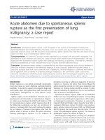
báo cáo khoa học: "Acute abdomen due to spontaneous splenic rupture as the first presentation of lung malignancy: a case report" ppsx
Bạn đang xem bản rút gọn của tài liệu. Xem và tải ngay bản đầy đủ của tài liệu tại đây (639.78 KB, 4 trang )
CAS E REP O R T Open Access
Acute abdomen due to spontaneous splenic
rupture as the first presentation of lung
malignancy: a case report
Angelos Kyriacou
1
, Nolan Arulraj
1*
and Haren Varia
2
Abstract
Introduction: Spontaneous splenic rupture is well recognized in the context of hematological malignancies
(lymphoproliferative and myeloproliferative disorders); a few case reports have also linked solid tumors, such as
pancreatic and liver cancer, with the occurrence of spontaneous splenic rupture. This is the first case report of lung
cancer as a likely cause of spontaneous splenic rupture.
Case presentation: A 61-year-old Caucasian woman presented to our hospital with non-specific symp toms. She
developed an ‘acute’ abdomen and went into a state of shock within twelve hours of her presentation. She was
diagnosed with spontaneous splenic rupture with radiology and following a laparotomy. She made an uneventful
recovery postoperatively and was simultaneously found to have a bronchial adenocarcinoma.
Conclusion: Spontaneous splenic rupture is a potentially fatal but often unrecognized cause of acute abdomen. It
should be routinely considered in the differential diagnosis of acute (’surgical’) abdomen and when present it
should be promptly dealt wi th, most commonly with a laparotomy. Once the diagnosis is confirmed there should
be an aggressive drive to identify an underlying etiology; malignancy is the commonest culprit. Solid tumors
should be considered as underlying causes despite being less common than hematological neoplasms. This case
report demonstrates lung malignancy as an underlying precipitating cause of spontaneous splenic rupture.
Introduction
Splenic rupture is a rare albeit potentially catastrophic
event that causes an ‘acute’ abdomen and hemodynamic
instability. It should be urgently investigated, diagnosed
and treated, often with splenectomy and less often with
conservative management or with splenic artery
embolization.
Splenic rupture can be divided into traumatic (or non-
spontaneous) and atraumatic (or spontaneous). Diagnos-
tic criteria were developed by Orloff and Peskin in 1958
[1] for spontaneous rupture which requires that all of
the following conditions are met: (a) no history of
trauma or unusual effort that could rupture the spleen;
(b) no evidence of disease in the organs, o ther than the
spleen, which is known to affect the spleen adversely
causing pathological rupture; (c) no evidence of
perisplenic scarring or adhesions s uggestive of previous
rupture or trauma; (d) other than hemorrhage the
spleen should be normal on both gross inspection and
histology; (e) clotting studies should be normal; (f) other
criteria including no significant rise in viral antibody
titers in acute or convalescent sera.
Spontaneous splenic rupture (SSR) can be subdivided
further into true and pathological rupture corresponding
to normal and pa thological appearance s of the spleen
on histologica l examination. Therefore, in essence, the
original Orloff and Pes kin criteria describe ‘true’ SSR. A
systematic literature review [2] of 845 cases of atrau-
matic splenic rupture between 1980 and 2008 found
that the former was much rarer than the latter (7% t rue
versus 93% pathological splenic rupture). The causes of
pathological rupture are shown in Figure 1.
Hematological malignancies (mainly non-Hodgkin’ s
lymphoma , chronic myeloid leukemia and acute lym-
phoblastic l eukemi a) comprise the majority of the neo-
plastic causes, with solid tumo rs rarely re ported in the
* Correspondence:
1
Department of Medicine, Whinney Heys Road, Blackpool Victoria Hospital,
Blackpool, FY3 8NR, UK
Full list of author information is available at the end of the article
Kyriacou et al. Journal of Medical Case Reports 2011, 5:444
/>JOURNAL OF MEDICAL
CASE REPORTS
© 2011 Kyriacou et al; licensee BioMed Central Ltd. This is an Open Access artic le distributed under the terms of the Creative
Commons Attribution License ( which permits unrestri cted use, distribution, and
reproduction in any medium, provided the original work is properly cited.
literature (such as hepatocellular [3] and pancreatic car-
cinoma [4]).
The investigation of choice for confirming the diagno-
sis is computed tomography (CT) of the abdomen [5],
which h as a sensitivity and specificity of 90-95%. Sple-
nectomy remains the treatment of choice for splenic
rupture with hemodynamic instability, and should be
urgently performed once the diagnosis is confirmed.
Proximal splenic artery embolization can be performed
to treat SSR [6] in a hemodynamically stable patient but
requires an interventional radiology department with
expertise in the relevant procedure and close monitoring
of vital signs and hemoglobin concentration, ideally in
an intensive care setting. The potential benefits of this
technique include a reduced risk of repeated he mor-
rhage with preservation of the splenic tissue and
reduced levels of postoperative sepsis [6,7].
InasystematicreviewbyRenzulliet al. [2] s plen ect-
omy was performed in 84.1% of cases; the rest were
either treated conservatively (14.7%) or with organ- pre-
serving surgery (1.2%). Whenever possible, the underly-
ing cause should be treated. Factors that increase
mortality include underlying neoplastic disease, spleno-
megaly and increasing age [2].
Case Presentation
A 61-year-old Caucasian woman, previously in good
health, presented with a three-day history of feeling gen-
erally unwell with dizziness, vomiting, abdominal, left
lower chest pain and shoulder pain. She denied any sore
throat, feeling feverish or other symptoms suggestive of
an i nfluenza-like i llness. There was no cough or sputum
production. There was nothing in the hist ory to suggest
a recent viral or other infective process including
human immunodeficiency virus or acquired immune
deficiency state and no history of any trauma or injury.
She had no significant previous medical or surgical his-
tory and was not taking any medications. There was no
background or family history of cancer, hematologic or
clotting disorders. She was a smoker of approximately
40 pack years and had unlimited exercise tolerance. She
worked as a nurse and there was no history of previous
exposure to asbestos or other occupational hazards.
On examination she was ill-looking, conscious and
orientated with a blood pressure 89/49 mmHg, heart
rate of 72 beats per minute, saturations 97% on a non-
rebreather mask and temperature of 36.9°C. The admis-
sion e xamination revealed normal cardiac examination
and bibasal inspiratory crepitations with left upper
quadrant and epigastric tenderness on abdominal
examination.
Her blood work-up on admission showed a hemoglo-
bin level of 11.4 g/L, white cell count of 19 × 10
9
/L
with neutrophils of 17 × 10
9
/L and a C-reactive protein
of 7 mg/L. Urea and electrolytes, liver function and coa-
gulation tests were in the normal range. A bloo d film
examination d id not reveal any atypical lymphocyte s or
other abnormalities. A posteroanterior and lateral chest
radiograph was performed (Figure 2).
Hemodynamic stability was achieved following rehy-
dration with intravenous fluids. However, eight hours
after admission she developed an acute abdomen with
clinical signs of shock. Her blood pressure was 88/52
mmHg and heart rate 105 beats per minute. Further to
that her oxygen saturations were 91% on a non-
rebreather mask with a Glasgow coma scale of 14/15 (E
= 4, V = 4, M = 6). Inspection revealed skin and con-
junctival pallor. Clinical examination revealed new
abdominal signs of generalized abdominal tenderness
with guarding, rigidity and absent bowel sounds, consis-
tent with an acute abdomen. A repeat test showed her
hemoglobin level had dropped to 5.0 g/L.
She was stabilized with multiple blood transfusions
and underwent an urgent computer tomography scan of
her thorax and abdomen, which showed an 8.5 × 3.6 cm
left hilar mass with extensive mediastinal adenopathy,
bibasal small effusions and consolidation, and a large
splenic hematoma of 15 × 12 cm with high attenuation
suggestive of active bleeding (Figure 2, Figure 3 and 4).
There was a lytic area affecting her T9 vertebra, which
likely represented metastases rather than wedge fracture,
but there were no abnormalities or neoplastic disease
affecting the intra-abdominal organs.
ȋʹǤ͵ΨȌ
ȋǤͺΨȌ
Ƭ
ȋͻǤʹΨȌ
ȀǦ
ȋʹͲΨȌ
ȋͻ͵ΨȌ
ȋΨȌ
ȋ͵ͲǤ͵ΨȌ
Figure 1 The causes of pathological rupture, adapted from
Renzulli et al. [2].
Figure 2 Large left anterior mediastinal mass with prominent
left hilum.
Kyriacou et al. Journal of Medical Case Reports 2011, 5:444
/>Page 2 of 4
Pneumococcal and meningococcal vaccines were
administered and our patient was then promptly taken
to theater for laparotomy. On examination of her inter-
nal organs at laparotomy, other than hemorrhage and
rupture of her spleen, there was no other gross abnorm-
ality and no evidence of disease in her other intra-
abdominal organs. She spent two days being ventilated
in our intensive care unit and su bsequently made a full
recovery from the surgery. She was commenced on life-
long penicillin V and was subsequently discharged home
a week after admission, fully mobile and independent in
terms of her activities of daily living.
Her splenic histology was negative for hematologic or
other malignancy and no other path ology was identified.
She then underwent a bronchoscopy; transbronchial
needle aspiration revealed numerous malignant nodes
consistent with non-small cell carcinoma. Histology of
bronchial biopsies confirmed invasive adenocarcinoma.
This was consistent with likely stage IV in view o f the
likely bone metastases found on CT. She was referred to
the local oncologist and her case was discussed at the
lung cancer multidisciplinary team meeting. In view of
the diagnosis and staging, a palliative treatment pathway
was agreed from the outset and our patient received pal-
liative chemotherapy. She died five months after her
presentation.
Discussion
To the best of our knowledge, this is the first case
report of SSR in association with lung cancer. Lung can-
cer with splenic rupture has been reported either in the
context of splenic metastasis [8], after the initiation of
chemotherapy in a patient with a splenic hamartoma
(splenoma) [9] or after pegfilgrastim was given to pre-
vent neutropenia [10]. Few case reports have previously
described the occurrence of splenic rupture in patients
with known lung cancer; usually in widel y diss eminated
metastatic disease. The incidence of isolated splenic
metastasis ranges from 0-26% of all patients with splenic
metastasis from a literature review [11]. The length of
time until diagnosis of splenic metastasis from the diag-
nosis of a primary lung cancer ranges between 0-8 years
[11]. However, our case is of interest because there were
no metastatic deposits and the lung cancer presented
itself with SSR, as opposed to respiratory symptoms.
The mortality rate for conservati vely managed sponta-
neous sp lenic rupture was 22% at 30 days; mortality was
30% versus 5% for malignant and benign underlying dis-
ease, respectively [12]. The traumatic splenic rupture
postoperative mortality rate was 18% [12].
It is possible that our patient had two concurrent,
unrelated p athologies: bronchial adenocarcinoma and a
true SSR. However, given the well-described association
between neoplastic disease and SSR, the above case
reports linking the two conditions and the rarity of true
spontaneous splenic rupture, it is likely that the two
conditions are linked with the former precipitating the
latter.
Conclusion
Our case illustrates the need for extra vigilance to make
the diagnosis of splenic rupture in the context of a lack
of trauma. The exact pathophysiology that links lung
cancer with SSR (in the absence of splenic metastasis or
use of chemotherapy or pegfilgrastim) remains unclear.
The possible explanations include a hypercoagulable
state secondary to the underlying malignancy.
In summary, this case demonstrates that l ung cancer
could potentially precipitate SSR; it could even present
itself as SSR as in our patient. This association is of rele-
vance to a wi de variety of health care professionals
including acute and general physicians, general sur-
geons, oncologists, radiologists and others. The other
Figure 3 Left hilar mass measuring 5 cm with associated
significant mediastinal adenopathy and a 3 cm upper right
paratracheal node.
Figure 4 A and B. Large splenic hematoma. Inferomedial areas of
high attenuation suggestive of active bleeding. Compressed and
slit-like inferior vena cava indicative of hemodynamic instability.
Extensive ascites also present.
Kyriacou et al. Journal of Medical Case Reports 2011, 5:444
/>Page 3 of 4
important learning point from this case is that although
SSR is a rare cause of acute abdomen, we should bear in
mind that it is fatal if misdiagnosed and left untreated; it
shouldthereforebeconsideredinthedifferentialdiag-
nosis of a wide range of medical and surgical conditions.
Consent
Written informed consent was obtained from the patient
for p ublication of this manuscript and any accompany-
ing images. A copy of the written consent is available
for review by the Editor-in-Chief of this journal.
Acknowledgements
The authors would like to thank Mr Ross Jones at the Department of
General Surgery at Blackpool Victoria Hospital for assistance in the
preparation of this manuscript.
Author details
1
Department of Medicine, Whinney Heys Road, Blackpool Victoria Hospital,
Blackpool, FY3 8NR, UK.
2
Department of Radiology, Whinney Heys Road,
Blackpool Victoria Hospital, Blackpool, FY3 8NR, UK.
Authors’ contributions
AK contributed to the medical care of this patient, the manuscript
preparation and revision, and also performed the literature review. NA
contributed to the medical care of this patient, the manuscript preparation
and revision. HV contributed to the radiological review of the case report
and the manuscript revision. All authors read and approved the final
manuscript.
Competing interests
The authors declare that they have no competing interests.
Received: 16 February 2011 Accepted: 7 September 2011
Published: 7 September 2011
References
1. Orloff MJ, Peskin GW: Spontaneous rupture of the normal spleen: a
surgical enigma. Int Abstr Surg 1958, 106(1):1-11.
2. Renzulli P, Hostettler A, Schepfer AM, Gloor B, Candinas D: Systematic
review of atraumatic splenic rupture. Br J Surg 2009, 96(10):1114-1121.
3. Sugahara K, Togashi H, Aoki M, Mitsuhashi H, Matsuo T, Watanabe H, Abe T,
Ohno S, Saito K, Saito T, Shinzawa H, Tanida H, Ito M, Takahashi T:
Spontaneous splenic rupture in a patient with large hepatocellular
carcinoma. Am J Gastroenterol 1999, 94(1):276-278.
4. Smith WM, Lucas JG, Frankel WL: Splenic rupture: a rare presentation of
pancreatic carcinoma. Arch Pathol Lab Med 2004, 128(10):1146-1150.
5. Rabushka LS, Kawashima A, Fishman EK: Imaging of the spleen: CT with
supplemental MR examination. Radiographic 1994, 14(2):307-332.
6. Stephenson JT, DuBais JJ: Nonoperative management of spontaneous
splenic rupture in infectious mononucleosis: a case report and review of
the literature. Pediatrics 2007, 120(2):e432-e435.
7. Pachter HL, Grau J: The current status of splenic preservation. Adv Surg
2000, 34:137-174.
8. Massarweh S, Dhingra H: Unusual sites of malignancy: case 3. Solitary
splenic metastasis in lung cancer with spontaneous rupture. J Clin Oncol
2001, 19(5):1574-1575.
9. Ballardini P, Incasa E, Del Noce A, Cavazzini L, Martoni A, Piana E:
Spontaneous splenic rupture after the start of lung cancer
chemotherapy. A case report. Tumouri 2004, 90(1):144-146.
10. Watring NJ, Wagner TW, Stark JJ: Spontaneous splenic rupture secondary
to pegfilgrastim to prevent neutropenia in a patient with non-small-cell
lung carcinoma. Am J Emerg Med 2007, 25(2):247-248.
11. Schmidt BJ, Smith SL: Isolated splenic metastasis with primary lung
adenocarcinoma. South Med J 2004, 97(3):298-300.
12. Görg C, Cölle J, Görg K, Zugmaier G: Spontaneous rupture of the spleen:
ultrasound patterns, diagnosis and follow-up. Br J Radiol 2003,
76(910):704-711.
doi:10.1186/1752-1947-5-444
Cite this article as: Kyriacou et al.: Acute abdomen due to spontaneous
splenic rupture as the first presentation of lung malignancy: a case
report. Journal of Medical Case Reports 2011 5:444.
Submit your next manuscript to BioMed Central
and take full advantage of:
• Convenient online submission
• Thorough peer review
• No space constraints or color figure charges
• Immediate publication on acceptance
• Inclusion in PubMed, CAS, Scopus and Google Scholar
• Research which is freely available for redistribution
Submit your manuscript at
www.biomedcentral.com/submit
Kyriacou et al. Journal of Medical Case Reports 2011, 5:444
/>Page 4 of 4