báo cáo khoa học: "Two successful natural pregnancies in a patient with severe uterine prolapse: A case report" ppsx
Bạn đang xem bản rút gọn của tài liệu. Xem và tải ngay bản đầy đủ của tài liệu tại đây (496.5 KB, 3 trang )
CAS E REP O R T Open Access
Two successful natural pregnancies in a patient
with severe uterine prolapse: A case report
Davide De Vita
1
and Salvatore Giordano
2*
Abstract
Introduction: Uterine prolapse is a common gynecologic condition that is rare during or before pregnancy. We
report an exceptional case of two pregnancies in a totally prolapsed uterus.
Case presentation: A 36-year-old Caucasian woman with a history of uterine prolapse presented with pregnancy.
A vaginal pessary was applied to keep her uterus inside the pelvis after manual reposition. The pessary was
removed at the 24th week. The gravid uterus persisted in the abdominal cavity because of its increased volume.
Conclusion: Our case shows that pregnancy during uterine prolapse is possible and that careful assessment is
required to prevent complications during delivery. According to our experience, an elective caesarean section near
term could be the safest mode of delivery.
Introduction
Uterine prolapse is a common gynecologic condition but
it is extremely rare during pregnancy with an estimated
incidence of one per 10,000 to 15,000 deliverie s [1]. Few
cases are described in the literature, especially on its
correlation with subsequent pregnancy.
Women with prolapse may have a variety of pelvic
floor symptoms. Symptoms include pelvic heaviness, a
dragging sensation in the vagina, protrusion coming
down from the vagina and backache, but only some of
these symptoms are directly related to the prolapse.
Case presentation
A 36-year-old Caucasi an woman, gravida 3, para 2, pre-
sented to our antenatal outpatient clinic in the 10
th
week of gestation complaining of uterine prolapse and
amenorrhea. Five years earlier, at the age of 31 years,
she had her first spontaneous vaginal delivery, after 39
weeks of clinically unremark able gestation and after a
seven-hour labor. A living male baby weighing 2950 g,
with Apgar scores of 10/10, was delivered. After that, a
total uterine prolapse (POP-Q IV) was observed and,
therefore, a pelvic reconstruction operat ion was
scheduled. However, she missed the appointment and
she was lost to follow-up.
Four years later, at the age of 35 years, the patient had
her first pregnancy in a prolapsed uterus and the deliv-
ery was performed by an elective caesarean section after
38 weeks of gestation. During this second pregnancy fol-
low-up she experienced symptoms of heaviness, but no
pelvic pain or urinary incontinence. Pelvic examination
showed that the uterus persisted in the pelvis because of
increased volume. The cervical os was closed, while the
entire cervix was lyi ng outside the vulva duri ng the first
three months and after week 18 it appeared completely
inside. When the cervix was outside the vulva, it
appeared enlarged a nd edematous w ith marked ectro-
pion but it was not ulcerated. A live male baby weighing
3150 g, with Apgar scores of 10/10, was delivered with
elective caesarian section. After that, a total uterine pro-
lapse persisted but she refused any procedure for pelvic
reconstruction; neither was a vaginal pessary used.
One year later, at the age of 36 years, she presented
again in our clinic with a 10-week pregnancy in a pro-
lapse d uterus. A vaginal pessary was applied to keep the
uterus inside the pelvis after manual reposition. The
pessary was removed at the 24
th
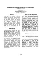
week. The gravid uterus
persisted in the abdominal cavity because it was
increased in volume (Figure 1). She did not show any
symptoms of heaviness or urinary incontinence. The
cervix was lying at the os of the vulva (POP-Q II)
* Correspondence:
2
Department of Surgery, Division of Plastic Surgery, Turku University Hospital,
OS 299, PL 52, 20521, Turku, Finland
Full list of author information is available at the end of the article
De Vita and Giordano Journal of Medical Case Reports 2011, 5:459
/>JOURNAL OF MEDICAL
CASE REPORTS
© 2011 De Vita and Giordano; licensee BioMed Central Ltd. This is an Open Ac cess article distributed under the terms of the C reative
Commons Attribution Licen se ( es/by/2.0), which permits unrestricted use, distributio n, and
reprodu ction in any mediu m, provided the original work is pro perly cited.
without signs of dessication or ulceration. It was
enlarged and edematous but showed no evidence of cer-
vical incompetence.
Serial transabdominal ultrasonograpic examinations
showed a normally developing fetus in longitudinal posi-
tion in the uterine cavity. Elect ive caesarean section was
performedatthe38
th
week. A living, healthy female
baby weighing 3030 g, with Apgar scores of 10/10, was
delivered.
The postnatal period was uneventful and she was dis-
charged home four days later in good health. Normal
postpartum uterine involution was observed. After that,
a total uterine prolapse (POP-Q IV) was still observed
(Figure 2).
She is scheduled for follow-up examinatio n and pelvic
reconstruction surgery.
Conclusion
Uterine prolapse is a common gynecologic condition but
is extremely rare during pregnancy as shown by the few
similar reports in the literature. Certainly the literature
before 1970, while it does not always specify the exact
degree of prolapse, suggests a much higher incidence in
more disadvantaged areas and where gr and multiparity
was more common. We found two reports of natural
term pregnancy with an initially procidencia uteri [2,3]
and one case of in vitro fertilization and embryo transfer
pregnancy with an initially complete uterine prolapse
[4].
In the classification of uterine prolapse using the POP-
Q evaluation, total uterine prolapse extending outside
the introitus with eversion of the entire vagina witho ut
standing or traction is calle d third-fourth degree pro-
lapse [5].
Multiple factors are usually involved in the genesis of
uterine prolapse but the most prominent cause is preg-
nancy, associated with prolonged labor, or difficult deliv-
ery. However, it may also occur spontaneously, although
very rarely, even in nulliparous women.
In our case, the patient had sexual intercourse without
any vaginal pessary.
Conservative management with close follow-up and
bed rest can alleviate clinical symptoms and reduce
potential complications correlated with this condition
[2,4]. We recommend a vaginal pessary application dur-
ing the first six months, until the volume of the uterus
volume is increased
Complications such as patient discomfort, cervical
dessication and ulceration, urinary tract infection, acute
urinary retention, abortion, pre-term labor and even
maternal death have been previously described [3,6]. We
did not observe any of these complications except
patient discomfort with light symptoms of heaviness
without pelvic pain.
Although in a very recent report Eddib et al. [3] man-
aged a sim ilar case with a vaginal d elivery, we believe
that elective Caesarean section near term could be the
safest delivery modality in order to avoid a progression
of the prolapse and uterine rupture or damage [1,6].
This procedure can be also effective in preventing organ
prolapse.
In conclusion, our case i llustrates that natural preg-
nancy during uterine prolapse is possible and the man-
agement of uterine prolapse during labor should be
individualized, depending on the severi ty of the
Figure 1 Resolution of the prolapse during the f inal period of
gestation because of the increased uterus volume.
Figure 2 The patient after elective Caesarean section with total
uterine prolapse.
De Vita and Giordano Journal of Medical Case Reports 2011, 5:459
/>Page 2 of 3
prolapse, gestational age, parity, and the patient’ s
preference.
A vaginal delivery can be expected, but, according to
our experience, an elective caesarean section ne ar term
could be a valid and safe delivery option.
Consent
Written informed consent was obtained from the patient
for publication of this case report and accompanying
images. A copy of the written consent is available for
review by the Editor-in-Chief of this journal.
Author details
1
Department of Obstetrics and Gynaecology, Santa Maria della Speranza
Hospital, via Fiorignano, Battipaglia, 84091, SA, Italy.
2
Department of Surgery,
Division of Plastic Surgery, Turku University Hospital, OS 299, PL 52, 20521,
Turku, Finland.
Authors’ contributions
DD analyzed and interpreted the patient data, performed clinical
examinations and revised the manuscript. SG designed the case and was a
major contributor in writing the manuscript. Both authors read and
approved the final manuscript.
Competing interests
The authors declare that they have no competing interests.
Received: 1 July 2011 Accepted: 14 September 2011
Published: 14 September 2011
References
1. Guariglia L, Carducci B, Botta A, Ferrazzani S, Caruso A: Uterine prolapse in
pregnancy. Gynecol Obstet Invest 2005, 60:192-194.
2. Jeng CJ, Lou CN, Lee FK, Tzeng CR: Successful pregnancy in a patient
with initially procidentia uteri. Acta Obstet Gynecol Scand 2006, 85:501-502.
3. Eddib A, Allaf MB, Lele A: Pregnancy in a woman with uterine
procidentia: a case report. J Reprod Med 2010, 55:67-70.
4. Chun SS, Park KS: Birth of a healthy infant after in vitro for fertilization
and embryo transfer in patient of total uterine prolapse. J Assist Reprod
Genet 2001, 18:346-347.
5. Bump RC, Mattiasson A, Bø K, Brubaker LP, DeLancey JO, Klarskov P,
Shull BL, Smith AR: The standardization of terminology of female pelvic
organ prolapse and pelvic floor dysfunction. Am J Obstet Gynecol 1996,
175:10-17.
6. Daskalakis G, Lymberopoulos E, Anastasakis E, Kalmantis K, Athanasaki A,
Manoli A, Antsaklis A: Uterine prolapse complicating pregnancy. Arch
Gynecol Obstet 2007, 276:391-392.
doi:10.1186/1752-1947-5-459
Cite this article as: De Vita and Giordano: Two successful natural
pregnancies in a patient with severe uterine prolapse: A case report.
Journal of Medical Case Reports 2011 5:459.
Submit your next manuscript to BioMed Central
and take full advantage of:
• Convenient online submission
• Thorough peer review
• No space constraints or color figure charges
• Immediate publication on acceptance
• Inclusion in PubMed, CAS, Scopus and Google Scholar
• Research which is freely available for redistribution
Submit your manuscript at
www.biomedcentral.com/submit
De Vita and Giordano Journal of Medical Case Reports 2011, 5:459
/>Page 3 of 3