báo cáo khoa học: "Persisting right-sided chylothorax in a patient with chronic lymphocytic leukemia: a case report" pot
Bạn đang xem bản rút gọn của tài liệu. Xem và tải ngay bản đầy đủ của tài liệu tại đây (1.47 MB, 6 trang )
JOURNAL OF MEDICAL
CASE REPORTS
Persisting right-sided chylothorax in a patient
with chronic lymphocytic leukemia: a case report
Scholz et al.
Scholz et al. Journal of Medical Case Reports 2011, 5:492
(3 October 2011)
CAS E REP O R T Open Access
Persisting right-sided chylothorax in a patient
with chronic lymphocytic leukemia: a case report
Godehard A Scholz
1
, Horia Sirbu
2
, Sabine Semrau
3
, Katharina Anders
4
, Andreas Mackensen
1
and
Bernd M Spriewald
1*
Abstract
Introduction: Chylothorax caused by chronic lymphocytic leukemia is very rare and the best therapeutic approach,
especially the role of modern immunochemotherapy, is not yet defined.
Case presentation: We present the case of a 65-ye ar-old male Caucasian patient with right-sided chylotho rax
caused by a concomitantly diagnosed chronic lymphocytic leukemia. As first-line treatment four cycles of an
immunochemothera py, consisting of fludarabine, cyclophosphamide and rituximab were administered. In addition,
our patient received total parenteral nutrition for the first two weeks of treatment. Despite the very good clinical
response of the lymphoma to treatment, the chylothorax persisted and percutaneous radiotherapy of the thoracic
duct was applied. However, eight weeks after the radiotherapy the chylothorax still persisted and our patient
agreed to a surgical intervention. A ligation of the thoracic duct via a muscle sparing thoracotomy was performed,
resulting in a complete cessation of the pleural effusion. Apart from the first two weeks our patient was treated on
an out-patient basis for nearly six months.
Conclusion: In this case of chylothorax caused by chronic lymphocytic leukemia, immunochemotherapy in
combination with conservative treatment, and even consecutive radiotherapy, were not able to stop pleural
effusion, despite the very good clinical response of the chronic lymphocytic leukemia to treatment.
Out-patient management using repetitive thoracocenteses can be safe as bridging until definitive surgical ligation
of the thoracic duct.
Introduction
Chylothorax is a ra re condition defined by chyle enter-
ing the pleural space, caused by a disruption or blockade
of the thoracic duct [1]. The pleural effusion is usually
of milky white appearance due to a high lipid concentra-
tion. To distinguish chylothorax from nonchylous effu-
sions, such as pseudochylot horax, the triglyceride level
is determined. A triglyceride level great er than 110 mg/
dL is highly suggestive of a chylous effusion. In cases
where triglycerides range be tween 50 mg/dL and 110
mg/dL, a d iagnosis of chylotho rax can be made using
lipid electrophoresis to detect the presence of chylomi-
crons [2,3].
Disruptionofthethoracicductmaybeduetotrau-
matic or non-traumatic causes. In adults, the incidence
of non-traumatic causes is reported between 50% and
70% of cases [4,5]. Among the no n-traumatic causes,
lymphoma and metastatic cancer are most common.
Chronic lymphocytic leukemia (CLL), however, is a rare
cause of chylothorax, with only a few cases reported in
the literature so far [6].
Since chylothorax is an overall infrequent condition
the best therapeutic approach is still under debate
[1,5,7,8]. In particular, the use of modern immunochem-
otherapy, including the anti-CD20 antibody rituximab,
in lymphoma-associated chylothorax has not y et been
described.
Here we present the course of a patient presenting
with chylothorax caused by a concomitantly diagnosed
CLL who received conservative treatment in conjunction
with immunochemotherapy, followed by radiotherapy
and finally surgery to control his persisting pleural
effusions.
* Correspondence:
1
Department of Internal Medicine 5 - Hematology/Oncology, University of
Erlangen-Nürnberg, Krankenhausstrasse 12, 91054 Erlangen, Germany
Full list of author information is available at the end of the article
Scholz et al. Journal of Medical Case Reports 2011, 5:492
/>JOURNAL OF MEDICAL
CASE REPORTS
© 2011 Scholz et al; licensee Bio Med Central Ltd . This is an Open Access article distribute d under the terms of the Creative Commons
Attribution License (http: //creativecommons.org/licenses/by/2.0), which permits unrestricted use, distribution, and reproduction in
any medium, provided the original work is properly cited.
Case presentation
A 65-year-old male Caucasian patient was admitted with
respiratory distress and suspected non-Hodgkin’slym-
phoma. His medical history revealed arterial hyperten-
sion, diabetes mellitus type 2 and cholecystolithiasis.
On clinical examination our obese patient (body mass
index 41.5) suffered from dyspnea at rest. He presented
with enlarged cervical and axillary lymph nodes, hepa-
tosplenomegaly and diminished breath sounds over his
right lung. A blood count revealed a leukocytosis of
35,000 leukocytes/μL of blood, with 80% par tial ly aty pi-
cal small lympho cytes and Gumprecht’ sshadowcells.
Hemoglobin concentration and thrombocytes were
within normal range. Immunophenotyping revealed that
67% of leukocyt es were CD19+ B-lymphocytes, with co-
expression of CD5, CD20, CD23 and a clonal restriction
for the lambda light chain. This established the diagno-
sis of CLL. Bone marrow puncture demonstrated a med-
ium degree of infiltration, with monoclonal B-cells
beginning to replace the normal hematopoiesis.
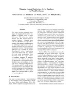
A chest X-ray was performed and showed a right-
sided complete opacity suggesting a pleural effusion
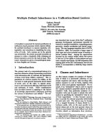
(Figure 1). T horacentesis produced a milky pleur al fluid
(Figure 2A). The cellular cont ent consisted of 80% lym-
phocyte s, two thirds of which expressed the B-CLL phe-
notype. Further analysis of the pleural fluid revealed
triglyceride levels over 700 mg/dL and cho lesterol levels
below 70 mg/dL, establishing the diagnosis of
chylothorax. Our patient received a pleural drainage,
which initially produced nearly 3 liters of chyle per 24
hours. A computed tomography (CT) scan depicted
enlarged lymph nodes in the cervical, axillary and med-
iastinal region, and suspected splenic involvement with
several hypodense lesions. Taking the findings into
account, our patient was diagnosed with a right-sided
chylothorax caused by a concomitantly diagnosed CLL,
stage Binet B or Rai II.
The chylothorax represented a major complication of
the CLL, and so immunochemotherapy consisting of flu-
darabine (25 mg/m
2
on days one to three), cyclopho-
sphamide (250 mg/m
2
on days one to three) and
rituximab (375 mg/m
2
on day one) was initiated. Our
patient received four courses, repeated every four weeks.
Figure 1 Initial posterior-anterior chest X-ray demonstrating a
complete right-sided opacity, later diagnosed as chylothorax.
$
$ %
Figure 2 Appearance of the pleural fluid before and after a
low dietary fat intake. (A) The high triglyceride content of over
700 mg/dL caused a milky appearance, characteristic of chylothorax.
(B) A low-fat diet and concomitant reduced triglyceride levels in the
pleural effusion resulted in a change towards a clear amber-colored
fluid.
Scholz et al. Journal of Medical Case Reports 2011, 5:492
/>Page 2 of 5
Since the therapeutic effect of reduced dietary intake on
chylothorax had been described previously, our patient
received total parenteral n utrition for two weeks, start-
ing with the first cycle of the immunochemotherapy.
The chylous effusion disappeared nearly completely, and
the chest drain could be removed after 10 days. After
two weeks an enteral low-fat diet enriched with med-
ium-chain triglycerides was started, to continue therapy
on an out-patient basis. Unfortunately the chylothorax
relapsed and thoracentesis of a volume of 1 L to 1.5 L
once to twice a week became necessary. Due to the low-
fat intake the appearance of the pleural effusion had
changed f rom milky-white to clear amber-colored (Fig-
ure 2B).
Our patient received four cycles of immunochem-
otherapy and regular thoracentesis on an out-patient
basis. S ince patients with protracted chylothorax are at
risk of malnutrition and immunosuppression, our
patient rec eived antifungal and antiviral prophylaxis in
addition to vita min supplementation. However, the chy-
lothorax persisted, despite a good clinical response of
the CLL, with normalized blood count s and complete
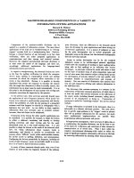
regression of the lymphadenopathy (Figure 3).
In light of this, percutaneous radiotherapy of his med-
iastinum and thoracic duct, with an overall dose of 24
Gy, was initiated. Radiation induces an inflammatory
response which can result in an obliteration of the dis-
rupted thoracic duct [9]. However, up to eight weeks
after completion of the radiotherapy the chyloth orax
still persisted with continued requirement for regular
pleural tapping.
Finally our patient agreed to a surgical intervention. A
supradiaphragmal ligation of the thoracic duc t via a
right muscle sparing thoracotomy was carried out. In
addition, a decortication of his right lung was necessary
because, d uring his surgery, a pleural fibrosis was diag-
nosed. The pleural fibrosis was most likely caused by
the long-term chylothorax with repetiti ve thoracenteses.
Our patient quickly recovered and the pleural effusions
ceased completely. The time from the first diagnosis of
chylothorax until the final surgical intervention was six
months. O ur patient is s till in complete remission after
24 months of follow-up.
Discussion
Our patient presented with pronounced dyspnea, which
was caused by a right-sided pleural effusion diagnosed
as chylothorax. Pleural effusion, although not uncom-
moninnon-Hodgkin’ s lymphoma, is less often seen in
CLL [10]. The differential diagnosis of pleural effusion
in a patient with CLL includes infection, pleural involve-
ment and lymphatic obstruction [10]. Chylothorax, how-
ever, is a rare complication of non-Hodgkin’s lymphoma
(especially CLL) and should be considered by analyzing
triglyceride and cholesterol concentrations in addition
to routine parameters [3]. The present case showed that
the chyle contained 80% B-CLL cells on immunopheno-
typing, replacing the normally present T-cells. This is in
accordance with fi ndings of two previous cases reported
by Doerr et al. and Zimhony et al. [11,12], whereas Rice
et al. found predominantly T-cells in the chyle of their
CLL patient [6]. Therefore immunophenotyping of chyle
may have limited value in diagnosing chylothorax in
CLL patients.
Despite numerous anatomic variations, the thoracic
duct usually arises from the cisterna chyli. From there it
ascendsthroughtheaortichiatusontherightsideof
the vertebral column and crosses t o the left side
between the sixth and fourth thoracic vertebra, before it
$
*
%
*
Figure 3 A CT scan demonst rates para-aortal
lymphadenopathy (A) before and (B) after two cycles of
immunochemotherapy. Para-aortal lymphadenopathy (arrow
heads) might have been the most probable cause of the thoracic
duct injury resulting in chylothorax (asterisk). Immunochemotherapy
reduced the lymphadenopathy after only two cycles. Nevertheless,
the pleural effusion (asterisk) still persisted, indicating that the
thoracic duct injury had not healed. The aorta is indicated (dotted
circle).
Scholz et al. Journal of Medical Case Reports 2011, 5:492
/>Page 3 of 5
empties into the ju nction of the left jugular and subcl a-
vian veins [5,7]. A right-sided chylothorax, therefore,
indicates an injury below the fifth thoracic vertebra,
which has to be taken into account when planning
radiotherapy or surgery.
As depicted in Figure 2A the pleural fluid was initially
milky white, which might already raise suspicion of its
origin. However, the gross appearance of the chy-
lothorax may be misleading in over half of the cases,
since nutrition has a strong influence on lipid content
and therefore on the appearance of the chyle [2,3] . With
a low dietary fat intake the chyle clears to a serous
appearance, as demonstrated in Figure 2B.
The amount of chyle produced per day correlates with
dietary fat intake and was reported to range from 10
mL/kg to over 100 mL/kg body weight [8]. Due to this
high amount of over 2 L on average per day, a rupture
of the thoracic duct c an result in rapid development of
extensive pleural effusion with consecutive impaired
breathing [1]. Therefore, immediate thoracentesis and a
pleural drainage may be necessary. In addition, diet
therapy, especially total parenteral nutrition in combina-
tion with pleural dra inage, has been shown to be able to
reduce chyle production and resolve chylothorax with-
out increasing mortality [13]. In our patient, total par-
enteral nutrition resulted in a dramatic decrease in
drained chyle from nearly 3 L to less than 100 mL per
24 hours and the pleural drainage could be removed
after 10 days. However, after initiation of a low fat oral
diet, the chylothorax recommenced, albeit at a lower
rate, and required regular thoracenteses over the next
three months.
In e arly reports, the mortality of chylothorax reached
50% for traumatic chylothorax and was fatal in non-
traumatic cases [14]. This has now been reduced to a
mortality of around 10% [1,7]. The amount of drained
chyle in our patient was about 3 L per week, less t han
500 mL per day, and so the risk of conservative treat-
ment to evaluate the bene fit of the anti-CD20 antibody,
rituximab, in combination with immuno chemotherapy
seemed acceptable. Furthermore, it has been suggested
that malignant chylothorax may not benefit from surgi-
cal in tervention [15]. However, the combined a pproach
of conservative treatment with a low fat diet and immu-
nochemotherapy had no measurable effect on the chy-
lothorax in this case.
Radiation may caus e damage to the thoraci c duct and
induce chylothorax, most likely by inducing inflamma-
tion and obstruction [5]. On the other hand, the same
mechanisms of radiation can be exploited to treat chy-
lothorax [9,16]. In the present case however, radiation
therapy had n o effect, despite the prior effective anti-
CLL therapy. A similar observation was reported by
Zimhony et al. [12]. The chylothorax of their patient
with CLL did not improve after chemotherapy and med-
iastinal irradiation, and required pleurodesis to resolve
pleural effusion. However, mediastinal irradiation can be
effective in CLL-associated chylothorax, as demonstrated
by Ampil et al., who reported the case of a female CLL
patient who developed chyloth orax under continuous
treatment with chlorambucil and prednisone. Following
mediastinal irradiation with 1000 cGy over five days, her
chylous effusion resolved during the nearly five years of
follow-up [17].
In our case, however, it was only surgical intervention
that was able to stop the chyle effusi on. One other case
of CLL-associated chylothorax reported in the literature
had also received successful ligation of the thoracic
duct, albeit in combination with pleurodesis [11].
Despite h er age of 9 3 years, that patient recovered well
after surgery, indicating that thoracic duct ligation is
well tolerated. Surgical ligation of the thoracic duct was
introduced in 1946 by Lampson [14]. Modern, less trau-
matic surgery, such as muscle sparing thoracotomy as
described by Bethencourt, allows a quick recovery and
discharge of the patient [18]. Surgical ligation of the
thoracic duct therefore seems a well tolerated therapeu-
tic option in non-traumatic chylothorax also.
Another therapeutic option for chylothorax is pleurod-
esis. Mares and colleagues reported a case series of talc
pleurodesis for chylothorax caused by lymphoma,
including one patient with CLL and colon carcinoma. In
contrast to our case, the patients in their series had
end-stage lymphoma. Although the CLL patient was not
specifically pointed out, p leurodesis was described as
successful in all cases. However, high short-term mortal-
ity due to the underlying disease was noted [19]. Simi-
larly the patient with CLL reported by Rice et al., who
was treated symptomatically by repeated thoracentesis
and total parenteral nutrition, died shortly after develop-
ing chylothorax [6]. The patient with CLL reported by
Aranda et al., who was started on chlorambucil and pre-
dnisone for CLL treatment and repeated thoracentesis
after developing chylothorax, died shortly thereafter
[20]. This indicates that CLL patients developing chy-
lothorax late in their disease course may have a limited
prognosis. Whether the prognosis for these patients
might improve with modern immunochemotherapy
remains to be seen. It is also interesting t o note that
patients with longer reported survival had either suc-
cessful thoracic duct ligation, mediastinal irradiation or
pleurodesis [11,12,17]. In o ur opinion, this allows for
the conclusion that treating physicians should aim for
definitive resolution of the chylothorax.
Conclusion
Pleural drainage and total parenteral nutrition were effi-
cient for initial emergency treatment of chylothorax
Scholz et al. Journal of Medical Case Reports 2011, 5:492
/>Page 4 of 5
caused by CLL. Addition of the anti-CD20 antibody
rituximab to the chemotherapy was effective as anti-
CLL therapy, but had no effect o n the chylothorax.
Whether this was due to individual features of this case,
or may represent more general characteristics, remains
to be seen. Under anti-infectious prophy laxis, regular
surveillanc e and a chyle production of less than 500 mL
per day, a prolonged treatment on an out-patient basis
with regular thoracenteses was safe as a bridging treat-
ment before definitive surgical intervention. Interdisci-
plinary case manageme nt of lymphoma-asso ciated
chylothorax, including hematologists, radiation oncolo-
gists and thoracic surgeons is desirable.
Consent
Written informed consent was obtained from the patient
for publicatio n of this case report and any accompany-
ing images. A copy of the written consent is available
for review by the Editor-in-Chief of this journal
Author details
1
Department of Internal Medicine 5 - Hematology/Oncology, University of
Erlangen-Nürnberg, Krankenhausstrasse 12, 91054 Erlangen, Germany.
2
Department of Thoracic Surgery, University of Erlangen-Nürnberg,
Krankenhausstrasse 12, 91054 Erlangen, Germany.
3
Department of Radiation
Oncology, University of Erlangen-Nürnberg, Krankenhausstrasse 12, 91054
Erlangen, Germany.
4
Department of Radiology, University of Erlangen-
Nürnberg, Krankenhausstrasse 12, 91054 Erlangen, Germany.
Authors’ contributions
All authors were directly involved in the care of the patient described in this
case report. GAS, AM and BMS were responsible for the oncological care of
the patient. HS performed the surgery. SS was responsible for application of
the radiation therapy and KA reviewed the radiological diagnostics. GAS and
BMS wrote the manuscript.
Competing interests
The authors declare that they have no competing interest s.
Received: 23 May 2011 Accepted: 3 October 2011
Published: 3 October 2011
References
1. McGrath EE, Blades Z, Anderson PB: Chylothorax: aetiology, diagnosis and
therapeutic options. Respir Med 2010, 104:1-8.
2. Staats BA, Ellefson RD, Budahn LL, Dines DE, Prakash UB, Offord K: The
lipoprotein profile of chylous and nonchylous pleural effusions. Mayo
Clin Proc 1980, 55:700-704.
3. Maldonado F, Hawkins FJ, Daniels CE, Doerr CH, Decker PA, Ryu JH: Pleural
fluid characteristics of chylothorax. Mayo Clin Proc 2009, 84:129-133.
4. Doerr CH, Allen MS, Nichols FC, Ryu JH: Etiology of chylothorax in 203
patients. Mayo Clin Proc 2005, 80:867-870.
5. Valentine VG, Raffin TA: The management of chylothorax. Chest 1992,
102:586-591.
6. Rice TW, Milstone AP: Chylothorax as a result of chronic lymphocytic
leukemia: case report and review of the literature. South Med J 2004,
97:291-294.
7. Doerr CH, Miller DL, Ryu JH: Chylothorax. Semin Respir Crit Care Med 2001,
22:617-626.
8. Nair SK, Petko M, Hayward MP: Aetiology and management of
chylothorax in adults. Eur J Cardiothorac Surg 2007, 32:362-369.
9. Gerstein J, Kofahl-Krause D, Fruhauf J, Bremer M: Complete remission of a
lymphoma-associated chylothorax by radiotherapy of the celiac trunk
and thoracic duct. Strahlenther Onkol 2008, 184:484-487.
10. Alexandrakis MG, Passam FH, Kyriakou DS, Bouros D: Pleural effusions in
hematologic malignancies. Chest 2004, 125:1546-1555.
11. Doerr CH, Staats BA, Markovic SN: Chylothorax in chronic lymphocytic
leukemia patient. Am J Hematol 2002, 70:237-240.
12. Zimhony O, Davidovitch Y, Shtalrid M: Chronic lymphocytic leukaemia
complicated by chylothorax. J Intern Med 1994, 235:375-377.
13. Marts BC, Naunheim KS, Fiore AC, Pennington DG: Conservative versus
surgical management of chylothorax. Am J Surg 1992, 164:532-534,
discussion 534-535.
14. Lampson RS: Traumatic chylothorax; a review of the literature and report
of a case treated by mediastinal ligation of the thoracic duct. J Thorac
Surg 1948, 17:778-791.
15. Ferguson MK:
Thoracoscopy for empyema, bronchopleural fistula, and
chylothorax. Ann Thorac Surg 1993, 56:644-645.
16. Johnson DW, Klazynski PT, Gordon WH, Russell DA: Mediastinal
lymphangioma and chylothorax: the role of radiotherapy. Ann Thorac
Surg 1986, 41:325-328.
17. Ampil FL, Burton GV, Hardjasudarma M, Stogner SW: Chylous effusion
complicating chronic lymphocytic leukemia. Leuk Lymphoma 1993,
10:507-510.
18. Bethencourt DM, Holmes EC: Muscle-sparing posterolateral thoracotomy.
Ann Thorac Surg 1988, 45:337-339.
19. Mares DC, Mathur PN: Medical thoracoscopic talc pleurodesis for
chylothorax due to lymphoma: a case series. Chest 1998, 114:731-735.
20. Aranda EA, Aguinaco R: Chylothorax complicating chronic lymphocytic
leukemia. Neth J Med 2001, 58:223-224.
doi:10.1186/1752-1947-5-492
Cite this article as: Scholz et al.: Persisting right-sided chylothorax in a
patient with chronic lymphocytic leukemia: a case report. Journal of
Medical Case Reports 2011 5:492.
Submit your next manuscript to BioMed Central
and take full advantage of:
• Convenient online submission
• Thorough peer review
• No space constraints or color figure charges
• Immediate publication on acceptance
• Inclusion in PubMed, CAS, Scopus and Google Scholar
• Research which is freely available for redistribution
Submit your manuscript at
www.biomedcentral.com/submit
Scholz et al. Journal of Medical Case Reports 2011, 5:492
/>Page 5 of 5