báo cáo khoa học: "Gross hematuria caused by a congenital intrarenal arteriovenous malformation: a case report" docx
Bạn đang xem bản rút gọn của tài liệu. Xem và tải ngay bản đầy đủ của tài liệu tại đây (989.48 KB, 5 trang )
CAS E REP O R T Open Access
Gross hematuria caused by a congenital
intrarenal arteriovenous malformation: a case
report
Gianpaolo Carrafiello
1*
, Domenico Laganà
1
, Gaia Peroni
1
, Monica Mangini
1
, Federico Fontana
1
, Davide Mariani
1
,
Gabriele Piffaretti
2
and Carlo Fugazzola
1
Abstract
Introduction: We report the case of a woman who presented with gross hematuria and was treated with a
percutaneous embolization.
Case presentation: A 48-year-old Caucasian woman presented with gross hematuria, left flank pain, and clot
retention. The patient had no history of renal trauma, hypertension, urolithiasis, or recent medical intervention with
percutaneous instrumentation. The patient did not report any bleeding disorder and was not taking any
medication. Her systolic and diastolic blood pressure values were normal at presentation. The patient had anemia
(8 mg/dL) and tachycardia (110 bpm). She underwent color and spectral Doppler sonography, multi-slice
computed tomography, and angiography of the kidneys, which showed a renal arteriovenous malformation pole
on top of the left kidney.
Conclusions: The feeding artery of the arteriovenous malformation was selectively embolized with a microcatheter
introduced using a right transfemoral approach. By using this technique, we stopped the bleeding, preserved renal
parenchymal function, and relieved the patient’s symptoms. The hemodynamic effects associated with the
abnormality were also corrected.
Introduction
Renal arteriovenous malformations (AVMs) are rare
lesions and may be acquired or congenital. Acquired
renal AVMs (arteriovenous fistulas [AVFs]) are relatively
rare, accounting for 3% to 5% of all renal AVMs [1].
Hematuria is the major and most common symptom;
other clinical manifestations, such as hypertension, left
ventricular hypertrophy, cardiac failure, and abdominal
pain are also usually associated with AVMs [2]. The
usual treatment of AVMs is nephrectomy [3,4], but
endovascular embolization can now be considered an
alternative [5-8]. We present a case of a congenital renal
AVM in a woman who presented t o our hospital with
gross hematuria and was treated with endovascular
embolization in an urgent setting.
Case presentation
A 48-year-old Caucasian woman was admitted to our
hospital with left flank pain and gross hematuria with
clot retention. The patient did not report any h istory of
renal trauma, hypertension, known urolithiasis, or recent
medical intervention in which percutane ous instrumen-
tation was used. The patient denied any bleeding disor-
der and was not taking any medication. Her physical
examination results were normal, and there was no
abdominal bruit on auscultation. The patient’s blood
pressure was normal at 90/60 mmHg, and her heart rate
was 110 bpm.
Her biochemical and coagulation parameters were
within normal limits. Urine analysis showed no evidence
of leukocytosis, but erythrocytes were present. Urinary
system ultrasonography revealed no kidney or bladder
lithiasis and no parenchymal or collecting system
abnormalities of either kidney.
Both computed tomography (CT) and Doppler sono-
graphy were performed. Doppler sonography was
* Correspondence:
1
Department of Radiology, Ospedale di Circolo e Fondazione Macchi,
University of Insubria, Varese, Italy
Full list of author information is available at the end of the article
Carrafiello et al. Journal of Medical Case Reports 2011, 5:510
/>JOURNAL OF MEDICAL
CASE REPORTS
© 2011 Carrafie llo et al; licensee BioMed Central Ltd. This is an Open Access arti cle distributed unde r the terms of the Creative
Commons Attribution License ( which permits unrest ricted use, distribution, and
reproduction in any medium, provided the original work is properly cited.
performed using an IU 22 scanner (Philips, Best, The
Netherlands) with a 2 MHz to 4 MHz convex probe.
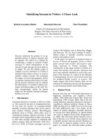
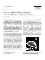
Both color a nd spectral Doppler sono grams were
obtained, which showed turbulent flow with an
increased flow velocity of 59.2 cm/second (Figure 1).
The patient underwent multi-slice CT (MSCT) (Aqui-
lion 64; Toshiba Medical Systems, Tokyo, Japan). After
unenhanced CT was performed, 120 mL of iopromide
(370 Iomeron; Bracco Imaging SpA, Milan, Italy) was
administered using a mechanical injector at a f low rate
of 4 mL/second. Biphasic CT was then performed in the
arterial phase, and delayed venous phase scanning was
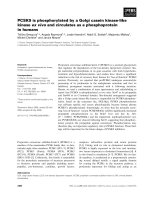
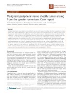
performed at a fixed delay of 90 secon ds. The CT scan
showed the presence of tortuous blood arterial opacified
vessels with thin arterial r amifications of spiral form
located next to the ileum on the upper pole of the left
kidney (Figure 2).
The patient was immediately carried into the angio-
graphy room for endovascular treatment. Selective left
renal artery angiography was performed using a right
transfemoral approach and a 5-French sheath (Terumo
Corp., Tokyo , Japan) with a 0.036-inch hydrophilic
guidewire coupled with a 5-French c obra-shaped cathe-
ter (Cordis, Warren, NJ, USA).
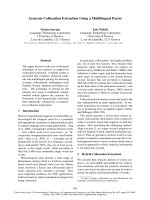
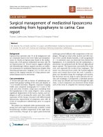
Digital subtraction arteriography (DSA) demonstrated
the feeding artery to the AVM. The lesion was selectively
catheterized with a microcatheter (Progreat; Terumo
Corp.) and e mbolized with 4 mm and 3 mm microcoils
(Vortex; Boston Scientific, Natick, MA, USA) and micro-
particles of polyvinyl alcohol 300μ to 500μ and 700μ to
900μ (Bead Block; Terumo Corp.) (Figure 3). No compli-
cations occurred during or after the procedure.
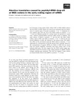
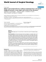
At the end of the procedure, comp lete excision of the
AVM was detected using DSA (Figure 4). The patient’s
hemodynamic parameters, such as blood pressure, were
monitored. The patient was discharged seven days later
with no signs of hematuria.
Discussion
AVFs, first described by Varela in 1928 [9], are relatively
uncommon lesions with considerable clinical impact. They
may cause hypertension, l ocal thrombosis, peripheral embo-
lization, high-output cardiac failure, and h ematuria [10].
AVFs can be congenital, acquired, or idiopathic.
About 70% to 80% of all AVFs are acquired and may
occur as a result of renal biopsy, blunt or penetrating
trauma, inflammation, malignancy, or renal surgery
[11,12].
Figure 1 Spectral Doppler sonogram showing the arteriovenous malformation (AVM). The image aliasing area highlights the AVM.
Carrafiello et al. Journal of Medical Case Reports 2011, 5:510
/>Page 2 of 5
AVFs are a congenital condition in 20% to 30% of
cases. It is usually located on the kidney upper pole
(45%), but it also can be detected in the mid-point or in
the kidney lower pole in an equal ratio [13]. The left
kidney is more frequently involved, and women are
affected twice as often as men. The peak incidence is in
patients ages 30 to 40 years, and AVFs are rare in the
pediatric population [2].
Acquired fistulas are usually caused by iatrogenic inju-
ries. A fistula can appear after renal needle biopsy, often
in kidney transplant patients, and sometimes these
fistulas are a post-operative complication after nephrost-
omy or nephrectomy, particularly in cases of intra-
operative injuries of the renal pedicle [14,15].
A fistula caused by angioplasty in a segmental renal
artery branch has also been reported in the literature
[16]. Malignant tumors of the kidney and metastases
can cause fistulas as a result of v ein erosion. Other pos-
sible causes are penetrat ing or b lunt abdom inal trauma,
fibromuscular dysplasia, and aneurysm of the renal
artery [3,4].
Congenital renal arteriovenous fistulas are the most
uncommon form, but their incidence may be underesti-
mated because patients are usually asymptomatic [5,6].
There are two types of congenital AVMs: (1) crisoid, a
malformed lesion characterized by multiple varix-like
vascular communications and a major incidence of gross
hematuria [13], and (2) aneurysmal, which typically
occur in elderly patients when a pre-existing arterial
aneurysm erodes into an adjacent vein [1].
This kind of malformation has been treated to date
with surgical therapy, such as nephrectomy, which is
still considered as the first-choice treatment by some
authors for patients who present with alterations in the
cardiovascular system, such as renin-mediat ed hyperten-
sion caused by fistula-related relative ischemia or high-
output cardiac f ailure caused by an increase in venous
return [17]. Endovascular approaches to treating AVMs
are now increasingly performed [13].
In our patient, typical diagnostic criteria of the dis-
ease were met. The patient was immediately referred
to the Dep artment of Radiology for imaging assess-
ments because of her age; moreover, she had received
only liquid re-infusions, a nd neither plasma nor solu-
tion of succinylated gelatine (Gelofusine Braun Medi-
cal, Milan, Italy) had been administered. It is
Figure 2 Computed tomography with iodinated contrast
enhancement shows the presence of tortuous blood arterial
vessel with thin arterial ramifications of spiral form. The
arrowhead indicates the renal vein, the arrows indicate the renal
artery, and the asterisk indicates the renal AVM.
Figure 3 Selective digital subtraction arteriography of t he left
kidney showing dynamic images of the AVM. The black arrow
indicates the renal artery, the white arrow indicates the AVM, and
the arrows indicate the renal vein.
Figure 4 Digital subtraction arteriography performed after
selective embolization of the lesion with the use of microcoils.
Complete excision of the lesion is shown. The renal pelvis is
opacified by contrast medium used for arteriography.
Carrafiello et al. Journal of Medical Case Reports 2011, 5:510
/>Page 3 of 5
remarkable that our patient had not undergone any
surgical intervention before her presentatio n to our
hospital, which is in contrast to what has been pre-
sented in the literature [13].
Our aim was to immediately treat the AVM by per-
forming endovascular embolization to stop the bleeding,
preserve renal parenchymal function, and eradicate the
symptoms and hemody namic effects associated with the
abnormality that we have seen in our patient, who had a
reduction in hemoglobin and an increase in heart rate.
It is truly important to preserve renal function in
patients who have just one functioning kidney or renal
insufficiency [13]. Indications for treating an AVM are a
progressive increase in the size of the fistula, recurrent
or persisten t hematuria, and hemodynami c effects asso-
ciated with the abnormality, especially decompensation,
hypertension, and high-output cardiac failure. Recently,
endovascular techniques have also been used to treat
giant aneurysms with AVFs. For small renal AVFs,
macroparticles or methyl cyanoacrylate glue should be
used [5-7]; for larger fistulas, however, coils or detach-
able balloons are preferable. If there is concern regard-
ing systemic and pulmonary emboli, a high-flow AVF
should be managed by performing a n open resection or
ligation [5-7].
The benefits of percutaneous treatment are avoidance
of nephrectomy, reduction of peri-operative risk and
post-operative morbidity, reduced surgical time and hos-
pital stay, and decreased incidence of renal ischemia [7].
Post-embolization syndrome (PES) may occur some-
times after transcatheter arterial embolization. PES con-
sists of fever, loin pain, nausea, and vomiting, but
selective embolization of renal AVMs allows for the pre-
servation of the renal parenchyma and therefore leads to
minimal PES [8].
Conclusions
Congenital AVMs are uncommon and Color Doppler
ultrasonography, MSCT, angiography, and DSA are the
most important tools for making the diagnosis in an
urgent setting. The therapeutic decis ion must be made
by considering the genera l condition of the patient and
his or her symptoms. The only therapy considered in
the past was nephrectomy, but embolization by selective
catheterization can be considered safe and effective.
However, many studies need to be done t o confirm the
role of embolization.
Consent
Written informed consent was obtained from the patient
for publication of this case report and any accompany-
ing images. A copy of the writ ten consent is available
for review by the Editor-in-Chief of this journal.
Abbreviations
AVM: arteriovenous malformation; CT: computed tomography; DSA: digital
subtraction arteriography; MSCT: multi-slice computed tomography.
Author details
1
Department of Radiology, Ospedale di Circolo e Fondazione Macchi,
University of Insubria, Varese, Italy.
2
Department of Vascular Surgery,
Ospedale di Circolo e Fondazione Macchi, University of Insubria, Varese, Italy.
Authors’ contributions
GC, DL, and FF carried out the diagnostic studies and performed the
percutaneous embolization. DM and GP reviewed the literature. MM and GP
wrote the case report. CF checked and edited the manuscript. All authors
read and approved the final manuscript.
Competing interests
The authors declare that they have no competing interests.
Received: 24 October 2009 Accepted: 8 October 2011
Published: 8 October 2011
References
1. Sountoulides P, Zachos I, Paschalidis K, Asouhidou I, Fotiadou A, Bantis A,
Palasopoulou M, Podimatas T: Massive hematuria due to a congenital
renal arteriovenous malformation mimicking a renal pelvis tumor: a case
report. J Med Case Reports 2008, 2:144.
2. Dönmez FY, Coşkun M, Uyuşur A, Hunca C, Tutar NU, Başaran C, Cakir B:
Noninvasive imaging findings of idiopathic renal arteriovenous fistula.
Diagn Interv Radiol 2008, 14:103-105.
3. Cho KJ, Stanley JC: Nonneoplastic congenital and acquired renal
arteriovenous malformations and fistulas. Radiology 1978, 129:333-343.
4. Lupattelli T, Garaci FG, Manenti G, Belli AM, Simonetti G: Giant high-flow
renal arteriovenous fistula treated by percutaneous embolization.
Urology 2003, 61:837.
5. Saliou C, Raynaud A, Blanc F, Azencot M, Fabiani JN: Idiopathic renal
arteriovenous fistula: treatment with embolization. Ann Vasc Surg 1998,
12:75-77.
6. Campbell JE, Davis C, Defade BP, Tierney JP, Stone PA: Use of an
Amplatzer Vascular Plug for transcatheter embolization of a renal
arteriovenous fistula. Vascular 2009, 17:40-43.
7. Trocciola SM, Chaer RA, Lin SC, Dayal R, Scherer M, Garner M, Coll D,
Kent KC, Faries PL: Embolization of renal artery aneurysm and
arteriovenous fistula: a case report. Vasc Endovascular Surg 2005,
39:525-529.
8. Somani BK, Nabi G, Thorpe P, Hussey J, McClinton S: Therapeutic
transarterial embolisation in the management of benign and malignant
renal conditions. Surgeon 2006, 4:348-352.
9. Varela ME: Aneurisma arteriovenoso de los vasos renales y asistolia
consecutiva. Rev Med Latino-Am 1928, 14:32-44.
10. Anomalies of the upper urinary tract: In Campbell’s Urology 8 edition.
Edited by: Walsh PC, Retik AB, Vaughan ED, Wein AJ, Kavoussi LR, Novick
AC, Partin AW. Peters CA: Amsterdam: Elsevier Science; 2002:3422-3423.
11. Takaha M, Matsumoto A, Ochi K, Takeuchi M, Sonoda T: Intrarenal
arteriovenous malformation. J Urol 1980, 124:315-318.
12. Abdel-Gawad EA, Housseini AM, Cherry KJ, Bonatti H, Maged IM, Norton PT,
Hagspiel KD: CT angiography of renal arteriovenous fistulae: a report of
two cases. Vasc Endovascular Surg 2009, 43:416-420.
13. Seitz M, Waggershauser T, Khoder W: Congenital intrarenal malformation
presenting with gross hematuria after endoscopic intervention: a case
report. J Med Case Reports 2008, 2:326.
14. Lacombe M: Renal arteriovenous fistula following nephrectomy. Urology
1985,
25:13-16.
15.
Bozgeyik Z, Ozdemir H, Orhan I, Cihangiroglu M, Cetinkaya Z:
Pseudoaneurysm and renal arteriovenous fistula after nephrectomy: two
cases treated by transcatheter coil embolization. Emerg Radiol 2008,
15:119-122.
16. Oleaga JA, Grossman RA, McLean GK, Rosen RJ, Freiman DB, Ring EJ:
Arteriovenous fistula of a segmental renal artery branch as a
complication of a percutaneous angioplasty. AJR Am J Radiol 1981,
136:988-989.
Carrafiello et al. Journal of Medical Case Reports 2011, 5:510
/>Page 4 of 5
17. Dean RH, Meacham PW, Weaver FA: Ex vivo renal artery reconstruction:
indications and techniques. J Vasc Surg 1986, 4:546-552.
doi:10.1186/1752-1947-5-510
Cite this article as: Carrafiello et al.: Gross hematuria caused by a
congenital intrarenal arteriovenous malformation: a case report. Journal
of Medical Case Reports 2011 5:510.
Submit your next manuscript to BioMed Central
and take full advantage of:
• Convenient online submission
• Thorough peer review
• No space constraints or color figure charges
• Immediate publication on acceptance
• Inclusion in PubMed, CAS, Scopus and Google Scholar
• Research which is freely available for redistribution
Submit your manuscript at
www.biomedcentral.com/submit
Carrafiello et al. Journal of Medical Case Reports 2011, 5:510
/>Page 5 of 5