báo cáo khoa học: "An osseous lesion in a 10-year-old boy with Hodgkin’s lymphoma: a case report" ppsx
Bạn đang xem bản rút gọn của tài liệu. Xem và tải ngay bản đầy đủ của tài liệu tại đây (1.19 MB, 3 trang )
CAS E REP O R T Open Access
An osseous lesion in a 10-year-old boy with
Hodgkin’s lymphoma: a case report
Machiel van den Akker
1*
, Vadiem Zudekov
2
, Asher Moser
3
and Joseph Kapelushnik
3
Abstract
Introduction: Osseous involvement of Hodgkin’s lymphoma is uncommon. When osteolytic lesions are seen on
imaging it is important to evaluate potential other causes.
Case presentation: We report the case of a 10-year-old Caucasian boy who presented to our facility with a bony
lesion of the right clavicle and enlarged cervical lymph nodes. A simultaneous biopsy of the lymph node and of
the osteolytic process of his right proximal clavicle was performed and revealed two different kinds of lesions: a
mixed cellularity Hodgkin’s lymphoma and an osteochondroma.
Conclusions: Since the latter is a common benign bone tumor, which should not interfere with the staging of the
lymphoma, we emphasize the importance of ensuring that all efforts are made to acquire a diagnostic biopsy of all
atypical lesions.
Introduction
Lymphoma is the third most common childhood malig-
nancy following leukemia and brain tumors, accounting
for approximately 12% of childhood cancers. Two-thirds
of lymphomas diagnosed in children are non-Hodgkin’s
lymphomas (NHL), with the remainder being Hodgkin’s
lymphomas (HL). Anatomic extent of disease and tumor
burden at presentation are s ignificant factors determin-
ing choice of therapy and prognosis. HL typically
involves the lymphatic system, and is usually supra-dia-
phragmatic. HL often follows a pattern of contiguous
spread from one nodal group to the next ana tomical
region. Extra-nodal involvement is more common in
NHL. Extra-nodal invasion of adjace nt tissues is se en in
up to 15% of cases and hematogenous spread in up to
10% of newly diagnosed cases. Osseous localizations
have been described in 10% to 20% of cases of relapsed
or refractory HL, but less than 2 % at the time of initial
presentation. Here, we descri be a case of an osteochon-
droma (OC) in a child with Hodgkin’ s disease not
affecting therapy or prognosis.
Case presentation
Due to enlarge d lymph nodes in his right neck region, a
10-year-old Caucasian boy underwent ultrasonic investi-
gation and was treated with a short course of antibiotics
18 months prior to his presentation at our facility. Two
months before his current admissio n, our patient
reported local pain and enlargement of the same area in
the neck. No B symptoms were evident. A second anti-
biotic t reatment was prescri bed, presuming a diagnosis
of lymphadenitis at that time, and all laboratory tests
were within the normal range except a slight microcytic
hypochromic anemia (hemoglobin 10.8 g/dL, mean cor-
puscular volume 72).
Plain X-rays of the chest showed no abnormal find-
ings. An ultrasonographic study of the neck showed
enlarged lymph nodes (measuring up to 3.2 cm in dia-
meter) on the right side, mostly in the posterior triangle.
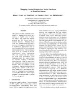
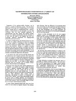
A computed tomography (CT) scan of the neck, thorax
and abdomen confirmed a heterogeneous mass of
enlarged lymph nodes on the right side of the neck and
an osteolytic process accompanied by a periosteal (and
soft-tissue) reaction in the right proximal clavicle, con-
spicuous for a tumor or chronic osteomyelitis (Figure
1). Radionuclide imaging with gallium-67 citrate showed
pathologic absorption on the right side of the neck, in
accordance with the enlarged lymph nodes, but not in
the right proximal clavicle. A comparable study with
* Correspondence:
1
Department of Paediatric Haematology Oncology, Queen Paola Children’ s
Hospital, 2020, Antwerpen, Belgium
Full list of author information is available at the end of the article
van den Akker et al. Journal of Medical Case Reports 2011, 5:511
/>JOURNAL OF MEDICAL
CASE REPORTS
© 2011 van den Akker et al; licensee BioMed Centra l Ltd. This is an Open Access article distributed under the terms of the Creative
Commons Attribution License ( which permits unrestricte d use, distribution , and
reproduction in any medium, provided the ori ginal work is properly cited.
fluorodeoxyglucose positron emission tomography
(FDG-PET) was not available.
Biopsies of a lymph node in the posterior triangle on
the right side of the neck and from the right proximal
clavicle were taken. The biopsy of the lymph node co n-
firmed the diagnosis of Hodgkin’ slymphomawith
mixed cellularity and a focal inter-follicular pattern.
Immunohistochemistry stains were positive for CD15
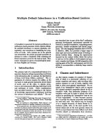
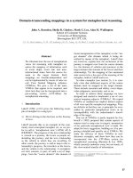
and CD30. The clavicle biopsy showed bone tissue with
exoph ytic cartilagi nous tissue (Figure 2), consistent with
osteochondroma without evidence of involvement with
HL. Our patient underwent four courses of ABVD che-
motherapy protocol (doxorubicin, bleomycin, vinblastine
and dacarbazine) for stage IIa disease and is currently
five years on from cessation of all treatment and in
complete remission.
Discussion
Osteocartilaginou s exostoses (osteochondromas (OC))
are the commonest of all bone tumors, compromising
about one-third of benign bone tumors. They may be
sporadic, genetic or secondary (for example, from radia-
tion). The majority are solitary, mostly located around
the knee and in the proximal humerus, while about 20%
arise in the axial skeleton. Typically, they a re metaphy-
seal or metadiaphyseal in origin, oriented away from the
adjacent joint. OC may occur at multiple sites, particu-
larly in cases of hereditary multiple exostoses synd rome
(HMES) with an autosomal dominant inheritance. Most
children are asymptomatic. OC usually presents as a
solitary mass, as a fracture or as a mechanical osteoarti-
cular complication (deformity or joint dysfunction).
Plain radiography is the mainstay of imaging for OC,
while a CT scan can be helpful when planning resection.
An MRI scan is needed when there are concerns of a
malignancy. Bone scans are highly sensitive, but with
low specificity, and are in general not useful. Histology
is necessary to confirm a diagnosis of OC, showing a
cartilage-capped exophyti c, ses sile or pedunculated pro-
jection that has a continuous peri-osteum, cortex and
marrow connecting with the underlying bone. The carti-
lage cap is the site of active growth and the degree of
maturity is parallel with the host bone. It is not com-
mon for OC to grow beyond skeletal maturity. The risk
of malignant transformation to chondrosarcoma of a
solitary OC is 1% to 5%, but for HMES the reported
incidence is up to 9% [1]. Frequent sites are the pelvis,
proximal femur and the shoulder girdle. The presence
of an enlarging mass (especially after skeletal maturity)
and recent onset of pain should arouse suspicion of
malignant transformation. Treatment of OC is usually
accomplished by resection or curettage, and rarely
requires bone grafting.
Although presenting symptoms due to o sseous invol-
vement rarely occur in HL, osseous involvement is seen
at radiography in approximately 20% of affected patients
throughout the course of the disease [2]. Osseous invol-
vement may be indicative of widespre ad, aggressive dis-
ease with a relative poor prognosis, associated with
unfavorable histological subtypes. Primary osseous HL is
very rare and must be distinguished from systemic HL
with diffuse bone and bone marrow involvement and
from osseous metastases in advan ced stage of disease.
Due to t he nature of HL, most cases are associated with
synchronous lymph node involvement. Bone scintigra-
phy has a sensitivity and accuracy of 95% in detecting
osseous involvement, but in most cases bone HL is
revealed on plain X-rays and CT scanning [3]. The
roentgen graphic features of bony involvement of HL
are non-specific, and may be solitary (33%) or polyosto-
tic (66%) [4]; the edge is usually wide and ill defined,
Figure 1 Computed tomography scan showing an osteolytic
process with periosteal reaction of the right proximal clavicle.
Figure 2 Irregular fragment of a bone lesion showing
cartilaginous cup and exophytic bone (hematoxylin and eosin
staining, magnification ×250).
van den Akker et al. Journal of Medical Case Reports 2011, 5:511
/>Page 2 of 3
there may be a periosteal reaction with bone destruc-
tion, and the lesions are predominantly osteolytic with
blurred borders [2]. Fractures are rarely the first mani-
festations. Soft-tissue tumors are often seen adjacent to
the bone lesions. Differential diagnosis included primary
sarcoma of the bone, NHL, leukemia, metastasis, and
the most frequent (histopathologic and radiographic)
misdiagnosis, osteomyelitis. In general, routine bone
scintigraphy seems to be of limited value in the clinical
assessment of chil dren with malignant lymphoma unless
there are specific osseous symptoms. FDG-PET has
replaced the 67 Ga scintigraphy for evaluating c hildren
and younger adults with newly diagnosed HD [5].
Experience of OC is limited and is also not uniformly
informative with regards to diagnostic investigation for
malignant transformation.
Conclusions
We describe a case of coinciding osseous lesion and HL.
We conclude that all efforts should be taken to make an
exact diagnosis by biopsy of all suspicious locations,
including bony structures, in order to make an accurate
diagnosis and subsequently start the appropriate
treatment.
Consent
Written informed consent was obtained from the
patient’ s legal guardian for publication of this case
report and any accompanying images. A copy of the
written consent is available for review by the E ditor-in-
Chief of this journal.
Author details
1
Department of Paediatric Haematology Oncology, Queen Paola Children’ s
Hospital, 2020, Antwerpen, Belgium.
2
Radiology Department, University
Medical Center Soroka, Beer Sheva, Israel.
3
Department of Paediatric
Haematology Oncology, University Medical Center Soroka, Beer Sheva, Israel.
Authors’ contributions
MA was responsible for the data collection, obtaining consent, and was the
author of the manuscript. VD was responsible for the imaging and part of
the Discussion section. AM was responsible for carefully reviewing the
article. JK was responsible for the medical care of our patient and for part of
the Discussion section. All authors read and approved the final manuscript.
Competing interests
The authors declare that they have no competing interests.
Received: 23 May 2011 Accepted: 8 October 2011
Published: 8 October 2011
References
1. Altay M, Bayrakci K, Yildiz Y, Erekul S, Saglik Y: Secondary chondrosarcoma
in cartilage bone tumors: report of 32 patients. J Orthop Sci 2007,
12:415-423.
2. Guermazi A, Brice P, de Kerviler EE, Ferme C, Hennequin C, Meignin V,
Frija J: Extranodal Hodgkin disease: spectrum of disease. Radiographics
2001, 21:161-179.
3. Sandrasegaran K, Robinson PJ, Selby P: Staging of lymphoma in adults.
Clin Radiol 1994, 49:149-161.
4. Edeiken-Monroe B, Edeiken J, Kim EE: Radiologic concepts of lymphoma
of bone. Radiol Clin North Am 1990, 28:841-864.
5. Hines-Thomas M, Kaste SC, Hudson MM, Howard SC, Liu WA, Wu J, Kun LE,
Shulkin BL, Krasin MJ, Metzger ML: Comparison of gallium and PET scans
at diagnosis and follow-up of pediatric patients with Hodgkin
lymphoma. Pediatr Blood Cancer 2008, 51:198-203.
doi:10.1186/1752-1947-5-511
Cite this article as: van den Akker et al.: An osseous lesion in a 10-year-
old boy with Hodgkin’s lymphoma: a case report. Journal of Medical Case
Reports 2011 5:511.
Submit your next manuscript to BioMed Central
and take full advantage of:
• Convenient online submission
• Thorough peer review
• No space constraints or color figure charges
• Immediate publication on acceptance
• Inclusion in PubMed, CAS, Scopus and Google Scholar
• Research which is freely available for redistribution
Submit your manuscript at
www.biomedcentral.com/submit
van den Akker et al. Journal of Medical Case Reports 2011, 5:511
/>Page 3 of 3