báo cáo khoa học: "Diverticulitis complicated by pylephlebitis: a case report" ppsx
Bạn đang xem bản rút gọn của tài liệu. Xem và tải ngay bản đầy đủ của tài liệu tại đây (463.96 KB, 4 trang )
CAS E REP O R T Open Access
Diverticulitis complicated by pylephlebitis:
a case report
Mahesh Gajendran
1*
, Thiruvengadam Muniraj
2
and Mohamed Yassin
2
Abstract
Introduction: Pylephlebitis is defined as septic thrombophlebitis of the portal venous system, usually secondary to
infection or inflammation in the abdomen. In the current report, we pres ent a case of pylephlebitis that
complicated the course of a very common pathology, diverticulitis.
Case presentation : A 62-year-old Caucasian woman with a history of sigmoid diverticulitis presented to our facility
with a three-week history of abdominal pain, fevers, chills, loss of appetite and fatigue. Her laboratory test results
showed leuko cytosis and elevated alkaline phosphatase. A computed tomography scan revealed portal vein
thrombosis and a sigmoid diverticulitis with an abscess. Our patient was given pipercillin-tozabactam followed by
sigmoid colectomy and loop transverse colostomy. A peritoneal fluid sample culture grew Escherichia coli. Our
patient had an uneventful post-operative course and the leukocytosis resolved in the next four days. She improved
clinically and was discharged home on ertapenem and enoxaparin. A follow-up computed tomography scan two
weeks later showed a new pelvic abscess that was drained by a pigtail catheter but there was no change in the
portal venous thrombus. A repeat computed tomography scan one month later revealed resolution of the pelvic
abscess but persistence of portal vein thrombus, for which enoxaparin was continued.
Conclusions: This is a classic case of pylephlebitis that demonstrates the importance of recognizing that the portal
vein thrombus is infected and treating the condition appropriately.
Introduction
Pylephlebitis is defined as septic thrombophlebitis of the
portal venous system, usually secondary to infection or
inflammation in the abdomen. The common causes
include diverticulitis, appendicitis or cholangitis [1].
Pylephlebitis has to be differentiated from the bland
portal vein thrombus. Bland portal vein thrombosis is
more common than pylephlebitis and the management
is different. Here, we present a case of pylephlebitis that
complicated the course of a very common pathology,
diverticulitis.
Case presentation
A 62-year-old Caucasian woman with a history of sig-
moid diverticulitis (seven months prior to admission) was
admitted for three weeks of sharp intermittent left lower
quadrant abdominal pain, low-grade fever, chills, loss of
appetite and fatigue. She denied diarrhea, bloody stools,
nausea, or vomiting. The only abnormal finding on physi-
cal examination was tenderness in the left lower quad-
rant. Her initial laboratory test results showed a white
cell count of 17,700 cells/mm
3
, hemoglobin 13.7 gm/dL
and elevated alkaline phosphatase two times the normal
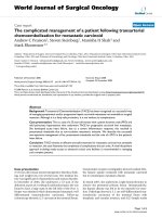
level. A computed tomography (CT) scan of the abdo-
men revealed portal vein thrombosis, low attenuation
liver lesions (Figures 1, 2, 3) and extensive sigmoid diver-
ticulitis with a 4 × 1.8 cm abscess. This was a new throm-
bus compared to a previous CT scan, performed two
months previously. The color doppler confirmed the pre-
sence of portal vein thrombus (Figure 4). An MRI scan of
the abdomen did not reveal any additional i nformation.
Our patient was given pipercillin-tozabactam followed by
exploratory laparotomy, sigmoid colectomy and loop
transverse colostomy. An intra-operative ultrasonography
of the liver was suggestive of early liver abscesses, but we
were not able to aspirate. A peritoneal fluid sample cul-
ture grew Escherichia coli. Our patient had an uneventful
post-operative course and her leukocytosis resolved in
* Correspondence:
1
University of Pittsburgh Medical Centre, Department of Medicine, 200
Lothrop Street, Pittsburgh, PA 15213, USA
Full list of author information is available at the end of the article
Gajendran et al. Journal of Medical Case Reports 2011, 5:514
/>JOURNAL OF MEDICAL
CASE REPORTS
© 2011 Gajendran et al; licensee BioMed Central Ltd. T his is an Open Acc ess article distributed under the terms of the Creative
Commons Attribution License ( .0), which permits unrestricted use, distribution, a nd
reproduction in any medium, provided the original work is properly cited.
Figure 1 Computed tomography (CT) scan showing right portal vein thrombosis.
Figure 2 Computed tomography (CT) scan showing multiple low attenuation liver lesions.
Gajendran et al. Journal of Medical Case Reports 2011, 5:514
/>Page 2 of 4
the n ext four days. She improved clinically and was dis-
charged home on ertapenem and enoxaparin. A follow-
up CT scan two weeks later showed a new pelvic abscess
7.5 × 6 cm that was drained by a pigtail catheter, but
there was no change in the portal venous thrombus. Her
hypercoagulable profile was negative. A repeat CT scan
one month later revealed resolution of the pelvic abscess
but persistence of portal vein thrombus for which enoxa-
parin was continued.
Conclusions
Unlike bland portal vein thro mbosis, pylephlebitis is
more commonly associated with liver abscesses and bac-
teremia [2]. Escherichia coli and Bacteroides fragilis are
the most common isolates in blood [3]. Doppler ultra-
sound, CT scanning and MRI scan ning of t he abdomen
has improved the ability to diagnose pylephlebitis [4].
CT scanning demonstrates portal vein thrombus as a
non-enhancing, low-density thrombus within the vessel
lumen with non-homogeneous enhancement of the
hepatic parenchyma [5]. MRI can help to distinguish
acute fr om chronic portal vein thrombosis [6]. Manage-
ment of pylephlebitis is best achieved by treating the
primary sou rce using broad-spectrum intravenous anti-
biotics and surgical intervention (appendectomy or
colectomy with abscess drainage) [1,2]. Early diagnosis
Figure 3 Computed tomography (CT) scan scout view showing
right portal vein thrombus and liver abscess.
Figure 4 Color Doppler showing no flow in right portal vein.
Gajendran et al. Journal of Medical Case Reports 2011, 5:514
/>Page 3 of 4
and treatment is critical. The role of anticoagulation in
the treatment of pylephlebitis is controversial [7].
Consent
Written informed consent was obtained from the patient
for publication of this case report and any accompany-
ing images. A copy of the written consent is available
for review by the Editor-in-Chief of this journal.
Author details
1
University of Pittsburgh Medical Centre, Department of Medicine, 200
Lothrop Street, Pittsburgh, PA 15213, USA.
2
University of Pittsburgh Medical
Centre - Mercy, Department of Medicine, 1400 Locust Street, Pittsburgh, PA
15219, USA.
Authors’ contributions
All authors equally contributed to the writing of the manuscript. All authors
reviewed the final manuscript and approved it for submission.
Competing interests
The authors declare that they have no competing interests.
Received: 3 June 2011 Accepted: 10 October 2011
Published: 10 October 2011
References
1. Kasper DL, Sahani D, Misdraji J: Case records of the Massachusetts
General Hospital. Case 25-2005. A 40-year-old man with prolonged fever
and weight loss. N Engl J Med 2005, 353:713-722.
2. Plemmons RM, Dooley DP, Longfield RN: Septic thrombophlebitis of the
portal vein (pylephlebitis): diagnosis and management in the modern
era. Clin Infect Dis 1995, 21:1114-1120.
3. Kanellopoulou T, Alexopoulou A, Theodossiades G, Koskinas J,
Archimandritis AJ: Pylephlebitis: an overview of non-cirrhotic cases and
factors related to outcome. Scand J Infect Dis 2010, 42:804-811.
4. Harch JM, Radin RD, Yellin AE, Donovan AJ: Pylethrombosis. Serendipitous
radiologic diagnosis. Arch Surg 1987, 122:1116-1119.
5. Balthazar EJ, Gollapudi P: Septic thrombophlebitis of the mesenteric and
portal veins: CT imaging. J Comput Assist Tomogr 2000, 24:755-760.
6. Zirinsky K, Markisz JA, Rubenstein WA, Cahill PT, Knowles RJ, Auh YH,
Morrison H, Kazam E: MR imaging of portal venous thrombosis:
correlation with CT and sonography. AJR Am J Roentgenol 1988,
150:283-288.
7. Duffy FJ Jr, Millan MT, Schoetz DJ Jr, Larsen CR: Suppurative pylephlebitis
and pylethrombosis: the role of anticoagulation. Am Surg 1995,
61:1041-1044.
doi:10.1186/1752-1947-5-514
Cite this article as: Gajendran et al.: Diverticulitis complicated by
pylephlebitis: a case report. Journal of Medical Case Reports 2011 5:514.
Submit your next manuscript to BioMed Central
and take full advantage of:
• Convenient online submission
• Thorough peer review
• No space constraints or color figure charges
• Immediate publication on acceptance
• Inclusion in PubMed, CAS, Scopus and Google Scholar
• Research which is freely available for redistribution
Submit your manuscript at
www.biomedcentral.com/submit
Gajendran et al. Journal of Medical Case Reports 2011, 5:514
/>Page 4 of 4


![Tài liệu Báo cáo khoa học: The stereochemistry of benzo[a]pyrene-2¢-deoxyguanosine adducts affects DNA methylation by SssI and HhaI DNA methyltransferases pptx](https://media.store123doc.com/images/document/14/br/gc/medium_Y97X8XlBli.jpg)