Báo cáo y học: " Renal cell carcinoma metastasizing to solitary fibrous tumor of the pleura: a case report" ppt
Bạn đang xem bản rút gọn của tài liệu. Xem và tải ngay bản đầy đủ của tài liệu tại đây (2.7 MB, 4 trang )
CAS E REP O R T Open Access
Renal cell carcinoma metastasizing to solitary
fibrous tumor of the pleura: a case report
Christopher Kragel and Shi Wei
*
Abstract
Introduction: A tumor metastasizing to another malignancy is an uncommon phenomenon. Since it was first
described in 1902, there have been fewer than 200 cases reported in the literature, with lung cancer metastasizing
to renal cell carcinoma being the most frequently described pattern. Here we report a case of a solitary fibrous
tumor of the lung acting as the recipient for a renal cell carcinoma. To our knowledge, this is the first reported
case of such a combination and the second case involving a solitary fibrous tumor.
Case presentation: A 58-year-old Caucasian man who developed a persistent dry cough presented to our
hospital. Imaging studies revealed a large pleural-based mass in the left lung. A biopsy of the mass showed a
spindle-cell lesion consistent with a solitary fibrous tumor. The patient underwent surgical excision of the 13 cm
mass. The pathological examination confirmed the diagnosis of a solitary fibrous tumor but also demonstrated
discrete foci of metastatic renal cell carcinoma. Until that point, a primary renal cell carcinoma tissue diagnosis had
not been made and the initial radiological work-up was inconclusive.
Conclusion: Awareness of the unusual phenomenon of tumor-to-tumor metastasis is important for practicing
surgical pathologists, particularly in the evaluation of a mass lesion showing bimodal histology. This case also
highlights the importance of careful examination of surgical specimens, as minute and un usual findings can direct
patient care.
Introduction
The coexistence of two primary neoplasms in one
patient is not uncommon, and these tumors may even
arise at the same anatomic site ("collision tumor”). How-
ever, tumor-to-tumor metastasis is an extremely rare
but interesting phenomenon. Since first described by
Berent in 1902 [1], fewer than 200 cases have been
reported in the English-language literature. The most
frequent donor tumor site is the lung, while renal cell
carcinoma is by far the most common recipient [2,3].
This combination constitutes approximately one-third of
all reported cases. However, renal cell carcinoma acting
as a donor tumor is extraordinarily rare, with only nine
cases reported to date [4-12]. Interestingly, meningiomas
are the most frequent recipients of donor r enal cell car-
cinoma, followed by papillary carcinoma of the thyroid.
Here we report the first case of a solitary fibrous tumor
of the lung acting as the recipient of a donor renal cell
carcinoma.
Case presentation
A 58-year-old Ca ucasian man who developed a pe rsis-
tent dry cough and hemoptysis presented to our hospi-
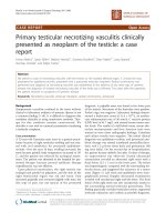
tal. Computed tomography (CT) revealed a large,
pleural-based mass in the left lung (Figure 1). A needle
biopsy showed a spindle-cell neoplasm which was
immunoreactive with CD34 and thus m ostly consistent
with a solitary fibrous tumor. The patient underwent
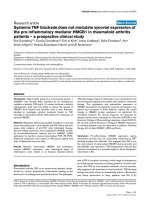
further radiological w ork-up. Whole-body positron
emission tomography (PET) showed diffuse, low-level
fluorodeoxyglucose (
18
F-FDG) uptake of the large,
biopsy-proven, solitary fibrous tumor of pleura in the
left hemithorax. However, there was a focus of moderate
18
F-FDG uptake in the superior aspect of the lesion,
which was worrisome for malignancy (Figure 2). In addi-
tion, subcarinal necrotic lymphadenopathy was noted,
which raised suspicions of metastasis. Multiple non-
18
F-
* Correspondence:
Department of Pathology, University of Alabama at Birmingham,
Birmingham, AL 35249-7331, USA
Kragel and Wei Journal of Medical Case Reports 2011, 5:248
/>JOURNAL OF MEDICAL
CASE REPORTS
© 2011 Kragel and Wei; licensee BioMed Central Ltd. T his is an Open Access article distributed under the terms of the Creative
Commons Attribution License ( which permits unrestri cted use, distribution, and
reproduction in any medium, provided the original work is properly cited.
FDG-avid large bilateral renal cysts were evident. The
evaluation of other organ systems was unrevealing.
The patient underwent surgical excision of the tumor,
including left thoracotomy, partial pleurectomy, wedge
resection of left upper and lower lobes and thoracic lym-
phadenectomy. Grossly, the tumor was homogeneously
tannish-white and solid, measuring 13.0 cm × 9.0 cm × 6.0
cm. Microscopic examination revealed a cellular mesench-
ymal neoplasm composed of bland spindled cells with a
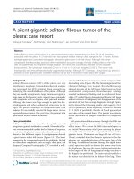
patternless architecture. The lesion possessed “staghorn”
vessels and a hyalinized stroma, especially in the peri-vas-
cular regions (Figure 3a). The lesional cells were strongly
immunoreactive with CD34 (Figure 3d). Thus, the features
were characteristic of a solitary fibrous tumor.
Within the solitary fibrous tumor, there were two
microscopic foci of nested epithelioid cells with clear
cell features in the background of a delicate vascular
network (Figures 3b and 3c). To further explore the nat-
ure of these cells, a battery of immunohistochemical
staining was performed. The cells of interest were posi-
tive for broad-spectrum cytokeratin (Figure 3e) and
vimentin (Figure 3f) and were also immunoreactive with
CD10 (Figure 3g) and paired box gene 2 (PAX2) (Figure
3h). Thus, these cells most likely represented metastatic
clear cell renal cell carcinoma. One lymph node showed
necrotizing granulomata, but all thoracic nodes were
negative for malignancy.
Post-operatively, a multidiscip linary team weighed the
treatment options. However, in the coming months,
further imaging analysis revealed additional metastases
to the liver, spine and brain. The patient underwent
Figure 1 Computed tomographic scan revealing a large
pleural-based mass in the left hemithorax.
Figure 2 Positron emiss ion tomographic scan. The left pleural
mass showed only diffuse low-level fluorodeoxyglucose (
18
F-FDG)
uptake of the mass. However, there was a focus of moderate
18
FFDG activity in the superior aspect of the lesion, which was
worrisome for malignancy.
Figure 3 Histologic and immunophenotypic characteristics of
the tumor. (a) Sections of pleural-based solitary fibrous tumor
showing a cellular spindle-cell neoplasm with a patternless
architecture and “staghorn” vessels. (b and c) A nodular collection
of epithelioid clear cells was incidentally found within the tumor.
(d) These cells are negative for CD34 (in contrast to the solitary
fibrous tumor on the left), but immunoreactive with (e) broad-
spectrum cytokeratin, (f) vimentin, (g) CD10 and (h) paired box
gene 2 (PAX2).
Kragel and Wei Journal of Medical Case Reports 2011, 5:248
/>Page 2 of 4
chemotherapy, spinal radiation therapy and gamma
knife radiosurgery for brain metastasis. With metastatic
disease causing increased morbidity and no further
treatment options available, t he patient was placed in
hospice care and died within six months of the initial
diagnosis.
Discussion
In 1968, Campbell et al.[13]reviewedpreviously
reported cases and asserted the criteria fo r tumor-to-
tumor metastasis as follows: (1) the existence of more
than one primary tumor, (2) the recipient tumor is a
true neoplasm, (3) the donor tumor is a true metastasis
with established growth in the host tumor that is not
the result of contiguous growth ("collision tumor” )or
embolization of tumor cells and (4) tumors that have
metastasized to the lymphatic system, where a lymphor-
eticular malignant tumor already exists, are excluded.
Thus, our present case meets these criteria.
Virtually any tumor may b ecome a potential recipient
of a donor metastatic tumor, but renal cell carcinoma is
by far the most common one [2,3]. This is likely because
kidneys receive significant blood flow and renal cell car-
cinoma is typically vascularly rich and thus easily har-
bors circulating tumor emboli [2,3]. It has also been
suggested that the high glycogen and lipid content of
carcinoma cells may serve as a suitable environment for
metastatic deposits [14] and thus may reflect the “ seed
and soil” hypothesis of cancer metastasis [15]. A solitary
fibrous tumor is extraordinary rare as a recipient tumor,
and our present case report r epresents only the second
reported such case. The tumor typically has alternating
hypercellular and hypocellular areas and characteristic
branching, staghorn vessels which may captivate blood-
borne metastases, as in the case of renal cell carcinomas.
As a donor tumor, however, renal cell carcinoma is
extremely uncommon, with only nine cases reported in
the literature to date. All four patients with known fol-
low-up died of the disease [4,6-8], which is compatible
with the significantly unfavorable prognosis of other
stage IV renal cell carcinomas. Interestingly, tumors of
central nervous system [4,5,7,9,12] and thyroid carcino-
mas [8,10,11] represent frequent recipient t umors for
donor renal cell carcinomas, suggesting that these
organs and tumoral tissues may provide a f ertile sub-
strate or are some way predisposed targets for secondary
growth of renal cell carcinoma.
The diagnosis of renal cell carcinoma metastasizing to
solitary fibrous tumor is paramount in this case as the
metastasis was the first confirmation of renal cell carci-
noma in this patient. Retrospectively, the renal cysts
identified in the initial radiological work-up may repre-
sent cystic r enal cell carcinoma. This case exemplifies
the importance of careful scrutiny of the pathologic
specimens because rare or unusual pathologic findings
may be of utmost clinical importance. In addition, our
present case report also emphasizes the need for a de-
quate sampling (that is, one section per centimeter of
tumor mass), as only one of the 14 sections of the
tumor possessed small metastatic foci.
Conclusions
Awareness of the unusual phenomenon of tumor-to-
tumor metastasis is important for practicing surgical
pathologists, particularly in the evaluation of a mass
lesion showing bimodal histology. This case also
highlights the importance of careful examination of
surgical specimens, as minute and unusual findings
can direct patient care. Moreover, the relative fre-
quency of specific neoplasms involved in tumor-to-
tumor metastasis may shed light on the pathogenesis
of tumor metastasis.
Consent
Written informed consent was obtained from the patient
for publication of this case report and any accompany-
ing images. A copy of the written consent is available
for review by the Editor-in-Chief of this journal.
Authors’ contributions
CK and SW were the responsible pathology resident and attending
pathologist, respectively, of this patient, and both authors were major
contributors to the manuscript. Both authors read and approved the final
manuscript.
Competing interests
The authors declare that they have no competing interests.
Received: 6 December 2010 Accepted: 29 June 2011
Published: 29 June 2011
References
1. Berent W: Seltene metastasenbildung. Zentralbl Allg Pathol 1902, 13:5.
2. Petraki C, Vaslamatzis M, Argyrakos T, Petraki K, Strataki M, Alexopoulos C,
Sotsiou F: Tumor to tumor metastasis: report of two cases and review of
the literature. Int J Surg Pathol 2003, 11:127-135.
3. Sella A, Ro JY: Renal cell cancer: best recipient of tumor-to-tumor
metastasis. Urology 1987, 30:35-38.
4. Osterberg DH: Metastases of carcinoma to meningioma. J Neurosurg 1957,
14:337-343.
5. Breadmore R, House R, Gonzales M: Metastasis of renal cell carcinoma to
a meningioma. Australas Radiol 1994, 38:144-3.
6. Ozenc A, Ruacan S, Baykal A: Renal cell carcinoma and ipsilateral
pheochromocytoma with neoplasm-to-neoplasm metastasis. J Urol 1997,
157:1831-1832.
7. Franke FE, Altmannsberger M, Schachenmayr W: Metastasis of renal
carcinoma colliding with glioblastoma. Carcinoma to glioma: an event
only rarely detected. Acta Neuropathol 1990, 80:448-452.
8. Baloch ZW, LiVolsi VA: Tumor-to-tumor metastasis to follicular variant of
papillary carcinoma of thyroid. Arch Pathol Lab Med 1999, 123:703-706.
9. Han HS, Kim EY, Han JY, Kim YB, Hwang TS, Chu YC: Metastatic renal cell
carcinoma in a meningioma: a case report. J Korean Med Sci 2000,
15:593-597.
10. Ryska A, Cáp J: Tumor-to-tumor metastasis of renal cell carcinoma into
oncocytic carcinoma of the thyroid: report of a case and review of the
literature. Pathol Res Pract 2003, 199:101-106.
Kragel and Wei Journal of Medical Case Reports 2011, 5:248
/>Page 3 of 4
11. Bohn OL, De las Casas LE, Leon ME: Tumor-to-tumor metastasis. Renal cell
carcinoma metastatic to papillary carcinoma of thyroid: report of a case
and review of the literature. Head Neck Pathol 2009, 3:327-330.
12. Kimiwada T, Motohashi O, Kumabe T, Watanabe M, Tominaga T:
Lipomatous meningioma of the brain harboring metastatic renal-cell
carcinoma: a case report. Brain Tumor Pathol 2004, 21:47-52.
13. Campbell LV Jr, Gilbert E, Chamberlain CR Jr, Watne AL: Metastases of
cancer to cancer. Cancer 1968, 22:635-643.
14. Ottosson L, Berge T: Metastasis from carcinoma to carcinoma. Acta Pathol
Microbiol Scand 1968, 73:481-488.
15. Paget S: The distribution of secondary growths in cancer of the breast.
Lancet 1889, 133:571-573.
doi:10.1186/1752-1947-5-248
Cite this article as: Kragel and Wei: Renal cell carcinoma metastasizing
to solitary fibrous tumor of the pleura: a case report. Journal of Medical
Case Reports 2011 5:248.
Submit your next manuscript to BioMed Central
and take full advantage of:
• Convenient online submission
• Thorough peer review
• No space constraints or color figure charges
• Immediate publication on acceptance
• Inclusion in PubMed, CAS, Scopus and Google Scholar
• Research which is freely available for redistribution
Submit your manuscript at
www.biomedcentral.com/submit
Kragel and Wei Journal of Medical Case Reports 2011, 5:248
/>Page 4 of 4