Báo cáo y học: "De Garengeot’s hernia in a 60-year-old woman: a case report" pot
Bạn đang xem bản rút gọn của tài liệu. Xem và tải ngay bản đầy đủ của tài liệu tại đây (471.27 KB, 3 trang )
CAS E REP O R T Open Access
De Garengeot’s hernia in a 60-year-old woman:
a case report
Petros Konofaos
*
, Eleftherios Spartalis, Anastasios Smirnis, Konstantinos Kontzoglou and Grigorios Kouraklis
Abstract
Introduction: De Garengeot first described the presence of the appendix within a femoral hernia in 1731.
Case presentation: We report the case of a 66-year-old Caucasian woman who presented with acute appendicitis
within an incarcerated femoral hernia. This is the first reported case of de Garengeot’s her nia in the Balkan area.
Conclusions: Appropriate management without incurring any delay for radiological imaging can be promising for
an uneventful postoperative course. The treatment of choice of this disease entity is emergency surgery and
consists in simultaneous appendectomy through the hernia incision and primary hernia repair. In patie nts with
large hernia defects or in older people the use of mesh for repairing the hernia defect can be an excellent choice.
Introduction
From 1731, when Rene Jacques Croissant de Garen geo t
first described the presence of the appendix within a
femoral hernia [1], to date there have been fewer than
90 cases reported in the literature. de Garengeot’s hernia
is an incidental finding occurring in 0.9% of femoral
hernia repairs [2], and appendicitis is rarer still, with an
incidence of 0.08-0.13% [3]. There is a female predispo-
sition (13:1, 93% in women), probably in keeping with
the increased incidence of femoral hernia in women [3].
We report the case of a female patient with acute
appendicitis within an incarcerated femoral hernia. This
is the first reported case of de Garengeot’sherniainthe
Balkan area.
Case presentation
A previously healthy 66-year-old Caucasian woman
presented with a 24-hour history of sudden onset pain-
ful right-sided groin swelling. On clinical examination,
there was a fixed, round, tender mass about 5 × 3 cm
in size in the right groin, above the inguinal crease.
Her temperature was 38.7°C and she did not appear to
be in distress. She did not have any bowel obstruction
revealed by clinical examination or on the abdominal
X-ray. Her past medical history was insignificant.
Her laboratory findings were within normal limits
except an increased WBC count (13.00 K/μL) with
80% neutrophils.
A presumptive diagnosis of a chronically incarcerated
femoral or inguinal hernia versus a strangulated hernia
or an inguinal abscess was made with plans for a right
groin exploration using a more curved low inguinal inci-
sion under general anesthesia (Figure 1). When the he r-
nia sac was opened, an inflamed appendix was seen.
The appendix was thickened and inflamed, but there
was no perforation. Intraoperative findings were consis-
tent with an inflamed and gangrenous appendix p ro-
truding through the femoral hernial sac (Figure 2).
Rou tine appendectomy was perfor med throu gh the her-
nialsac.Themouthoftheherniawaswideandthe
senior surgeon was even able to pass a finger through
the hernia into the peritoneal cavity. The hernial sac
was closed using a V-shaped polypropylene mesh. A
broad-spectrum antibiotic cover was provided at induc-
tion. The postoperative course was uneventful and the
patient was discharged home on the third day after the
procedure. The histological examination was consistent
with acute appendicitis.
Discussion
Although femoral hernias account for 4% of all groin
hernias, a hernia sac can contain any of the intraabdom-
inal contents such as omentum. A pelvic appendix has
the highest risk of entering a femoral hernial sac [4].
The evolution of inflammation in the appendix is
* Correspondence:
2
nd
Department of Propedeutic Surgery, ‘LAIKO’ General Hospital, 36,
Megistis Str, Athens 11364, Greece
Konofaos et al . Journal of Medical Case Reports 2011, 5:258
/>JOURNAL OF MEDICAL
CASE REPORTS
© 2011 Konofaos et al ; licensee BioMed Central Ltd. This is an Open Access article distributed under the terms of the Creative
Commons Attribution License ( which permits unrestricted use, distribution, and
reproduction in any mediu m, provided the original work is properly c ited.
thought to be secondary to its engagement in the her-
nial sac. Although there are occasional case s diagnosed
preoperatively, typically the appendix is found inciden-
tally during repair without any preoperative signs or
symptoms [5].
De Garengeot’ s hernia is often misdiagnosed as an
incarcerated or strangulated femoral hernia. The inci-
dence of an appendix in a femoral hernia is reported to
be 0.5-5% [2,6-8]; the reason for this wide variation is
the paucity of cases and no published large case series.
The clinical picture of this entity is that of incarcerated
femoral or inguinal hernia and includes vague
abdominal pain and tenderness and an erythematous
groin lump [7]. The signs of appendicitis are oversha-
dowed by a tight femoral hernia neck and pelvic rigidity;
this anatomical feature prevents the spread of inflamma-
tion to the peritoneal cavity [9].
Abdominal X-ray does not aid in the diagnosis of de
Garengeot’ s hernia. Computed tomography (CT) and
ultrasound have been succ essfully used for preoperative
evaluation [10]. The presence of a low-p ositi oned cecum
along with tubular structure within the hernial sac and
stranding of nearby fat on CT have been reported to have
98% specificity and sensitivity for diagnosing or ruling out
appendicitis within a hernial sac. In our case, further preo-
perative radiological refinement (with either CT and/or
ultrasound) would not have changed the decision to oper-
ate as this patient had a clinically strangulated hernia,
The treatment of choice of this disease entity is emer-
gency surgery [6] and consists in simultaneous appen-
dectomy through the hernia incision and primary hernia
repair. Although alternative approaches have been
described in the literature, the low curved inguinal
approach adopted in t his case pro vided adequate expo-
sure for both the femoral canal exploration and intraab-
dominal access. Alternative approaches such as Cooper’s
ligament repair and a preperitoneal approach [6] have
been described in the literature, but the low inguinal
approach adopted in t his case pro vided adequate expo-
sure for both the femoral canal exploration and intraab-
dominal access.
Choice of repair in a femoral hernia containing a
pathological appendix is debatable. Generally prosthetic
material is not preferred in a contaminated field due to
the risk of infection [10], but a few reports have men -
tioned mesh repair even in the presence of an inflamed
appendix with no postoperative infection [11].
Even though there is at least one report of infection
with the use of mesh, even in the absence of acute
appendicitis [6], this reconstructive option has to be
adopted by the surgeon especially in cases with large
hernia defects or in older patients (in order to avoid
hernia recurrence). The presence of perforation of the
appendix is a contraindication for the use of mesh for
repairing the hernia defect. In recent studies, the con-
sensus is that if there are no signs of abscess formation
or perforati on, repair by prost hetic mesh is poss ible
without infection or rec urren ce [12]. Nguyen et al [13]
pointed out that the factor contributing to the increased
incidence of infection is the delay in diagnosis.
In this case, the operation was performed immediately
and no abscess was found in the hernial sac. There was
no evidence of perforation and the patient was more
than 60-years-old.
The most common complication of the de Garengeot’s
hernia repair is wound infection with a rate reaching
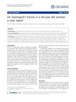
Figure 1 Preoperative frontal view that demonstrates a red,
round bulge in the groin area. The black dotted line shows how
the curved low inguinal incision was performed
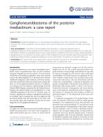
Figure 2 Intraoperative image of the inflamed ga ngrenous
appendix protruding through the femoral hernial sac.
Konofaos et al . Journal of Medical Case Reports 2011, 5:258
/>Page 2 of 3
29%. S ome cases of necrotizing fasciitis and even death
have been reported [5], probably related to the delay in
diagnosis and the older age of the patients.
Conclusions
Although the incidence of de Garengeo t’ sherniais
extremely low, the surge on has always to keep it in
mind in cases with femoral hernias and regional symp-
toms of inflammation due to the lack of abdominal
signs o f appendicitis. Ap pr opriate management without
incurring any delay for radiological imaging can be pro-
mising of an uneventful postoperative course. In patients
with large hernia defects or in older patients the use of
mesh for repairing the hernia defect can be an excellent
choice.
Consent
Written informed consent was obtained from the patient
for publication of this case report and any accompanyi ng
images. A copy of the written consent is available for
review by the Editor-in-Chief of this journal.
Authors’ contributions
All authors read and approved the final manuscript. PK was a major
contributor in writing the manuscript. ES was involved in acquisition of data
and review of the literature. AS was involved in acquisition of data and
review of the literature. KK was involved in drafting the manuscript and
revising it critically for important intellectual content. GK was involved in
drafting the manuscript, revising it critically for important intellectual content
and gave final approval of the version to be published.
Competing interests
The authors declare that they have no competing interests.
Received: 14 December 2010 Accepted: 30 June 2011
Published: 30 June 2011
References
1. De Garengeot RJC: Traite des operations de chirurgie. 2 edition. Paris: Huart;
1731, 369-371.
2. Tanner N: Strangulated femoral hernia appendix with perforated sigmoid
diverticulitis. Proc Roy Soc Med 1963, 56:1105-1106.
3. Rajan SS, Girn HR, Ainslie WG: Inflamed appendix in a femoral hernial sac:
de Garengeot’s hernia. Hernia 2009, 13(5):551-553.
4. Carey LC: Acute appendicitis occurring in hernias: a report of ten cases.
Surgery 1967, 61:236-238.
5. Zissin R, Brautbar O, Shapiro-Feinberg M: CT diagnosis of acute
appendicitis in a femoral hernia. Br J Radiol 2000, 73:1013-1014.
6. Akopian G, Alexander M: De Garengeot hernia: appendicitis within a
femoral hernia. Am Surg 2005, 71:526-527.
7. Isaacs LE, Felsenstein C: Acute appendicitis in a femoral hernia: an
unusual presentation of a groin mass. J Emerg Med 2002, 23:15-18.
8. Gurer A, Ozdogan M, Ozlem N, Yildirim A, Kulacoglu H, Aydin R:
Uncommon content in groin hernia sac. Hernia 2006, 10:152-155.
9. Fukukura Y, Chang SD: Acute appendicitis within a femoral hernia:
multidetector CT findings. Abdom Imaging 2005, 30:620-622.
10. Cordera F, Sarr MG: Incarcerated appendix in a femoral hernia sac.
Contemp Surg 2003, 59:35-37.
11. Barbaros U, Asoglu O, Seven R, Kalayci M: Appendicitis in incarcerated
femoral hernia. Hernia 2004, 83:281-282.
12. Nguyen ET, Komenaka IK: Strangulated femoral hernia containing a
perforated appendix. Can J Surg 2004, 47:68-69.
13. Priego P, Lobo E, Moreno I, Sanchez-Picot S, Gil Olarte MA, Alonso N,
Fresneda V: Acute appendicitis in an incarcerated crural hernia: analysis
of our experience. Rev Esp Enferm Dig 2005, 97:707-715.
doi:10.1186/1752-1947-5-258
Cite this article as: Konofaos et al.: De Garengeot’s hernia in a 60-year-
old woman: a case report. Journal of Medical Case Reports 2011 5:258.
Submit your next manuscript to BioMed Central
and take full advantage of:
• Convenient online submission
• Thorough peer review
• No space constraints or color figure charges
• Immediate publication on acceptance
• Inclusion in PubMed, CAS, Scopus and Google Scholar
• Research which is freely available for redistribution
Submit your manuscript at
www.biomedcentral.com/submit
Konofaos et al . Journal of Medical Case Reports 2011, 5:258
/>Page 3 of 3