Báo cáo y học: "Progressive obtundation in a young woman with bilateral corpus striatum infarction: a case report" potx
Bạn đang xem bản rút gọn của tài liệu. Xem và tải ngay bản đầy đủ của tài liệu tại đây (560.14 KB, 5 trang )
CAS E REP O R T Open Access
Progressive obtundation in a young woman with
bilateral corpus striatum infarction: a case report
Osama SM Amin
1*
,Sa’ad Seud Shwani
1
, Hero M Zangana
1
and Nawa A Ameen
2
Abstract
Background: Bilateral ischemic infarction involving the corpus striatum is a rare event which usually results from
global cerebral hypoxia, intoxications, and drug abuse.
Case presentation: We report a 28 year old Caucasian woman who presented with progressive obtund ation and
later development of severe expressive dysphasia and Parkinsonism after sustaining ischemic stroke of both
corpora striata. Hemorrhagic transformation developed on day four of admission.
Conclusion: This is a rare case of bilateral basal ganglia infarction with hemorrhagic transformation in a young
patient. Our patient’s work up did not reveal any cause behind this stroke; however, advanced investigations (such
as genetic testing and conventional angiography) were not done. The damage resulted in motor dysphasia and
Parkinsonism. Neither dystonia nor other involuntary movements developed, and cognitive function was not
assessed because of the language disorder.
Background
The human basal ganglia, which have a complex anat-
omy and physiology, are supplied by several blood ves-
sels on either side. Bilateral ischemic infarction
involving the corpus striatum is a rare event which
usually results from global cerebral hypoxia, intoxica-
tions, and drug abuse.
Case report
A 28 year old Caucasian woman was brought to our
emergency department with a five hour history of pro-
gressive impairment in consciousness and slurred
speech. Her past history was unremarkable, and she
neither smoked nor drank alcohol. Her older brother
said that she took no medications and she did not use
illicit drugs as far as he knew. No history of head
trauma was obtained. At the time of admission, her
blood pressure was 1 40/70 mmHg with a pulse rate of
90 beats/minute, respiratory rate of 12 cy cles/minut e,
and a temperature 37.1°C. Our patient was stuporous
and there were no lateralizing signs or neck stiffness.
Both planter reflexes were down. Our patient underwent
a battery of investigations with the following results:
hemoglobin 13.6 g/L; total white cell count 9100/mL
3
;
platelets 270,000/mL
3
; mean corpuscular volume 84fL;
mean corpuscular h emoglobin concentration 33 g/dL;
erythrocyte sedimen tation rate 19 mm/hour; blood urea
35 mg/dL; serum creatinine 0.9 mg/dL; serum sodium
139 mEq/L; serum potassium 4.1 mEq/L; serum calcium
8.9 mg/dL; ser um total bilirubin 0.8 m g/dL; aspartate
transaminase 21 u/L; alanine transaminase 19 u/L; alka-
line phosphatase 190 u/L; serum total protein 7.3 g/dL;
serum albumin 4.4 g/dL; thyroid stimulating hormone
2.9 u/L; serum total triiodothyronine 1.3 nmol/L; serum
total thyroxin 89 nmol/L; serum total cholesterol 177
mg/dL; serum triglyceride 100 mg/dL; low density lipo-
protein cholesterol 128 mg/dL; very low density lipopro-
tein cholesterol 20 mg/dl; high density lipoprotein
cholesterol 38 mg/dl; prothrombin time 12 seconds;
activated partial thromboplastin time 31 seconds; a
serum Venereal Disease Research Laboratory test was
negative; and general urine examination and microscopy
were unremarkable. Blood and urinary screening for
cocaine, opioids and amphetamines was negative. A 12
lead electrocardiogram (ECG) was normal. A non con-
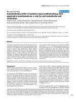
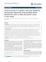
trast brain computed tomography (CT) scan showed
bilateral hypodensities in her corpus striatum (Fi gure 1).
In addition, there was a small hyper dense area at the
* Correspondence:
1
Department of Neurology, Sulaimaniya General Teaching Hospital,
Sulaimaniya City, Iraq
Full list of author information is available at the end of the article
Amin et al. Journal of Medical Case Reports 2011, 5:324
/>JOURNAL OF MEDICAL
CASE REPORTS
© 2011 Amin et al; licensee B ioMed Central L td. This is an Ope n Access article distributed un der the terms of t he Creative Commons
Attribution License ( nses/by/2 .0), which permits u nrestrict ed use, distribution, and reproduction in
any medium, provided the original work is properly cited.
anterior part of her right globus pallidus. The physician
suspected encephalitis, and managed our patient accord-
ingly. He ordered serology for toxoplasma and human
immunodeficiency virus, and a lumber puncture was
done: all of these te sts turned out to be negative. At day
four of admission, our patient became comatose, and
our neurology de partment was consulted. The Glasgow
coma scale was 3/15, no neck stiffness was detected,
and both planter reflexes were up. On day five, a brain
magnetic resonance imaging (MRI) scan with gadoli-
nium revealed hemorrhagic infarctions involving both
basal ganglia (Figure 2). Brain magnetic resonance
angiography and magnetic resonance venography (MRV)
were normal. Serum anti nuclear and rheumatoid factors
as well as anti phospholipid antibodies were negative.
Transthoracic and transesophageal echocardiographic
examinations were normal, as was th e carotid D oppler
study.
Our patient was managed as ischemic stroke with sec-
ondary hemorrhagic transformation. Anti hypertensives
and a statin were prescribed. Anti platelets and anticoa-
gulation were not given. Gradually over a period of
three weeks, our patient’s consciousness improved to a
degree of mild drowsiness. As for her language a ssess-
ment, comprehension was intact but there was no
speech output; she uttered few sounds, however, but no
comprehensible words. She had generalized rigidity and
hypokinesia. No abnormal movements were found an d
dystonic posturing was absent.
Discussion
The corpus striatum (which forms the bulk of the basal
ganglia) is composed of the neostriatum (made up of
putamen, caudate nucleus, and nucleus accumbens) and
the paleostriatum (with its internal and external seg-
ments of globus pallidus as well as the ventral pallidum)
Figure 1 Non contras t brain CT scan of our patient at the time of admission . Note the bilateral hypodensities, which fit the area of the
lenticular nucleus (putamen and globus pallidus) on both sides. There is also small hyperdensity at the right globus pallidus (black arrow). This
prompted the physician to suspect an infectious process instead of a vascular one. Our patient had bilateral infarction of the lenticular nucleus
with early hemorrhagic transformation inside the right one.
Amin et al. Journal of Medical Case Reports 2011, 5:324
/>Page 2 of 5
[1] In their study, Feekes and Cassell [2] found that the
human corpus striatum’s blood supply comes principally
from the medial and lateral lenticulostriate branches of
M1 and M2 segments of the middle c erebral artery a nd
from the recurrent artery of Huebner (which stems
from the A2 segment of the anterior cerebral artery).
The anterior choroidal and anterior communicating
arteries have a minor contribution. The middle cerebral
arter y also gives off direct small perforators to the stria-
tum, but these blood vessels contribute very little to the
overall blood supply [3].
Therefore, acute and extensive ischemic damage of
both corpora striata mainly requires occlusion of deep
perforating lenti culostriate branches of both middle cer-
ebral arteries and the arteries of Huebner.
Theoretically, multiple emboli to these blood vessels
can produce bilateral basal ganglia infarction. Russmann
et al. [4] found that eight out of their 13 patients with
extensive lenticular infarction had an embolic cause
(artery to artery in four p atients, cardioembolism in
three patients and one undetermined source). The unre-
markable ECG as well as echoca rdiograph ic and carotid
Doppler studies ruled out an embolic source.
Stam [5] suggested that cerebral venous sinus throm-
bosis should be suspected in patients with brain CT evi-
dence of hemorrhagic infarctions, especially if these
infarctions were multiple and did not follow a specific
arterial territory (as in our patient). The patient’sbrain
MRV was normal, however. We reviewed the brain MRI
and MRV with two radiologists; they disagreed with
Figure 2 Coronal T2 FLAIR (fluid attenuat ion inversion recovery) brain MRI film of our patient on the fifth day of hospital stay. Note
the bilateral heterogeneous hyper intensities at the right and left corpus striatum. These areas (which were not suppressed on this film)
represent ischemic infarction with hemorrhagic transformation.
Amin et al. Journal of Medical Case Reports 2011, 5:324
/>Page 3 of 5
cerebral venous sinus thrombosis as an etiology. The
sensitivity of combined brain MRI/MRV in the diagnosis
of cerebral venous sinus thrombosis is high [6,7]. In
addition, s ystemic lupus erythematosus and anti phos-
pholipid syndrome were on the differential diagnosis
list. The negative clinical and laboratory work u p can-
celled out these options.
Bilateral infarction of the corpus striatum is a well
documented event as an aftermath of pan cerebral hypo-
perfusion [8], intoxications and poisoning (such as cya-
nide [9] and carbon monoxide [10]), illicit drug use (for
example cocaine) [11], head trauma [12], and supraten-
torial neurosurgical procedures [13]. None of these fac-
tors was operative in our patient.
Hawker and Lange [ 8] found that pancerebral hypoxia
and ischemia are more likely to damage the globus palli-
dus; the putamen ranks second. The overall clinical pic-
ture also varies, ranging from akinetic rig id syndrome to
pure dystonia. According t o Grandas et al.[9],cyanide
poisoning destroys the putamen and external segments
of the globus pallidus; this combination results in severe
Parkinsonism and progressive dystonia. Approximately
13% of patients with carbon monoxide poisoning
develop delayed motor disorders, according to Quinn et
al. [ 10]; a variable combination of Parkinsonism, dysto-
nia, chorea, and myoclonus ensue.
Renard et al. [11] concluded that bilateral hemorrha-
gic infarction of basal ganglia usually occurs when
cocaine is co-administered with heroin, rather than after
cocaine abuse alone. Ishihara et al. [12] reported a case
of bilateral basal ganglia infarction in an 11-month-old
child who sustained a mild head trauma to his forehead.
Von Eckardstein an d his neurosurgic al team [13] per-
formed an operation on a 68 year old woman and
removed a right parietal parasagittal dural tumor with
reconstruction of the right wall of the superior sagittal
sinus; postoperatively, the patient remained unrespon-
sive and brain imaging revealed bilateral basal ganglia
infarction.
Due to the lack of expertise in our radiology depart-
ment, conventional cerebral angiography was not done.
The negative evaluation of the cause behind this
patient’s stroke would categorize our patient as having a
“ stroke of undetermined etiology,” according to the
TOAST classification [14]. However, as our work up
lacks several advanced investigations (such as genetic
testing and cerebral angiography) this categorization
cannot be done [15].
Isolated and discrete lesions involving various struc-
tures of corpus striatum usually result in specific clinical
features. For instance, damage to the anterioventral cau-
date can cause contra lateral choreoathetosis [16]. It
should b e noted that the ischemic infarction rarely con-
fines itself strictly to the corpus striatum; it usually
involves nearby structures, such as thalamus, hypothala-
mus, and internal capsule and other white matter pro-
jection fibers. Therefore, th e precise correlation between
bilateral lesions of corpus striatum and the resulting
cognitive, language, and motor dysfunction is usually
blurred.
Our patient’ s presentation of progressive obtundation
can be explained by the bilateral deep hemispheric dys-
function. During her recovery, our patient de monstrated
severe expressive dysphasia (rather than abulia). Mega
and Alexander [17] suggested that this form of subcorti-
cal dysphasia results from damage to the frontocaudate
functi onal system and the connecting deep white matter
fibers. According to Bhatia and Mar sden [18], h er Par-
kinsonism can be ascribed to bilateral lesions in the
putamen and/or globus pallidus. Cognitive and beha-
vioral abnormalities are very common in basal ganglia
lesions, especially bilateral ones [19]. Our patient’ s
severe language dysfunction rendered cognitive function
assessment virtually impossible.
Our patient was discharged five weeks after admis-
sion. She came back for a scheduled follow up visit
after one month. She still had severe expressive dys-
phasia and moderate hypokinesia and rigidity. After
careful questioning, the family denied any form of
involuntary movements or dystonia. Giroud et al.[20]
found that dystonia was the commonest consequence
of lenticular damage (whether acute or chron ic). On
the other hand, Russmann et al.[4]concludedthat
dystonia was a rare sequela to lenticular (putamen and
globus pallidus) lesions, a finding that is consistent
with ours.
Conclusion
This is a rare case of bilateral basal ganglia infarction
with hemorrhagic transformation in a patient. The
patient’s work up did not reveal any cause behind this
stroke; however, advanced investigations (such as
genetic testing and conventional angiography) were not
done. The damage resulted in motor dysphasia and Par-
kinsonism. Neither dystonia nor other involuntary
movements developed, and cognitive function was not
assessed because of the language disorder.
Consent
Written informed consent was obtained from the patient
for publicatio n of this case report and any accompany-
ing images. A copy of the written consent is avail able
for review by the Editor-in-Chief of this journal.
Author details
1
Department of Neurology, Sulaimaniya General Teaching Hospital,
Sulaimaniya City, Iraq.
2
Department of Medicine, Sulaimaniya General
Teaching Hospital, Sulaimaniya City, Iraq.
Amin et al. Journal of Medical Case Reports 2011, 5:324
/>Page 4 of 5
Authors’ contributions
Clinical work up was made by OSMA and SSS. SSS took the photos of the
brain imaging. The literature search was done by OSMA. HMZ and NAA
undertook patient follow up. OSMA wrote the manuscript; all authors read
and approved the final manuscript.
Authors’ information
OSMA is a board certified neurologist and a Fellow of the American College
of Physicians. SSS is a registrar in clinical adult neurology. HMZ is a
neurology trainee. NAA is an intern at the department of internal medicine
and neurology.
Competing interests
The authors declare that they have no competing interests.
Received: 10 December 2010 Accepted: 25 July 2011
Published: 25 July 2011
References
1. Brazis PW, Masdeu JC, Biller J: Localization in clinical neurology. 5 edition.
Philadelphia: Lippincott Williams & Wilkins; 2007, 422.
2. Feekes JA, Cassell MD: The vascular supply of the functional
compartments of the human striatum. Brain 2006, 129:2189-201.
3. Feekes JA, Hsu SW, Chaloupka JC, Cassell MD: Tertiary microvascular
territories define lacunar infarcts in the basal ganglia. Ann Neurol 2005,
58(1):18-30.
4. Russmann H, Vingerhoets F, Ghika J, Maeder P, Bogousslavsky J: Acute
infarction limited to the lenticular nucleus: clinical, etiologic, and
topographic features. Arch Neurol 2003, 60:351-5.
5. Stam J: Thrombosis of the cerebral veins and sinuses. N Engl J Med 2005,
352:1791-98.
6. Sajjad Z: MRI and MRV in cerebral venous thrombosis. J Pak Med Assoc
2006, 56(11):523-26.
7. Lafitte F, Boukobza M, Guichard JP, Hoeffel C, Reizine D, Ille O, Woimant F,
Merland JJ: MRI and MRA or the diagnosis and follow up of cerebral
venous thrombosis. Clin Radiol 1997, 52:672-79.
8. Hawker K, Lang AE: Hypoxic-ischemic damage of the basal ganglia. Mov
Disord 1990, 5:219-24.
9. Grandas F, Artieda J, Obeso JA: Clinical and CT scan findings in a case of
cyanide intoxication. Mov Disord 1989, 4:188-93.
10. Quinn DK, McGahee SM, Politte LC, Duncan GN, Cusin C, Hopwood CJ,
Stern TA: Complications of carbon monoxide poisoning: a case
discussion and review of the literature. Prim Care Companion J Clin
Psychiatry 2009, 11(2):74-9.
11. Renard D, Brunel H, Gaillard N: Bilateral haemorrhagic infarction of the
globus pallidus after cocaine and alcohol intoxication. Acta Neurol Belg
2009, 109:159-61.
12. Ishihara C, Sawada K, Tateno A: Bilateral basal ganglia infarction after mild
head trauma. Pediatr Int 2009, 51(6):829-31.
13. von Eckardstein KL, Youssef F, Hoch HH, Kiwit JC: Bilateral basal ganglia
infarction following resection of a right parietal parasagittal dural
tumour requiring sinus repair - an atypical complication of an atypical
venous drainage. Neurol Neurochir Pol 2006, 40(1):62-5.
14. Adams HP Jr, Bendixen BH, Kappelle LJ, Biller J, Love BB, Gordon DL,
Marsh EE: Classification of subtype of acute ischemic stroke. Definitions
for use in a multicenter clinical trial. TOAST. Trial of Org 10172 in Acute
Stroke Treatment. Stroke 1993, 24(1):35-41.
15. Ferro JM, Massaro AR, Mas JL: Aetiological diagnosis of ischaemic stroke
in young adults.
Lancet Neurol 2010, 9(11):1085-96.
16. Liles SL, Davis GD: Athetoid and choreiform hyperkinesias produced by
caudate lesions in the cat. Science 1969, 164:195-97.
17. Mega MS, Alexander MP: Subcortical aphasia: the core profile of
capsulostriatal infarction. Neurology 1994, 44:1824-29.
18. Bhatia KP, Marsden CD: The behavioral and motor consequences of focal
lesions of the basal ganglia in man. Brain 1994, 117:859-76.
19. Stout JC, Johnson SA: Cognitive impairment and dementia in basal
ganglia disorders. Curr Neurol Neurosci Rep 2005, 5(5):355-63.
20. Giroud M, Lemesle M, Madinier G, Billiar T, Dumas R: Unilateral lenticular
infarcts: radiological and clinical syndromes, aetiology, and prognosis. J
Neurol Neurosurg Psychiatry 1997, 63:611-15.
doi:10.1186/1752-1947-5-324
Cite this article as: Amin et al.: Progressive obtundation in a young
woman with bilateral corpus striatum infarction: a case report. Journal
of Medical Case Reports 2011 5:324.
Submit your next manuscript to BioMed Central
and take full advantage of:
• Convenient online submission
• Thorough peer review
• No space constraints or color figure charges
• Immediate publication on acceptance
• Inclusion in PubMed, CAS, Scopus and Google Scholar
• Research which is freely available for redistribution
Submit your manuscript at
www.biomedcentral.com/submit
Amin et al. Journal of Medical Case Reports 2011, 5:324
/>Page 5 of 5