Báo cáo y học: "Congenital aplasia of the optic chiasm and esophageal atresia: a case report" pptx
Bạn đang xem bản rút gọn của tài liệu. Xem và tải ngay bản đầy đủ của tài liệu tại đây (366.83 KB, 5 trang )
CASE REP O R T Open Access
Congenital aplasia of the optic chiasm and
esophageal atresia: a case report
Stefano Pensiero
1
, Paolo Cecchini
2
, Paola Michieletto
2
, Gloria Pelizzo
2
, Maurizio Madonia
1
and Fulvio Parentin
1,3*
Abstract
Introduction: The complete absence of the chiasm (chiasmal aplasia) is a rare clinical condition. Hypoplasia of the
optic nerve and congenital nystagmus are almost invariably associated characteristics. Microphthalmos or
anophthalmos are common features in chiasmal aplasia, while central nervous system abnormalities are less
frequent. Esophageal atresia can be isolated or syndromic. In syndromic cases, it is frequently associated with
cardiac, limb, renal or vertebral malformations and anal atresia. More rarely, esophageal atresia can be part of
anophthalmia-esophageal-genital syndrome, which comprises anophthalmia or microphthalmia, genital
abnormalities, vertebral defects and cerebral malformations. Here, a previously unreported case of chiasmal aplasia
presenting without microphthalmos and associated with esophageal atresia is described.
Case presentation: Aplasia of the optic chiasm was identified in a Caucasian Italian 8-month-old boy with
esophageal atresia. An ultrasound examination carried out at 21 weeks’ gestation revealed polyhydramnios.
Intrauterine growth retardation, esophageal atresia and a small atrial-septal defect were subsequently detected at
28 weeks ’ gestation. Repair of the esophageal atresia was carried out shortly after birth. A jejunostomy was carried
out at four months to facilitate enteral feeding. The child was subsequently noted to be visually inattentive and to
be neurodevelopmentally delayed. Magnetic resonance imaging revealed chiasmal aplasia. No other midline brain
defects were found. His karyotype was normal.
Conclusion: If achiasmia is a spectrum, our patient seems to depict the most severe form, since he appears to
have an extremely severe visual impairment. This is in contrast to most of the cases described in the literature,
where patients maintain good–or at least useful– visual function. To the best of our knowledge, the association of
optic nerve hypoplasia, complete chiasmal aplasia, esophageal atresia and atrial-septal defect, choanal atresia,
hypertelorism and psychomotor retardation has never been described before.
Introduction
Complete absence of the chiasmal struc ture, often asso-
ciated with optic nerve aplasia, is termed chiasmal apla-
sia, while the term achiasmia is used to identify the
abnormality of crossing fibers [1]. In fact, while in albin-
ism the temporal retinal fibers erroneously decussate at
the optic chiasm (OC), in achiasmia the majority of
fibers fail to cross at the O C and project ipsilaterally .
This condition is als o termed ‘non-decussating retinal-
fugal fiber syndrome’ (NDRFFS) [2,3]. Both albino and
achiasmatic anatomical and developmental abnormalities
can be functionally demonstrated by means of flash
visual evoked potentials (F-VEPs) [4,5]. In achiasmia, F-
VEPs show a higher positive component ipsilateral to
the stimulated eye, while in albinos the response is
greater contralaterally [4,1].
Congenital nystagmus is a consistent feature in achias-
mia, as well as in certain cases of optic nerve hypoplasia
[2,6]. Central nervous system (CNS) abnormalities, such as
septo-optic dysplasia, hypopituitarism, encephalocele or
corpus callosum agenesia are associated with achiasmia
[1].
The complete absence of the chiasm (chiasmal aplasia)
is a rarer clinical condition. Hypoplasia of the optic
nerve and congenital nystagmus are almost invariably
associated characteristics [6,7]. Microphthalmos or
anophthalmo s are common features in chiasmal aplasia,
while CNS abnormalities are less frequent [6,7].
* Correspondence:
1
Ophthalmology Unit, Department of Surgery, Institute for Maternal and
Child Health, Burlo Garofolo Trieste, Via dell’Istria 65/1, I-34100 Trieste, Italy
Full list of author information is available at the end of the article
Pensiero et al. Journal of Medical Case Reports 2011, 5:335
/>JOURNAL OF MEDICAL
CASE REPORTS
© 2011 Pensiero et al; licensee BioMed Central Ltd. This is an Open Access article distributed under the terms of the Creative
Commons Attribution License (http://creativecommons. org/licenses/by/2.0), which permits unrestricted use, distribut ion, and
reproduction in any mediu m, provided the original work is pro perly cited.
A c ase of chiasmal aplasia in a Caucasian baby, asso-
ciated with esophageal atresia (EA), is here described.
Case presentation
Polyhydramnios was detecte d in a 35-year-old primigra-
vida at 21 weeks’ gestation. Esophageal atresia was diag-
nosedat28weeks’ gestation on the basis of a small
stomach and polyhydramnios on an ultrasound exami-
nation. Other abnormalitie s detected include d a dilated
upper esophageal pouch and an atrial-septa l defect. Our
patient underwent periodic therapeutic amniocentesis
(1500-1900 ml of fluid per procedure) from 28 weeks’
gestation for relief of polyhydramnios and to prevent
premature onset of labor. A male fetus with a birth
weight of 1980 g was delivered by caesarean section at
37 weeks’ gestation. Apgar scores were six and eight at
one and five minutes respectively. A clinical examination
showed type III EA, right choanal atresia, atrial-septal
defect, telecanthus and hypertelorism without any
obvious strabismus. His mother’s history was negative
for familial visual anomalies and there was no evidence
of maternal infection or drug abuse during pregnancy.
His karyotype was normal.
Uncomplicated esophageal anastomosis with closure of
the tracheo-esophageal fistula was performed on the
second day of his life. Due to the persistence of suction
weakness, our patient was discharged two months later
with a naso-gastric tube and enteral nutrition. The baby
was referred for pediatric consultation at four months of
age. A failure to thrive (length and weight, head circum-
ference on third centile) due to gastroesophageal reflux
was documented. A laparoscopic gastri c fundoplication
was performed. A jejunostomy was also performed to
allow continuation of enteral feeding.
Thebabyunderwentacompleteophthalmicevalua-
tion at the age of four months: our patient showed erra-
tic eye movements and was unable to fixate on a light.
Horizontal slow nystagmus was observed. His pupil
reactivity to light was absent bilaterally. A slit-lamp bio-
microscopy of the anterior segment of both eyes was
unremarkable. Funduscopy revealed severe bilateral
opticdischypoplasiaandmild tortuosity of the retinal
blood vessels. His optic discs appear ed greyish and oval.
F-VEPs were repeatedly non-recordable bilaterally.
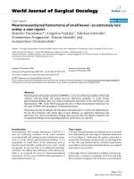
Brain magnetic resonance imaging (MRI) scans per-
formed at the age of nine months showed normal ey e
bulb structures, absence of the OC and optic radiations,
consistent with extreme hypoplasia or aplasia of those
structures. His optic nerves were bilaterally traced only
in the intraorbital portion and were of small appearance.
There was no evidence of his optic nerves more poster-
iorly (Figure 1). His other cerebral structures were
normal.
An evaluation at 10 months revealed delayed social
skills and language development.
General examination at 18 months of life showed
reduced stature, persistence of food aversion, and
delayed social contacts and language development.
Discussion
EA can be isolated or syndromic [8]. In syndromic
cases, EA is frequently associated with cardiac, limb,
renal and vertebral malformations and anal atresia [8].
In our case, his karyotype was normal, so chromosomal
anomalies responsible of syndromic EA (for example
trisomy 21, 18, 13 and 17q21.3-q23 deletion) were not
involved [8]. Other conditions frequently associated with
EA include VACTERL (vertebral anomalies, anal atresia,
cardiovascular anomalies, trachea-esophageal fistula,
esophageal atresia, renal and/or radial anomalies and
limb defects) syndrome, Feingold (oculo-digito-eso-
phago-duodenal) s yndrome and Rogers (anophthalmia-
esophageal-genital or AEG) syndrome [8]. Other condi-
tions occasionally associated with EA, and characterized
by an ocular involvement, are shown in Table 1. Mater -
nal diabetes and phenylketonuria, rarely associated with
EA, were excluded due to the normality of blood tests
during pregnancy. AEG syndrome comprises EA,
anophthalmia or microphthalmia, genital abnormalities,
vertebral defects and cerebral malformations [9]. There
is evidence of a genetic mechanism for this syndrome.
In a number of cases, deletion and/or mutation of the
transcripti onal regulator gene, Sox2, has been proved to
be involved [10,11]. The phenotypic variability may be
remarkable, ranging from anophthalmia to normal ocu-
lar development [12,13]. Sox2 also seems to play a cau-
sative role in isolated anophthalmia and microphthalmia
[11]. However, S Sox2 deletion or malfunction can
hardly have been involved in our patient, as his eyes
were fully developed and the only eye abnormality was
the optic nerve hypoplasia. Moreover he presented with
no genital or vertebral anomalies, while he did have a
heart malformation. He did not present ear anomalies,
typical of CHARGE (coloboma of the eye, heart defects,
atresia of the choanae, retardation of growth, genital
abnormalities, and ear abnormalities) and OAVS (oculo-
auriculo-vertebral spectrum) syndromes, but the first is
theonlysyndromepresentingwithchoanalatresia
(Table 1). Our patient could not therefore be classified
in any of the syndromes associated with EA.
In syndromes associated with optic nerve and/or OC
hypoaplasia, some genes responsible for the molecular
mechanisms of neural routing have been related to the
achiasmia spectrum. In the animal model, the lack of
the transcription factor Foxd1 induces chiasmal malfor-
mation and misprojection of retinal fibers [14]. Foxd1
Pensiero et al. Journal of Medical Case Reports 2011, 5:335
/>Page 2 of 5
Figure 1 Axial Tx and Ty MRI scans showing complete absence of the optic chiasm (black arrow); the optic nerves can be identified
only in the intraorbital portion (white arrow).
Table 1 Comparison among the clinical characteristic of our case and the syndromic form of EA
Features Our Case AEG (Rogers) VACTERL Feingold CHARGE OAVS Bartsocas-Papas
Esophageal atresia +++++++
Microcephaly -+-++-+
Optic chiasm aplasia +
Nystagmus +
Optic nerve hypoplasia +
Eyes anomalies -+-++++
Optic tract aplasia +
Anophthalmia/microphthalmia -+ +
Telecanthus/hypertelorism + +
Cerebral malformation +-+
Heart malformation +-++++-
Vertebral defects/other bone anomalies +++++
Genital/renal anomalies -++++++
Facial/visceral problems ++
Visual impairment +-
Mental retardation ++-++- -
Facial cleft +
Limb anomalies +-+-+
Ear deformities ++-
Choanal atresia +- +
Pensiero et al. Journal of Medical Case Reports 2011, 5:335
/>Page 3 of 5
misexpression leads to a cascade of other gene products
misregulation, which interferes with the normal develop-
ment of the OC and with the ratio of ipsilateral to con-
tralateral chiasmal nerve fiber routing [14]. Foxd1 has
not been described to play a role in EA and it is ques-
tionable how it could be involved in the multiple mal-
formations observed in the present repo rt. It seems
unlikely to consider EA a merely incidental finding. Pax
family genes have also been suggested to play an impor-
tant role in the correct development of the OC [15,16].
In the murine Pax mutant model, the OC fails to
develop, and retinal axons enter the ipsilateral optic
tract [15,16 ]. Again, the association with EA in our case
would be hard to explain.
Sami et al. [1] have suggested a classification system
for patients affected by achiasmia: type A, presenting
with isolated achiasmia and often nystagmus, with possi-
ble MRI evidence of a small chiasm; type B, presenting
with chiasmal hypoplasia and midline defects (septo-
optic dysplasia); and type C, presenting with chiasmal
hypoplasia and possible clefting disorders, encephalocele,
and agenesia of the corpus callosum. In Sami’sseries,
one patient did not fit into one of the three suggested
groups. The child (a boy of six years) suffered from mul-
tiple facial, visceral and developmental problems, includ-
ing EA. An MRI scan showed an isolated small chiasm.
The child exhibited horizontal nystagmus. To the best
of our knowledge, this is the only previous case of
achiasmia in a patient affected by EA, and appears
somehow similar to ours: OC aplasi a, horizontal nystag-
mus and esophageal atresia are shared features between
Sami’s case and ours. However, other traits are remark-
ably different. The main discrepancy is the presence of
the chiasm–even if hypoplasic–in the MRI scans in
Sami’s report, while no chiasm structure was detectable
on MRI scanning and no facial abnormalities were pre-
sent in our patient. The F-VE Ps results and the lack of
fixation and of pupil reactivity to light suggest a severe
visual impairment in this child, unlike the Sami case,
who exhibited fairly good visual function. Moreover our
child showed an atrial septal defect and choanal atresia,
which was not been described in the Sami case.
One could speculate that these two cases may have the
same etiology with different phenotypes. The rarity of
this condition and the relevant differences betwee n the
two cases, however, suggest grea t caution in attempting
to group them in a single clinical entity.
If achiasmia is a spectrum, our child seemed to depict
the most severe form, since he appeared to have an
extremely severe visual impairm ent, in contrast to most
of the cases described in literature that maintain a good–
or at least useful–visual function. The lack of fixation
and reactivity to light or structured stimuli and the
presence of roving eye movements were highly suggestive
of poor or no residual visual function. The lack of F-VEP
response–which is very unusual in achiasmia–confirmed
the OC aplasia suggested by MRI findings; moreover we
could consider the OC aplasia of our patient to be sec-
ondary to a primary bilateral severe optic nerve hypopla-
sia. Pomeranz [17] descri bed an 18-month-old boy with
bilateral optic nerve hypoplasia and OC not identifiable
at the MRI who showed profo undly abnormal F-VEP in
his right eye. However, left eye stimulation demonstrated
a typical VEP occipital asymmetry of the response, con-
sistent with NDRFFS. VEP results showed the presence
of a hypoplasic and not aplasic OC (OC was not detect-
able using MRI scans), with the characteristics of achias-
mia. On MRI scans no other major brain abnormalities
were detected. In contr ast to our case, no other visceral
malformations were noticed, and the baby showed a good
visual interaction with the environment.
Finally, OC aplasia has o ften been described in asso-
ciation with other major CNS abnormalities and unilat-
eral or bilateral anophthalmos or microphthalmos was
always present. This is in contrast to our case. The
genes that are involved in EA and in achiasmia are all
located in different chromosomes, so it is difficult to
predict simultaneous involvement of multiple genes. In
fact Pax is located in chromosome 11p13, Foxd11
on 5q12-13, Mycn (p roducing Feingold syndrome) on
2p24.1, Chd7 (CHARGE) on 8q12.2 and Sox2 (AEG) on
3q26. For this reason the syndrome here described, like
VACTERL and OAVS, is probably to be considered of
malformative origin.
Conclusion
We believe that our child does not fit into one of the
previously reported achiasmia or OC aplasia types or
reports. To the best of our knowledge, the association of
optic nerve hypoplasia, chiasmal and optic tracts aplasia
(confirmed by VEP and light pupil reactivity absence),
with telecanthus and/or hypertelorism, esophageal atre-
sia, atrial septal defect, choanal atresia, and developmen-
tal and language delays has never been described before.
Consent
Written informed consent was obtained from the par-
ents of the patient for publication of this case report
and any accompanying images. A copy of the written
consent is available for review by the Editor-in-Chief of
this journal.
Abbreviations
AEG: anophthalmia-esophageal-genital; CNS: central nervous system; EA:
esophageal atresia; F-VEPs: flash visual evoked potentials; MRI: magnetic
resonance imaging; NDRFFS: non-decussating retinal fugal fiber syndrome;
OC: optic chiasm.
Pensiero et al. Journal of Medical Case Reports 2011, 5:335
/>Page 4 of 5
Author details
1
Ophthalmology Unit, Department of Surgery, Institute for Maternal and
Child Health, Burlo Garofolo Trieste, Via dell’Istria 65/1, I-34100 Trieste, Italy.
2
IRCCS E Medea, Via Cialdini 5, I-33037 Pasian di Prato (UD), Italy.
3
Paediatric
Surgery Unit, Department of Surgery, Institute for Maternal and Child Health,
Burlo Garofolo Trieste, Via dell’Istria 65/1, I-34100 Trieste, Italy.
Authors’ contributions
SP was a major contributor in writing the manuscript. GP performed surgical
intervention.
PC and PM performed clinical and instrumental examinations. MM and FP
made a review of the literature and were involved in the diagnosis and
management of the patient. All authors have read and approved the final
manuscript.
Competing interests
The authors declare that they have no competing interests.
Received: 13 August 2010 Accepted: 1 August 2011
Published: 1 August 2011
References
1. Sami DA, Saunders D, Thompson DA, Russell-Eggitt IM, Nischal KK, Jeffrey G,
Dattani M, Clement RA, Liasis A, Taylor DS: The achiasmia spectrum:
congenitally reduced chiasmal decussation. Br J Ophthalmol 2005,
89(10):1311-1317.
2. Korff CM, Apkarian P, Bour LJ, Meuli R, Verrey JD, Roulet Perez E: Isolated
absence of optic chiasm revealed by congenital nystagmus, MRI and
VEPs. Neuropediatrics 2003, 34(4):219-223.
3. Apkarian P, Bour LJ, Barth PG, Wenniger-Prick L, Verbeeten B Jr: A unique
achiasmatic anomaly detected in non-albinos with misrouted retinal-
fugal projections. Eur J Neurosci 1994, 6(3):501-507.
4. Thompson DA, Kriss A, Chong K: Visual-evoked potential evidence of
chiasmal hypoplasia. Ophthalmology 1999, 106(12):2354-2361.
5. Leitch RJ, Thompson D, Harris CM, Chong K, Russell-Eggitt I, Kriss A:
Achiasmia in a case of midline craniofacial cleft with seesaw nystagmus.
Br J Ophthalmol 1996, 80(11):1023-1024.
6. Biega TJ, Khademian ZP, Vezina : Isolated absence of the optic chiasm: a
rare cause of congenital nystagmus. Am J Neuroradiol 2007, 28(2):392-393.
7. Sanjari MS, Ghasemi Falavarjani K, Parvaresh MM, Kharazi HH, Kashkooli MB:
Bilateral aplasia of the optic nerve, chiasm, and tracts in an otherwise
healthy infant. Br J Ophthalmol 2006, 90(4):513-514.
8. Geneviève D, de Pontual L, Amiel J, Sarnacki S, Lyonnet S: An overview of
isolated and syndromic oesophageal atresia. Clin Genet 2007,
71(5):392-399.
9. Shah D, Jones R, Porter H, Turnpenny P: Bilateral microphthalmia,
esophageal atresia, and cryptorchidism: the anophthalmia-esophageal-
genital syndrome. Am J Med Genet 1997, 70(2):171-173.
10. Hill CJ, Pilz DT, Harper PS, Castle B, Williams TH: Anophthalmia-esophageal-
genital syndrome: a further case to define the phenotype. Am J Med
Genet 2005, 132(1):57-59.
11. Zhou J, Kherani F, Bardakjian TM, Katowitz J, Hughes N, Schimmenti LA,
Schneider A, Young TL: Identification of novel mutations and sequence
variants in the SOX2 and CHX10 genes in patients with anophthalmia/
microphthalmia. Mol Vis 2008, 14:583-592.
12. Chassaing N, Gilbert-Dussardier B, Nicot F, Fermeaux V, Encha-Razavi F,
Fiorenza M, Toutain A, Calvas P: Germinal mosaicism and familial
recurrence of a SOX2 mutation with highly variable phenotypic
expression extending from AEG syndrome to absence of ocular
involvement. Am J Med Genet 2007, 143(3):289-91.
13. Zenteno JC, Perez-Cano HJ, Aguinaga M: Anophthalmia-esophageal
atresia syndrome caused by an SOX2 gene deletion in monozygotic
twin brothers with markedly discordant phenotypes. Am J Med Genet
2006, 140(18):1899-1903.
14. Herrera E, Marcus R, Li S, Williams SE, Erskine L, Lai E, Mason C: Foxd1 is
required for proper formation of the optic chiasm. Development
2004,
131(22):5727-5739.
15. Alvarez-Bolado G, Schwarz M, Gruss P: Pax-2 in the chiasm. Cell Tissue Res
1997, 290(2):197-200.
16. Torres M, Gómez-Pardo E, Gruss P: Pax2 contributes to inner ear
patterning and optic nerve trajectory. Development 1996,
122(11):3381-3391.
17. Pomeranz HD, Agadzi AK, Ekesten B: Achiasmia and unilateral optic nerve
hypoplasia in an otherwise healthy infant. Acta Ophthalmol Scand 2006,
84(1):140-144.
doi:10.1186/1752-1947-5-335
Cite this article as: Pensiero et al.: Congenital aplasia of the optic
chiasm and esophageal atresia: a case report. Journal of Medical Case
Reports 2011 5:335.
Submit your next manuscript to BioMed Central
and take full advantage of:
• Convenient online submission
• Thorough peer review
• No space constraints or color figure charges
• Immediate publication on acceptance
• Inclusion in PubMed, CAS, Scopus and Google Scholar
• Research which is freely available for redistribution
Submit your manuscript at
www.biomedcentral.com/submit
Pensiero et al. Journal of Medical Case Reports 2011, 5:335
/>Page 5 of 5