Báo cáo y học: " Toxoplasmosis presenting as a swelling in the axillary tail of the breast and a palpable axillary lymph node mimicking malignancy: a case report" potx
Bạn đang xem bản rút gọn của tài liệu. Xem và tải ngay bản đầy đủ của tài liệu tại đây (2.02 MB, 4 trang )
CAS E REP O R T Open Access
Toxoplasmosis presenting as a swelling in the
axillary tail of the breast and a palpable axillary
lymph node mimicking malignancy: a case report
HP Priyantha Siriwardana
1*
, Louise Teare
2
, Dia Kamel
3
and E Reggie Inwang
1
Abstract
Introduction: Lymphadenopathy is a common finding in toxoplasmosis. A breast mass due to toxoplasmosis is
very rare, and only a few cases have been reported. We present a case of toxoplasmosis that presented as a
swelling in the axillary tail of the breast with a palpable axillary lymph node which mimicked breast cancer.
Case presentation: A 45-year-old otherwise healthy Caucasian woman presented with a lump on the lateral
aspect of her left breast. Her mother had breast cancer that was diagnosed at the age of 66 years. During an
examination, we discovered that our patient had a discrete, firm lump in the axillary tail of her left breast and an
enlarged, palpable lymph node in her left axilla. Her right breast and axilla were normal. The clinical diagnosis was
malignancy in the left breast. Ultrasound and mammographic examinations of her breast suggested a pathological
process but were not conclusive. She had targeted fine-needle aspiration cytology (FNAC) and core biopsy of the
lesions. FNAC was indeterminate (C3) but suggested a possibility of toxoplasmosis. The core biopsy was not
suggestive of malignancy but showed granulomatous inflammation. She had a wide local excision of the breast
lump and an axillary lymph node biopsy. Histopathology and immunohistochemical studies excluded carcinoma or
lymphoma but suggested the possibility of intramammary and axillary toxoplasmic lymphadenopathy. The results
of Toxoplasma gondii IgM and IgG serology tests were positive, supporting a diagnosis of toxoplasmosis.
Conclusions: Toxoplasmosis rarely presents as a pseudotumor of the breast. FNAC and histology are valuable tools
for a diagnosis of toxoplasmosis, and serology is an important adjunct for confirmation.
Introduction
Lymphadenopathy is the most frequent clinical manifes-
tation of acute infection with Toxoplasma gondii in the
immunocompetent individual. Toxoplasma lymphadeni-
tis typically involves a lymph node in the head and neck
region, presents with or without systemic symptoms or
extranodal disease, and runs a benign clinical course
[1,2]. A breast mass due to toxoplasmosis is rare, and
only a few cases have been reported [3-5]. We present a
case of toxoplasmosis that presented as an axillary tail
(breast) mass and a palpable axillary lymph node which
mimicked breast cancer.
Case presentation
A 45-year-old Caucasian woman with a left axillary tail
(breast) mass and left-sided chest pain presented to t he
breast clinic. She also compla ined that her left breast
had changed in appearance. She had a positive family
history: her mother had breast cancer and her father
had lung cancer. There was no nipple discharge, feve r,
or history of trauma to her breast. She had two children
and had undergone a hysterectomy for benign disease
two years before. Both of her ovaries were retained.
There was no other significant medical history or
known allergies. Her general health was good.
The result of a general examination was normal.
There were two palpable nodules, one in t he upper
outer quadrant in the axillary tail of her left breast (20
mm) and the other in the left axilla (10 mm). The result
of an e xamination of her right breast and a xilla, abdo-
men, and other systems was normal. The most likely
* Correspondence:
1
Department of Surgery, Broomfield Hospital, Court Road, Chelmsford, Essex,
CM1 7ET, UK
Full list of author information is available at the end of the article
Siriwardana et al. Journal of Medical Case Reports 2011, 5:348
/>JOURNAL OF MEDICAL
CASE REPORTS
© 2011 Siriwardana et al; licensee BioMed Central Ltd. This is an Open Access ar ticle distributed under the terms of the Creative
Commons Attribution License ( which permits unrestricted use, distribution, and
reproduction in any medium, provided th e origina l work is properly cited.
diagnosis was considered to be a malignant lesion in the
left breast with metastatic involvement of an axillary
lymph node.
She underwent ultrasound and mammographic exami-
nations of her breasts. The mammogram showed a
smooth-outlined, soft-density lesion in her left breast
with no microcalcifications and a few small lymph
nodes in her left axillary tail. Ultrasound revealed that
the palpable lump in the lateral part of her left breast
was a 2 cm solid lesion with reduced echogenicity. The
other nodule, in the upper part of the left axilla, was
also solid (1 cm) and suggestive of a lymph node (M4
U4; that is, suspicious abnormality according to the
Breast Imaging Reporting and Data System, or BIRADS).
The radiological appearance was highly suggestive of a
lymphoma. Then she underwent targeted fine-needle
aspiration cytology (FNAC) of the axillary lesion and
core needle biopsy of the breast lesion. The FNAC was
indeterminate (C3) but showed numerous monotonous
lymphocytes in a background containin g lymphogranu-
lar bodies suggestive of granulomatous inflammation
such as toxoplasmosis. There were no malignant cells.
The core biopsy showed a small aggregate of epitheleoid
histiocytes and multinuclear giant cells in keeping with
granulomatous inflammation. There was no evidence of
a malignancy.
Her case was discussed at the multidisciplinary meet-
ing, and the team recommended a wide local excision of
the breast lesion with palpable axillary lymph node
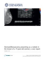
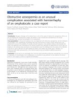
biopsy. The results of a histological examination (Fig-
ures 1 and 2) of the resected specimens of breast and
axillary lesions were suggestive of an intramammary and
axillary lymph node m ass with marked follicular
hyperplasia. In addition, there were prominent micro-
granulomas composed almost entirely of epithelioid cells
located within the hyperplastic follicles. Immunohisto-
chemical staining showed an anatomical distribution of
B- and T-cell markers. A Ziehl-Neelsen stain for acid-
fast bacilli and Grocott and PAS+D (periodic acid-Schiff
after diastase digestion) stains for fungi were negative.
The histological appearances were similar to those
described in toxoplasmosis, but the differential diag-
noses included other infectious diseases and lymphade-
nopathy-associated autoimmune or immunodeficiency
disorders. There were no features to suggest lymphoma
or other malignancy. Histological material was referred
for a second opinion that confirmed the above. The T.
gondii serology tests detected Toxoplasma IgG and IgM
antibodies suggestive of an acute or recently acquired
Toxoplasma infection. Our patient was treated sympto-
matically as there were no indications to treat her toxo-
plamosis with antiprotozoal drugs. She has been well for
the last two years since the diagnosis.
Discussion
Toxoplasmosis is caused by infection with T. gondii,an
obligate intracellular parasitic protozoa. The infection
produces a wide range of clinical syndromes in humans,
land and sea mammals, and various bird species. Toxo-
plasmosis passes from animals to humans, mainly via
infected cat feces. T. gondii infect s a large proportion of
the world’s population but rarely causes clinically signifi-
cant disease. Although infection does not normally
spread from person to person except t hrough preg-
nancy, toxoplasmosis can, in rare instance s, contaminate
blood transfusions and organs donated for transplanta-
tion. In most immunocompetent individuals, primary or
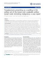
Figure 1 A microscopic examination of the specimens of
breast (axillary tail) lump and axillary lymph node shows
marked follicular hyperplasia with prominent small granulomas
composed almost entirely of epithelioid cells.
Figure 2 A microscopic examination of the specimens of
breast (axillary tail) lump and axillary lymph node shows
marked follicular hyperplasia with prominent small granulomas
composed almost entirely of epithelioid cells.
Siriwardana et al. Journal of Medical Case Reports 2011, 5:348
/>Page 2 of 4
chronic (latent) T. gondii infection is asymptomatic in
80% to 90% of healthy hosts [1].
Lymphadenopathy is the most frequent manifestation
of acute acquired infection in immunocompetent indivi-
duals. The typical presentation is a painless firm lym-
phadenopathy confined to one cha in of nodes, most
commonly cervical. Other physical manifestations
include low-grade fever, he patosplenomegaly, and skin
rash. Our patient did not have any such manifestations.
Toxoplasma lymphadenitis most frequently involves a
solitary lymph node in the head and neck region, pre-
sents with o r without systemic symptoms or extranodal
disease and runs a benign clinical course. However, ser-
ious extranodal disease does occur in a small percentage
of patients and includes myocarditis, pneumonitis, ence-
phalitis, chorioretinitis, and transmission of infection to
the fetus [2]. Individuals at risk for severe or life-threa-
tening toxoplasmosis include fetuses, newborns, and
immunologically impaired pat ients. In immunodeficient
individuals, toxoplasmosis most often occurs in those
with defects of T cell-mediated immunity, such as those
with hematologic malignancies, bone marrow and solid
organ transplants, or AIDS.
Both histological features of biopsy specimens or cytol-
ogy of needle aspirate [6] and serolo gical tests are impor-
tant in the diagnosis of toxoplasmosis and it was not
until both were available in this case that a diagnosis of
toxoplasmosis was made. The histological features have
been well described [2] but sometimes can be confused
with other disorders, particularly sarcoidosis, very early
tuberculosis, cat-scratch disease [7], and more benign
forms of Hodgkin disease, all of which may have a clinical
presentation similar to that of toxoplasmosis [2]. Immu-
nohistochemistry can help identify T. gondii within
pathology specimens. Molecular polymerase chain reac-
tion techniques have high specificity but low sensitivity
in lymph node specimens, and the role of molecular biol-
ogy in the diagnosis of toxoplasmosis has been reported
[8]. Serology tests are an important adjunct but, on their
own, must be interpreted with some care, as positive
tests wi th low titers are common, presumably because of
latent infection. In our case, however, serology testing
was strongly positive, supporting the histological findings.
In an otherwise healthy perso n who is not pregnant, as
in this case, treatment is no t indicated. Symptoms will
usually resolve within a few weeks [2]. If toxoplasmosis is
acquired in pregnancy, transplacental infection may lead
to severe disease in the fetus. Spiramycin may reduce the
risk of transmission of maternal infection to the fetus.
For people who have weakened immune systems, anti-
protozo al drugs such as a combination of pyrimethamine
and sulfadiazine are given for several weeks [2].
Conclusions
Toxoplasmosis rarely presents as a mass in the axillary
tail of the breast and may be considered as a differen-
tial diagnosis in p atients presenting with axillary lym-
phadenopathy. FNAC and histology are valuable tools
for a diagnosis of toxoplasmosis and serology i s an
important adjunct for confirmation. If the FNAC or
core biopsy suggests the possibility of toxoplasmosis,
serological investigations can confirm the diagnosis
and may help avoid further invasive procedures and
anxiety. Adult patients who are immunocompetent, are
not pregnant and do not have involvement of a vital
organ may be managed conservatively without antipro-
tozoal drugs.
Consent
Written informed consent was obtained from the patient
for publication of this case report and any accompany-
ing images. A copy of the writ ten consent is available
for review by the Editor-in-Chief of this journal.
Abbreviation
FNAC: fine-needle aspiration cytology.
Author details
1
Department of Surgery, Broomfield Hospital, Court Road, Chelmsford, Essex,
CM1 7ET, UK.
2
Department of Microbiology, Broomfield Hospital, Court Roa d,
Chelmsford, Essex, CM1 7ET, UK.
3
Department of Pathology, Broomfield
Hospital, Court Road, Chelmsford, Essex, CM1 7ET, UK.
Authors’ contributions
HPPS, the principal author, contributed to designing the report and writing
the introduction, case presentation, and discussion sections. LT and DK
contributed to the discussion. ERI collected the data, obtained consent from
the patient, supervised the project, and undertook the final revision before
submission. All authors read and approved the final manuscript.
Competing interests
The authors declare that they have no competing interests.
Received: 9 November 2010 Accepted: 4 August 2011
Published: 4 August 2011
References
1. Frankel JK: The Coccidia, Isospora, Toxoplasma and related genera.
Toxoplasmosis; parasite life cycle. In Pathology and Immunology. Edited
by: Hammond DM, Long PL. Baltimore: University Park Press; 1973:342-410.
2. McCabe RE, Remington JS: Toxoplasma gondii. In Principles and Practice of
Infectious Diseases. Part III 2 edition. Edited by: Mandell GL, Douglas RG,
Bennett JE. New York: John Wiley; 1985:154-1556.
3. Kouba K, Lobovská A, Kudrmann J, Lasovská J: Pseudotumours of
toxoplasmatic origin in female breast [in Czech]. Cesk Gynekol 1981,
46:365-372.
4. Pelikánová G, Pelikán A, Bolgác A, Sitár A: Toxoplasmosis as a cause of
pseudotumor of the breast in women [in Slovak]. Cesk Gynekol 1984,
49:737-740.
5. Turner JR: Toxoplasmosis presenting as a swelling in the axillary tail of
the breast. Postgrad Med J 1965, 41:39-40.
6. Shimizu K, Ito I, Sasaki H, Takada E, Sunagawa M, Masawa N: Fine needle
aspiration of toxoplasmic lymphadenitis in an intramammary lymph
node. A case report. Acta Cytol 2001, 45:259-262.
Siriwardana et al. Journal of Medical Case Reports 2011, 5:348
/>Page 3 of 4
7. Markaki S, Sotiropoulou M, Papaspirou P, Lazaris D: Cat-scratch disease
presenting as a solitary tumour in the breast: report of three cases. Eur J
Obstet Gynecol Reprod Biol 2003, 106:175-178.
8. Voglino G, Arisio R, Novero D, Marchi C, Fessia L: Lymphadenopathy
caused by Toxoplasma in an intramammary lymph node: role of
molecular biology in the diagnosis [in Italian]. Pathologica 1997,
89:446-448.
doi:10.1186/1752-1947-5-348
Cite this article as: Siriwardana et al.: Toxoplasmosis presenting as a
swelling in the axillary tail of the breast and a palpable axillary lymph
node mimicking malignancy: a case report. Journal of Medical Case
Reports 2011 5:348.
Submit your next manuscript to BioMed Central
and take full advantage of:
• Convenient online submission
• Thorough peer review
• No space constraints or color figure charges
• Immediate publication on acceptance
• Inclusion in PubMed, CAS, Scopus and Google Scholar
• Research which is freely available for redistribution
Submit your manuscript at
www.biomedcentral.com/submit
Siriwardana et al. Journal of Medical Case Reports 2011, 5:348
/>Page 4 of 4