báo cáo khoa học: "The margination propensity of spherical particles for vascular targeting in the microcirculation" pptx
Bạn đang xem bản rút gọn của tài liệu. Xem và tải ngay bản đầy đủ của tài liệu tại đây (513.45 KB, 9 trang )
BioMed Central
Page 1 of 9
(page number not for citation purposes)
Journal of Nanobiotechnology
Open Access
Research
The margination propensity of spherical particles for vascular
targeting in the microcirculation
Francesco Gentile
1
, Antonio Curcio
2
, Ciro Indolfi
2
, Mauro Ferrari
3,4
and
Paolo Decuzzi*
1,3
Address:
1
Center of Bio-/Nanotechnology and -/Engineering for Medicine University of Magna Graecia at Catanzaro, Viale Europa – Loc.
Germaneto, 88100, Catanzaro, Italy,
2
Division of Cardiology, University of Magna Graecia at Catanzaro Viale Europa – Loc. Germaneto, 88100,
Catanzaro, Italy,
3
The University of Texas Health Science Center Houston 1825 Pressler St, Houston, Texas, 77030, USA and
4
M.D. Anderson
Cancer Center and Rice University 1825 Pressler St, Houston, Texas, 77030, USA
Email: Francesco Gentile - ; Antonio Curcio - ;
Ciro Indolfi - ; Mauro Ferrari - ; Paolo Decuzzi* -
* Corresponding author
Abstract
The propensity of circulating particles to drift laterally towards the vessel walls (margination) in the
microcirculation has been experimentally studied using a parallel plate flow chamber. Fluorescent
polystyrene particles, with a relative density to water of just 50 g/cm
3
comparable with that of
liposomal or polymeric nanoparticles used in drug delivery and bio-imaging, have been used with a
diameter spanning over three order of magnitudes from 50 nm up to 10
μ
m. The number of
particles marginating per unit surface have been measured through confocal fluorescent
microscopy for a horizontal chamber, and the corresponding total volume of particles has been
calculated. Scaling laws have been derived as a function of the particle diameter d. In horizontal
capillaries, margination is mainly due to the gravitational force for particles with d > 200 nm and
increases with d
4
; whereas for smaller particles increases with d
3
. In vertical capillaries, since
the particles are heavier than the fluid they would tend to marginate towards the walls in
downward flows and towards the center in upward flows, with increasing with d
9/2
. However,
the margination in vertical capillaries is predicted to be much smaller than in horizontal capillaries.
These results suggest that, for particles circulating in an external field of volume forces (gravitation
or magnetic), the strategy of using larger particles designed to marginate and adhere firmly to the
vascular walls under flow could be more effective than that of using particles sufficiently small (d <
200 nm) to hopefully cross a discontinuous endothelium.
1 Introduction
In the early diagnosis, treatment and imaging of diseases,
as cancer and cardiovascular, the use of microparticles and
nanoparticles is emerging as a powerful tool [1,2]. These
are sufficiently small 'vectors' of therapeutic or/and imag-
ing agents to be systemically administered, transported by
Published: 15 August 2008
Journal of Nanobiotechnology 2008, 6:9 doi:10.1186/1477-3155-6-9
Received: 5 January 2008
Accepted: 15 August 2008
This article is available from: />© 2008 Gentile et al; licensee BioMed Central Ltd.
This is an Open Access article distributed under the terms of the Creative Commons Attribution License ( />),
which permits unrestricted use, distribution, and reproduction in any medium, provided the original work is properly cited.
n
s
V
s
V
s
V
s
V
s
Journal of Nanobiotechnology 2008, 6:9 />Page 2 of 9
(page number not for citation purposes)
the blood flow along the circulatory system and eventu-
ally recognize the diseased microenvironment (diseased
cells). A nanoparticle comprises an internal core with the
active agents and an external coating whit tailored phys-
ico-chemical properties. The interaction of the vectors
with the biological target (diseased cell) is generally gov-
erned by specific forces, mediated by the formation and
destruction of molecular bonds [3], and by non-specific
interactions regulated by short ranged forces as van der
Waals, electrostatic and steric [4].
Two different delivery strategies are currently under inves-
tigation and development: a passive targeting of the diseased
microenvironment relying on the permeability of the blood
vessels (enhanced retention and permeability effect), and an
active targeting of the diseased microvasculature relying on
the recognition of specific molecules overexpressed at the
site of interest [5]. It is known that tumor microvessels
exhibit a significant increase in permeability to large mol-
ecules with intercellular openings and intercellular gaps as
large as a micron [6], which could be crossed by suffi-
ciently small particles. However the level of permeability
is strongly dependent on the type of tumor, the site where
the tumor is developing, the state of the tumor and the
therapeutic treatment, and significant differences can be
observed between human and xenografts tumors [7]. In
addition to this, diseases other than cancer do no show
any significant vessel permeability, thus making a passive
targeting strategy non appropriate. On the other hand, a
growing body of evidences support the idea that specific
molecules are overexpressed at the surface of a diseased
vasculature [8], which could be used as 'docking sites' for
circulating particles. Following a microvas-culature target-
ing strategy could possibly be more effective than just rely-
ing on the matching between the size of the particles and
that of the vascular fenestrations. Evidently, the specific
recognition and firm adhesion of a circulating particle to
the vessel walls under flow is far from being an easy task.
For both delivery strategies, the systemically administered
particles should be designed to move in close proximity to
the vascular walls, 'sense' any significant biological differ-
ence between normal and abnormal endothelium and
seek for fenestrations in the case of a passive strategy, or
for specific vascular receptors, in the case of an active tar-
geting strategy. In other words, the nanoparticles should
be designed to spontaneously 'marginate', i.e. drift later-
ally towards the vessel walls, and interact with the blood
vessels rather than 'navigating' in the center of the capil-
lary as red blood cells (RBCs) do or, even worse, adhering
and being transported by the RBCs. It is here important to
recall that in physiology the term margination is referred
to the lateral drifting of leukocytes which have been
observed experimentally to collect near the walls of blood
vessels. This behavior has been mainly associated to the
interaction of leukocytes with RBCs which tend to push
the former away from the center of the capillary towards
the opposing wall [9], and it is no at all related to gravita-
tional forces, as clearly demonstrated by [10]. Systemi-
cally administered particles for the delivery of drugs and
other therapeutic agents have a characteristic size at least
one order of magnitude smaller than leukocytes and RBCs
(O (10)
μ
m), and, more importantly, than the thickness of
the cell free layer (O (10)
μ
m). As a consequence, the mar-
gination of nanoparticles can not only rely on the interac-
tion with other circulating cells, especially in the
microcirculation where RBCs are less abundant. The mar-
gination dynamics of nanoparticles has to be controlled
by their size, their shape and their possible interaction
with external long range force fields, as the gravitational
and electromagnetic fields.
In this work, the propensity to marginate of classical
spherical particles in a laminar flow and under the effect
of gravitational forces is studied. Particles with different
diameters spanning from 50 nm up to 10
μ
m are infused
within a parallel plate flow chamber mimicking the phys-
iological conditions of human microcirculation. The den-
sity of the particles relative to water is of just 50 g/cm
3
,
comparable with that of liposomal and polymeric based
particles used in such applications.
2 Materials and methods
The Flow Chamber System
The flow chamber system consists in a parallel plate flow
chamber (from Glycotech – Rockville, MD) installed
upon a 35 mm cell culture dish, where the particles are
injected by means of a Harvard Apparatus syringe pump.
The chamber is made up of a PMMA flow deck with an
inlet and an outlet holes connected through silastic tubing
to a syringe pump and a reservoir respectively (Fig. 1). Sit-
ting between the chamber and the dish, a silicon rubber
gasket defines the geometry of the channel where the par-
ticle solution is introduced. The gasket used in the present
experiments has a thickness h of 254
μ
m (0. 01 in), a
width w of 1 cm and a length L of 2 cm. The volumetric
flow rate Q defined through the syringe pump has been
fixed equal to 50
μ
l/min for all the experiments. Based on
these data, the mean velocity U (= Q/(wh)) within the
chamber is of about 0. 328 mm/s, a physiologically rele-
vant value for human capillaries; the shear rate S at the
substrate is given by the commonly used relation
S = 6Q/h
2
w = 7.75 s
-1
(1)
and the shear stress at the wall
η
S = 7. 75 × 10
-3
Pa being
η
= 10
-3
Pa s the viscosity of water. The channel Reynolds
number (=
ρ
Uh/
η
) is equal to about 8 × 10
-2
. The shear
rate and the shear stress are sufficiently small to allow for
the non-specific cell-particle adhesion. Experiments were
Journal of Nanobiotechnology 2008, 6:9 />Page 3 of 9
(page number not for citation purposes)
performed at room temperature (24°C) for a maximum
time of 10 min.
The Measurement Set-up
The flow chamber was mounted on the stage of a Leica
TCS-SP2
®
laser scanning confocal microscope system pro-
vided with a DM-IRB inverted microscope. The cells and
the particles in the chamber were imaged using a 20× dry
microscope objective – a field of view comprising 325 ×
325
μ
m
2
was mapped into 256 × 256 lines, a resolution
allowing for a line frequency of 800 Hz, and an acquisi-
tion rate derived as 800/256 Ӎ 3 fps, which is fast enough
to record continuously the dynamics of the particles. The
pinhole (~80
μ
m) and laser power (argon/krypton: 80%
power) were maintained throughout each experiment.
Confocal images of green fluorescence were collected
using a 488 nm excitation light. Both the Bright Field
images of the cell substrate and the fluorescent confocal
images of the nanoparticles were exported as tiff files into
MatLAB
®
and Mathematica
®
where were deconvoluted
using in-house developed software. The number of parti-
cles adhering to the whole substrate within the region of
interest and the number of particles adherent to the sole
cells within the region of interest were monitored for the
whole duration of the experiment. Fig. 2 presents the total
number of particles adhering within the region of interest
for three different particles sizes.
Cell Culture
Human umbilical vein endothelial cells (HUVECs) were
purchased from Cambrex, Inc. (East Rutherford, NJ). Cells
were maintained in EGMTM-2 – Endothelial Cell
Medium-2 (Cambrex Bio Science Walkersville Inc., MD)
supplemented with 2% FBS, 0. 04% hydrocortisone, 0.
4% hFGF-B, 0. 1% VEGF, 0. 1% rIGF-1, 0. 1% ascorbic
acid, 0. 1% hEGF, 0. 1% GA-1000, 0. 1% heparin, 100 U/
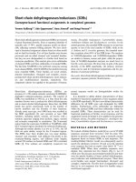
The PMMA flow deck of the flow chamber with the inlet and outlet tubings, and the gasket with a thickness of 254
μ
mFigure 1
The PMMA flow deck of the flow chamber with the inlet and outlet tubings, and the gasket with a thickness of 254
μ
m.
Journal of Nanobiotechnology 2008, 6:9 />Page 4 of 9
(page number not for citation purposes)
mL penicillin, and 100
μ
g/mL streptomycin and were
grown at 37°C with humidified 95% air/5% CO2.
For each experiment, cells were plated on a borosilicate
glass with a 0. 2 mg/cm
2
substratum of type A gelatine
(Sigma-Aldrich Corporation, MO). When HUVECs
reached 80% confluence, the borosilicate glass was
detached from the bottom of the plate and mounted in
the parallel plate flow chamber for particle-cell adhesion
analysis.
The Particles
Fluoresbrite
®
Microspheres from Polysciences were used.
These are Yellow Green fluorescent particles with an exci-
tation maximum at 441 nm and an emission maximum at
486 nm. Particles with different sizes were used namely 50
nm, 100 nm, 200 nm, 500 nm, 750 nm, and 1
μ
m, 6
μ
m, 10
μ
m.
3 Results and discussions
The lateral drifting of particulates in capillary flow has
been analyzed since the pioneering experiments of Pois-
uille in 1836 [11] who observed that RBCs do not distrib-
ute uniformly leaving a region devoid of particles in close
proximity to the walls: the cell free layer. More recently,
Segré and Silberberg [12] have showed that for small Rey-
nolds numbers a bolus of neutrally-buoyant particles
would preferentially migrate towards the walls of the tube
leading to a non uniform radial distribution with a peak
at about 0. 6 times the capillary radius. After Segré, many
authors experimentally investigated the behavior of non
neutrally buoyant rigid spheres [13-16], finding out that
the equilibrium position would depend on the relative
density of the particles to the fluid and the flow Reynolds
number. In 1994, Hogg [17] has presented a comprehen-
sive theoretical analysis for the migration of non-neutrally
buoyant spherical particles in two-dimensional shear
flows. Three different dimensionless parameters have
been introduced to describe the problem: the geometric
ratio
α
(= d/(2h)) between the particle diameter d and the
channel height h; the channel Reynolds number Re
c
(=
ρ
f
U
m
R
h
/
μ
) with
ρ
f
the density,
μ
the viscosity and U
m
the
mean flow velocity of the fluid, and the hydraulic radius
R
h
of the channel (R
h
= 2hw/(h + w)); and the buoyancy
number B = d
2
Δ
ρ
g
/(18
μ
U
m
), being Δ
ρ
the density of the
particle relative to the fluid. Different marginating behav-
The number n × 10
3
of marginating particles as a function of time during a typical flow chamber experiment for three different particle sizes (d = 50 nm; 750 nm and 10
μ
m)Figure 2
The number n × 10
3
of marginating particles as a function of time during a typical flow chamber experiment for three different
particle sizes (d = 50 nm; 750 nm and 10
μ
m).
Journal of Nanobiotechnology 2008, 6:9 />Page 5 of 9
(page number not for citation purposes)
iors have been identified depending on the values of the
combined parameters
α
2
/B and Re
c
B
2
compared to unity.
In the microcirculation, with U
m
of O (100)
μ
m/s, h and R
h
of O (100)
μ
m, and for d of O (10)
μ
m and smaller, it fol-
lows
having considered
μ
= 10
-3
Pa s, Δ
ρ
= 50 kg/m
3
and
ρ
f
= 10
3
kg/m
3
. For the flow chamber apparatus considered here
and d = 10
μ
m, it is
α
Ӎ 0. 04, Re
c
Ӎ 0. 16 and B Ӎ 0. 053
leading to
α
2
/B Ӎ 0. 03 and Re
c
B
2
Ӎ 4. 6 × 10
4
, much
smaller than unity.
3.1 Margination in Horizontal Capillaries
For
α
2
/B < 1 and Re
c
B
2
< 1, as observed in [17], the lateral
drift is mainly due to the gravitational force acting orthog-
onally to the flow direction and the drifting velocity is
only slightly different from that predicted by Stokes for
the settling of a particle in a quiescent fluid, that is to say
where Όh is the average separation distance of the particle
from the wall (see Fig. 3). Integrating (2) over an initial
separation distance ΌH
o
, and observing that when at ΌH
o
the particle would move longitudinally by the distance L
in a time Δt = L/(SΌH
o
), it follows that
where L is the length of the region of interest with surface
area A. Notice that in the case of a magnetic particle driven
towards the chamber substrate by an external magnetic
field, ΌH
o
would scale with d as in (3), being the magnetic
force F
mag
proportional to the volume of the particle, just
as the gravitation force. The separation distance ΌH
o
mul-
tiplied by A gives the volume of fluid within which are
comprised the particles candidate to sediment within the
region of interest (the sedimentation volume). If C is the
local concentration of the particles within the sedimenta-
tion volume, the number of depositing particles per unit
area A is readily given by
whereas the total volume of settling particles per unit
surface is defined as
Substituting in (4) and (5) for (3), it follows that under a
gravitational field (or magnetic field) the number of
a
m
r
rr
m
2
2
222
2
1
9
2
10
1
18
10/() ()B
U
hg
OReBRd
g
O
m
ch
f
=< = <
−−
Δ
Δ
and
v
dh
dt
g
dBU
G
m
=
〈〉
==
1
18
2
Δ
r
m
(2)
〈〉=
⎡
⎣
⎢
⎤
⎦
⎥
×H
gL
S
d
o
22
1
18
Δ
r
m
(3)
n
CV
A
CH C d
s
s
o
==〈〉∝×
(4)
V
s
VCH d Cd
so
=〈 〉× ∝ ×
p
34
6/
(5)
n
s
The margination trajectory of a spherical particle within a laminar flowFigure 3
The margination trajectory of a spherical particle within a laminar flow.
Journal of Nanobiotechnology 2008, 6:9 />Page 6 of 9
(page number not for citation purposes)
settling particles per unit area and their volume are
both proportional to the local volume concentration C of
particles and grows linearly with d ( ∝ d) and with the
fourth power of d ( ∝ d
4
), respectively.
The above 'back of the envelope' calculations, although
extremely simplified, are in decent agreement with the
experimental results obtained using the parallel plate flow
chamber apparatus. The total volume of the particles set-
tling per unit surface is shown as a function of the par-
ticle diameter in Fig. 4, ranging between 500 nm and 10
μ
m. These experiments have been performed keeping
fixed the total volume of the particles injected into the
chamber (i.e. 5. 2 × 10
7
μ
m
3
/ml) for each particle size, or
in other words, the total number of particles injected
decreases with d
-3.
In a double-logarithm diagram, the
average values of over five significant repetitions
(filled boxes) are well aligned around a straight line with
a nearly unit slope described by the relation
which gives almost the same scaling as predicted in (5),
assuming a fixed total volume of injected particles. In Fig.
5, the variation of the volume as a function of the total
number of particles injected n
tot
in the flow chamber is plot-
ted for a fixed particle size (d = 500 nm), showing a linear
increase of following the experimental relationship
which support the linear relationship between and C
as predicted in (5).
V
s
n
s
V
s
V
s
V
s
Vd R
s
==1454 6 0 976
12 2
., .
.
with
(6)
V
s
V
s
VnR
stot
=× =
−
196 10 0 981
6 0 956 2.
,.with
(7)
V
s
The volume of particles marginating per unit surface as a function of the particle diameter d ranging from 500 nm up to 10
μ
m
(fixed total volume of the injected particles V
tot
= 5.2 × 10
7
μ
m
3
and C
v
= 5.2 × 10
5
)
Figure 4
The volume of particles marginating per unit surface as a function of the particle diameter d ranging from 500 nm up to 10
μ
m (fixed total volume of the injected particles V
tot
= 5.2 × 10
7
μ
m
3
and C
v
= 5.2 × 10
5
).
0.5 0.6 0.7 1.1. 1.5 2. 3. 4. 5. 6. 7. 10.10.
d Μm
2
5
lO
3
2
5
lO
4
2
5
V Μm
3
mm
2
Fixed Total Volume
1454.6 d
1.25488
V
s
Journal of Nanobiotechnology 2008, 6:9 />Page 7 of 9
(page number not for citation purposes)
In Fig. 6, the behavior of the smaller particles is consid-
ered with a diameter ranging from 200 nm down to 50 nm.
However, differently from the previous case, shown in Fig.
4, the analysis has been performed for a fixed number of
injected particles (n
tot
= 10
8
), to limit the total amount of
particles to be used for a diameter of 50 nm. The - d
relation is different from that observed for the larger par-
ticles, and it is no more nearly linear in a double-loga-
rithm diagram being
whose scaling with d can not be predicted by just gravita-
tion or volume forces. For these small particles other
forces as colloidal forces (van der Waals, electrostatic) are
probably responsible for their margination, which arise
only with a small separation distance between the particle
and the substrate (tens to a hundred nanometers). An
ANOVA analysis has returned, for the data presented in
Fig. 4 to 6, p values much smaller than the critical value of
0. 05, being respectively p = 0. 0022, p = 0. 0035, and p =
0. 0047, thus implying a statistically significant difference
among the means.
3.2 Margination in Vertical Capillaries
For
α
2
/B and Re
c
B
2
smaller than unity, as observed in [17],
in vertical capillaries the lateral drift is modest with a
velocity scaling with , where R
p
is the particle Rey-
nolds number (R
p
=
ρ
p
U
m
d
2
/(
μ
R
ch
)). Therefore the lateral
drifting velocity would scale with d
3
rather with d
2
as in
horizontal capillaries (see eq.2), making the characteristic
size of the particles even more important. Following the
same reasonings as above for the horizontal capillaries, it
can be derived a ΌH
o
scaling with d
3/2
, and eventually a
number and a volume of settling particles per unit
area proportional to the local volume concentration C of
V
s
Vd R
s
==314 5 0 998
32 2
., .
.
with
(8)
BR
p
12/
n
s
V
s
The volume of particles marginating per unit surface as a function of the particle total number n
tot
(fixed diameter d = 500 nm)
Figure 5
The volume of particles marginating per unit surface as a function of the particle total number n
tot
(fixed diameter d = 500 nm).
10
7
10
8
810
8
n
tot
10
3
10
2
10
10
3
V Μm
3
mm
2
1.96075 10
6
n
0.95648
V
s
Journal of Nanobiotechnology 2008, 6:9 />Page 8 of 9
(page number not for citation purposes)
particles and scaling respectively with d
3/2
and d
9/2
( ∝
d
3/2
and ∝ d
9/2
).
The lateral drifting observed in vertical capillaries is again
associated with the difference in relative density between
the circulating particle and the fluid, being B, the buoy-
ancy parameter, different from zero. But more impor-
tantly, the sign of the velocity depends on the direction of
the flow: particles heavier than the fluid would drift
towards the wall for downward flows (margination) and
towards the capillary center line for upward flows (oppo-
site of margination). The opposite has been predicted and
observed to occur for particles less heavy than the fluid.
These behavior has been observed extensively in several
experiments [18].
4 Conclusion
The propensity of spherical nanoparticles to marginate
towards the vessel walls in the microcirculation has been
analyzed employing a parallel plate flow chamber. The
effect of the particle size and orientation of the capillary
with respect to external volume force fields (gravitation)
has been elucidated experimentally and supported by
simple scaling relations.
The number and total volume of particles margin-
ating per unit surface have been measured through confo-
cal fluorescent microscopy. Considering particles with a
density slightly larger than water (1050 kg/m
3
), and com-
parable with the density of liposomes and polymeric par-
ticles used in nanomedical applications, it has been
observed in horizontal channels that the lateral margina-
tion of particles with a diameter larger than 200 nm is
mainly governed by the gravitational force with and
scaling both proportionally to the volume concentra-
tion C of the particles and, respectively, to the diameter d
and the fourth power of the diameter d
4
. For smaller par-
ticles (d < 200 nm), the margination dynamics can not be
associated to gravitational forces being ∝ d
3.2
. Possi-
bly, in this case, colloidal interactions may govern particle
lateral drifting but this would already require the particle
n
s
V
s
n
s
V
s
n
s
V
s
V
s
The volume of particles marginating per unit surface as a function of the particle diameter d ranging from 50 to 200 nm (fixed
total number of the injected particles n
tot
= 10
8
and C = 10
14
m
-3
)
Figure 6
The volume of particles marginating per unit surface as a function of the particle diameter d ranging from 50 to 200 nm
(fixed total number of the injected particles n
tot
= 10
8
and C = 10
14
m
-3
).
5 6 7 8 9
lO
1
1.5 2
d Μm
lO
2
2
5
0.1
0.2
0.5
1.1.
2.
V Μm
3
mm
2
Fixed Total Number
314.47 d
3.20
V
s
Publish with BioMed Central and every
scientist can read your work free of charge
"BioMed Central will be the most significant development for
disseminating the results of biomedical research in our lifetime."
Sir Paul Nurse, Cancer Research UK
Your research papers will be:
available free of charge to the entire biomedical community
peer reviewed and published immediately upon acceptance
cited in PubMed and archived on PubMed Central
yours — you keep the copyright
Submit your manuscript here:
/>BioMedcentral
Journal of Nanobiotechnology 2008, 6:9 />Page 9 of 9
(page number not for citation purposes)
to be in sufficient close proximity of the wall, say tens up
to a hundred nanometer, in other words separation dis-
tances of the same order of magnitude of the particle size.
These results, although not exhaustive, are of interest in
the systemic delivery of nanoparticles designed to target
the vascular walls in the microcirculation. The experimen-
tal results and simple theoretical relations support the
idea of using large particles rather than small particles
with the same total volume. In fact, if the biological target
is the vascular wall and the particles are not required to
freely extravasate through the discontinuous endothe-
lium, then the larger spherical particles would more easily
sediment in horizontal capillaries and drift laterally in
vertical capillaries with downward flow. Also the larger
spherical particles would have a larger surface exposed to
the vascular cells increasing the likelihood of firm adhe-
sion once decorated with recognizing moieties [3]. The
separation between large and small particles would
depend on the relative density compared to the fluid,
however for the commonly used liposome and polymeric
particles sizes larger than 200 nm would perform better.
It should be noticed, in conclusion, that the present
results strictly apply when the interaction of the nanopar-
ticles with circulating blood cells can be disregarded,
which occurs in small capillaries and in the cell free layer
of arterioles and veins.
References
1. LaVan DA, McGuire T, Langer R: Small-scale systems for in vivo
drug delivery. Nat Biotechnol 2003, 21:1184-91.
2. Ferrari M: Cancer nanotechnology: opportunities and chal-
lenges. Nat Rev Cancer 2005, 5:161-71.
3. Decuzzi P, Ferrari M: The adhesive strength of non-spherical
particles mediated by specific interactions. Biomaterials 2006,
27(30):5307-14.
4. Decuzzi P, Lee S, Bhushan B, Ferrari M: A theoretical model for
the margination of particles within blood vessels. Ann Biomed
Eng 2005, 33:179-90.
5. Sakamoto J, Annapragada A, Decuzzi P, Ferrari M: Antibiological
barrier nanovector technology for cancer applications.
Expert Opin Drug Deliv 2007, 4(4):359-69.
6. Hashizume H, Baluk P, Morikawa S, McLean JW, Thurston G, Roberge
S, Jain RK, McDonald DM: Openings between defective
endothelial cells explain tumor vessel leakiness. Am J Pathol
2000, 156:1363-80.
7. Hobbs SK, Monsky WL, Yuan F, Roberts WG, Griffith L, Torchilin VP,
Jain RK: Regulation of transport pathways in tumor vessels:
role of tumor type and microenvironment. Proc Natl Acad Sci
USA 1998, 14(95):4607-12.
8. Neri D, Bicknell R: Tumor Vascular Targeting. Nature Cancer
Reviews 2005.
9. Goldsmith HL, Spain S: Margination of leukocytes in blood flow
through small tubes. Microvasc Res 1984, 27:204-222.
10. Lawrence MB, Kansas GS, Kunkel EJ, Ley K: Threshold levels of
fluid shear promote leukocyte adhesion through selectins
(CD62L,P,E). J Cell Biol 1997, 136(3):717-27.
11. Poiseuille JLM: "Recherches sur les Causes du Mouvement du
Sang Dans les. Vaisseaux Capillaires". Ann Sci Nat Ser 1836,
2(5):111-115.
12. Segré G, Silberberg A: Behavior of macroscopic rigid spheres in
Poiseuille flow: Part I. J Fluid Mech
1962, 14:115.
13. Matas J-P, Morris JF, Guazzelli E: Inertial migration of rigid spher-
ical particles in Poiseuille flow. J Fluid Mech 2004, 515:171-195.
14. Oliver DR: "Influence of Particle Rotation on Radial Migration
in the Poiseuflle. Flow of Suspensions". Nature 1962,
194:1269-1271.
15. Repetti RV, Leonard EF: Segre-Silberberg. annulus formation: a
possible explanation. Nature 1964, 203:1346-1350.
16. Jeffrey RC, Pearson JRA: Particle motion in laminar. vertical
tube flow. J Fluid Mech 1965, 22:721-735.
17. Hogg AJ: Inertial migration of a non-neutrally buoyant parti-
cle in a two-dimensional shear flow. J Fluid Mech 1994,
272:285-318.
18. Jeffrey RC, Pearson JRA: Particle motion in laminar vertical
tube flow. Journal of Fluid Mechanics 1965, 22:721-735.