Báo cáo khoa học: Molecular identification of monomeric aspartate racemase from Bifidobacterium bifidum pptx
Bạn đang xem bản rút gọn của tài liệu. Xem và tải ngay bản đầy đủ của tài liệu tại đây (201.63 KB, 6 trang )
Molecular identification of monomeric aspartate racemase from
Bifidobacterium bifidum
Tatsuyuki Yamashita
1
, Makoto Ashiuchi
1
, Kouhei Ohnishi
2
, Shin’ichiro Kato
2
, Shinji Nagata
1
and Haruo Misono
1,2
1
Department of Bioresources Science, Kochi University, Nankoku, Kochi, Japan;
2
Research Institute of Molecular Genetics,
Kochi University, Nankoku, Kochi, Japan
Bifidobacterium bifidum is a useful p robiotic agent exhibiting
health-promoting properties a nd contains
D
-aspartate a s an
essential component of the cross-linker m oiety i n t he pepti-
doglycan. To help understand
D
-aspartate biosynthesis in
B. bifidum NBRC 14252, aspartate racemase, which cata-
lyzes the r ac emization of
D
-and
L
-aspartate, was purified to
homogeneity and characterized. T he enzyme was a mono-
mer with a molecular mass of 27 kDa. This is the first report
showing th e presence of a m onomeric aspartate racemase.
Its e nzymologic properties, such as its lack of cofactor
requirement and susceptibility to thiol-modifying reagents
in catalysis, were similar to those of the dimeric aspartate
racemase from Streptococcus thermophilus. The monomeric
enzyme, however, showed a novel characteristic, namely,
that its thermal stability significantly increased in the pres-
ence of aspartate, especially the
D
-enantiomer. The gene
encoding the m onomeric as partate r acemase w as cloned and
overexpressed in Escherichia coli cells. The nucleotide
sequence of the aspartate racemase gene encoded a peptide
containing 241 amino acids with a calculated molecular mass
of 26 784 Da. T he recombina nt e nzyme w as purified to
homogeneity and i ts properties were almost t he same as
those of the B. bifidum enz yme.
Keywords: aspartate racemase; Bifidobacterium bifidum;
D
-aspartate; peptidoglycan; probiotic agent.
Bifidobacteria, including Bifidobacterium bifidum, have been
applied widely as probiotic agents exhibiting health-promo-
ting properties. Recent research suggested very interesting
functions of bifidobacterial peptidoglycans e.g. redu ction
of harmful bacteria and toxic compounds in the intestine,
antitumorigenic activities, and immunological enhancement
properties [1–3]. B acterial peptidogly cans contain several
kinds of
D
-amino acids [ 4] and a re thought to pro tect cells
from protease actions.
D
-Alanine and
D
-glutamate occur in
the main chains of bifidobacterial peptidoglycans [5]. The
cross-linker moieties of B. bifidum contain
D
-aspartate as
the essential component [5]. Two kinds of amino acid
racemases, alanine racemase [5] and glutamate racemase [6],
have been identified ubiquitously from bacteria, a nd it has
been assumed that the former, a pyridoxal 5¢-phosphate
(PLP)-dependent amino a cid racemase [5], is i nvolved in
D
-alanine biosynthesis and the latter, a PLP-independent
racemase [6], participates in the supply o f
D
-glutamate.
Most bacterial alanine racemases assemble in a dimer
structure [5], w hereas glutamate r acemases are m ainly
characterized as monome ric enzymes [6]. On the other
hand, aspartate racemase is found in limited organisms
[7–11], w hich e ncompass even peptidoglycan-less species,
such as archaea a nd mollusks. Recent studies showed two
distinct characteristics of aspartate racemases i n t he co-
enzyme requirement in catalysis [11,12]. Among them, the
PLP-independent aspartate racemase is considered to share
structural features and catalytic properties with the gluta-
mate racemase [12]. Nevertheless, aspartate racemases are
typically dimeric, and neither monomeric nor multimeric
aspartate racemase has been ide ntified yet.
This paper presents the first identification of
PLP-independent monomeric aspartate racemase from
B. bifidum NBRC 14252 and its enzymologic characteris-
tics, as well as cloning and overexpression of its gene in
Escherichia coli.
Materials and methods
Materials
N-tert-butyloxycarbonyl-
L
-cysteine (Boc-
L
-Cys) was pur-
chased from Novabiochem, La
¨
ufelfingen, Switzerland;
o-phthalaldehyde (OPA), from Nacalai Tesque, Kyoto,
Japan; a 4-lm Nova-Pack C18 column, from Waters,
Milford, MA, USA; a HiPrep Sephacryl S-200 column
(1.6 · 60 cm) and a HiTrap Butyl FF column (1.0 mL),
from Amersham Bioscience, Uppsala, Sweden; a Bio-Scale
Q20 column ( 20 mL) and a protein assay k it, from Bio-Rad,
Richmond, CA, USA; a TSK gel G3000SW column, from
Correspondence to M. Ashiuchi, Department of Bioresources Science,
Faculty of Agriculture, Kochi University, Nankoku, Kochi 783-8502,
Japan. Fax: +81 88 8645200, Tel.: +81 88 8645215,
E-mail:
Abbreviations:Boc-
L
-cys, N-tert-butyloxycarbonyl-
L
-cysteine;
BTP, bis-trispropane; IPTG, isopropyl thio-b-
D
-galactoside;
OPA, o-phthalaldehyde; PLP, pyridoxal 5¢-phosphate; Tes,
N-tris(hydroxymethyl)-methyl-2-aminoethansulfonic acid.
Enzymes: alanine racemase (EC 5.1.1.1); glutamate racemase
(EC 5.1.1.3); aspartate racemase (EC 5.1.1.13).
(Received 25 August 2004, revised 30 September 2004,
accepted 19 October 2004)
Eur. J. Biochem. 271, 4798–4803 (2004) Ó FEBS 2004 doi:10.1111/j.1432-1033.2004.04445.x
Tosoh, Tokyo, Japan; a PRISM kit, from PerkinElmer,
Fremont, CA, USA; restriction enzymes, T4 DNA ligase,
and i sopropyl thio-b-
D
-galactoside (IPTG), from Takara
Shuzo, Kyoto, Japan; and a plasmid pATE19, from
BioLeaders Corporation, Daejeon, Korea. All other chem-
icals were of analytical grade.
Bacteria and culture conditions
B. bifidum NBRC 14252 was cultured at 37 °Cfor48hin
GAM broth (pH 7 .1) comprising 1% peptone, 0.3% soy
peptone, 1% protease peptone, 1.35% digested serum,
0.5% yeast extract, 0.22% meat extract, 0.12% liver extract,
0.3% glucose, 0.25% KH
2
PO
4
, 0.3% NaCl, 0.5% soluble
starch, 0.03%
L
-cysteine/HCl, and 0.03% so dium thiogly-
colate (Nissui, Tokyo, Japan).
Enzyme and protein assays
The aspartate racemase activity was estimated by determin-
ation of t he antipode formed from either enantiomer of
aspartate by HPLC. The reaction mixture (200 lL) com-
posed of 0.1
M
bis-trispropane (BTP) buffer (pH 7.0),
50 m
ML
-aspartate, 4 m
M
dithiothreitol, 1 m
M
EDTA,
and enzyme was incubated at 45 °C for 10 h. The reaction
was terminated by a ddition of 50 lLof2
M
HCl. After
neutralization of the reaction mixture, it was incubated at
25 °C for 2 min with a 0.3
M
borate solution (pH 9.0)
containing 0.2% Boc-
L
-Cys and 0.2% OPA. A 2-lL aliquot
of the resulting mixture was subjected to a Shimadzu LC-10
HPLC system (Kyoto, Japan) composed of an LL-10AD
dual pump, a CBM-10 A control bo x, a n R F-10 A
spectrofluorometer, and a DGU-14 A degasser, with a
4-lm Nova-Pack C18 column (3.9 · 300 mm). Other
conditions were the same as those described by Hashimoto
et al . [13]. One unit of the enzyme was defined as the
amount of enzyme that catalyzes the formation of 1 lmol of
D
-aspartate from
L
-aspartate p er hour.
Protein concentrations were determined using a protein
assay kit with bovine serum albumin as a standard.
Enzyme purification
Harvested c ells of B. bifidum NBRC 14252 (wet weight,
104 g ) were suspended in 200 mL of a standard buffer
[10 m
M
N-tris(hydroxymethyl)-methyl-2-aminoethansulf-
onic acid (Tes) buffer (pH 6.5), 4 m
M
dithiothreitol, and
1m
M
EDTA] supplemented w ith 0.1
MD
-aspartate and
0.1 m
M
phenylmethanesulfonyl fluoride and then disrupted
by sonication on ice for 20 min. The suspension was
centrifuged at 12 000 g for 30 min, and the resulting
supernatant was dialyzed against the standard buffer
(pH 6 .5) and used as the cell extract. All the purification
procedures were performed at 4 °C, except heat treatment.
The cell extract (595 mL) was subjected to ammonium
sulfate fractionation. The 25–50% saturation fraction was
dissolved in the standard buffer (pH 6.5) and dialyzed
overnight against the same buffer. The enzyme s olution
was kept at 60 °C for 30 min in the presence of 0.1
M
D
-aspartate, and the formed p recipitate was r emoved by
centrifugation at 12 000 g for 30 min. The supernatant
(179 mL) was subjected to an AKTA prime FPLC system
(Amersham Bioscience, Uppsala, Sweden) equipped with a
Bio-Scale Q20 column (20 mL) that had been equilibrated
with the standard buffer (pH 6.5). After the column was
washed with the same buffer and a buffer containing 0.15
M
NaCl, the enzyme was eluted with t he buffer containing
0.3
M
NaCl. The active fractions were combined, d ialyzed
against the standard buffer (pH 6.5) overnight, and con-
centrated by ultrafiltration with an Amicon PM-10 mem-
brane. The enzyme solution was dialyzed against the
standard buffer (pH 6.5) containing ammonium sulfate
(15% saturation) and subjected to the FPLC system
equipped with a HiTrap Butyl FF column (1.0 mL) that
had b een equilibrated with the standard buffer ( pH 6.5)
containing ammonium sulfate (15% saturation). After the
column had been washed with the same buffer, the enzyme
was eluted with a linear g radient of ammonium sulfate
(15% to 0% saturation) in the buffer. T he active f ractions
were combined, dialyzed against the standard buffer
(pH 6 .5) overnight, and concentrated by ultrafiltration with
an Amicon PM-10 membrane. NaCl was a dded to the
enzyme solution (final concentration, 0.15
M
NaCl), and the
enzyme solution (2.2 mL) was subjected to the FPLC
system equipped with a HiPrep S ephacryl S-200 c olumn
(1.6 · 60 cm) that had been equilibrated with the standard
buffer (pH 6.5) containing 0.15
M
NaCl. The column was
developed at t he flow rate of 1.0 mLÆmin
)1
with the
standard buffer (pH 6.5) containing 0.15
M
NaCl. The
active fractions were combined, dialyzed against the stand-
ard buffer overnight, and concen trated by ultrafiltration
with an Amicon PM-10 membrane.
Electrophoresis
SDS/PAGE was carried out with 12.5% polyacrylamide by
the method of Laemmli [14].
Molecular mass determination
The molecular mass was determined by HPLC on a TSK gel
G300SW column (0.75 · 60 cm) at a flow rate of 0.7 mLÆ
min
)1
with the standard buffer ( pH 6.5) containing 50 m
M
D
-aspartate a nd 0.15
M
NaCl. A calibration curve w as
made with the following proteins: glutamate dehydrogenase
(290 kDa), lactate dehydrogenase (142 k Da), enolase
(67.0 kDa), adenylate kinase (32.0 kDa), and cytochrome c
(12.4 k Da). The molecular mass of the subunit was estimated
by SDS/PAGE. The following marker proteins (Amersham
Bioscience, Uppsala, Sweden) were used: rabbit muscle
phosphorylase b (97 kDa), bovin e serum albumin (66 kDa),
ovalbumin (45 kDa), carbonic anhydrase (30 kDa), trypsin
inhibitor (20.1 k Da), and a-lactalbumin (14.4 k Da).
Isolation of peptides obtained by lysyl endopeptidase-
digestion of the enzyme
The purified enzyme (1 nmol) was dialyzed against water
and lyophilized. The protein was dissolved in 20 lLof8
M
urea and incubated at 37 °C for 1 h. To the solution, 60 lL
of 0.2
M
Tris/HCl buffer (pH 9.0) and 5 pmol of lysyl
endopeptidase were added, and the m ixture was incubated at
37 °C for 12 h. The peptides were separated on a Shimadzu
HPLC system with a YMC-packed C4 colume (YMC,
Ó FEBS 2004 Bifidobacterial aspartate racemase (Eur. J. Biochem. 271) 4799
Kyoto, Japan) using a solvent system of 0.1% trifluoroacetic
acid and acetonitrile containing 0.7% trifluoroacetic acid. A
90-min linear gradie nt from 5 to 50% acetonitrile wa s used
to elute peptides at a flow rate of 1.0 mLÆmin
)1
.The
absorbance at 210 nm of the effluent was monitored
continuously. The peptides were isolated and lyophilized.
Amino acid sequence analysis
The N-terminal amino acid sequence of the enzyme and the
sequences of the isolated peptides were analyzed with an
Applied Biosystems (Forster, CA, USA) model 4 92-protein
sequencer linked to a phenylthiohydantoin d erivative ana-
lyzer.
Genetic manipulation
The chromosomal DNA of B. bifidum was prepared by the
method of Saito & Miura [15]. Two mixed primers were
designed from the N-terminal amino acid sequence of the
enzyme and a conserved region between the aspartate
racemase genes from Streptococcus thermophilus [16] and
Defulfurococcus strain SY [8]: the sense primer [5¢-GG(A,
T,G,C)GG(A,T,G,C)ATGGG(A,T,G,C)AC(A,T,G,C)(C,
T)T-3¢] and the a ntisense primer [5¢-A(A,G)TA(A,G)TG
(A,T,G,C)GC(A,T,G,C)GT(A,G)TT(A,G)CA-3¢]. PCR
was performed with ExTaq DNA polymerase (Takara
Shuzo, Kyoto, Japan). The nucleotide sequence of the
amplified DNA fragment (244 bp) was determined by means
of the PRISM kit with an Applied Biosystems 373A DNA
sequencer. Next, the inverse PCR of the B. bifidum chromo-
somal DNA was carried out as follows. The chromosomal
DNA was digested with the restriction enzyme SalI, and the
digested fragments were in cubated with T4 DNA ligase to
allow self-circulation (or self-ligation). Two single primers
were further d esigned from t he determined nucleotide
sequence of t he amplified DNA fragment: ASPR1 (5¢-TCCC
GATAATCGCCGACGACATCG-3¢)andASPR2(5¢-CG
GTTGACCAACCGGATATAGCTT-3¢). PCR was con-
ducted using the self-circulated frag ments (as a template
DNA) and the two primers, ASPR1 and ASPR2. The
nucleotide sequence of the 1-kb region containing a probable
structural gene of the enzyme was determined.
To establish the overproduction of the enzyme, we first
designed two single primers from the nucleotide sequences
at both t he immediate u p- and downstream e nds of the
structural gene of the enzyme: the sense primer AS-N
(5¢-CATG
CCATGGGACGACCATTTTTTGCG-3¢), in
which an NcoI site (underlined) is incorporated, and the
antisence primer AS-C (5¢-CCC
AAGCTTCTAGCGGC
GGATGGCCTTGGC-3¢), in which a HindIII site (under-
lined) i s included. The amplified fragment (726 bp) con-
taining the enzyme gene was digested with both restriction
enzymes NcoIandHindIII and ligated into the NcoI-
HindIII site of pATE19. The constructed plasmid was
named pBASPR. The sequence of the enzyme gene in
pBASPR was verified in both directions in the same way as
described above. Owing to t he re-cloning of the enzyme
gene into the Nco I-HindIII site of pATE19, the second
amino acid of the enzyme was changed from arginine to
glycine. The plasmid pBASPR was introduced into E. coli
cells. The E. coli JM109/pPASPR clone constructed was
used as the enzyme overproducer. The nucleotide sequence
data will appear in the DDBJ/EMBL/GenBank nucleotide
sequence databases under the accession number A B179841.
Isolation of recombinant aspartate racemase
Cells of the E. coli clone harboring pBASPR, an overpro-
ducer o f the enzyme, were inoculated into 1 L of Luria b roth
[5] containing ampicillin (50 lgÆmL
)1
)andIPTG(2m
M
)to
induce enzyme production. The c ulture was carried out at
37 °C for 16 h. Cells (5 g, wet weight) were suspended in
10 mL of the standard buffer (pH 6.5) supplemented with
0.1 m
M
phenylmethansulfonyl fluoride and then disrupted
by sonication at 4 °C. The cell debris was removed by
centrifugation at 12 000 g for 30 mins, and the supernatant
was dialyzed against the standard buffer (pH 6 .5) at 4 °C
overnight. The enzyme solution ( 19 mL) was used as the cell
extract. The recombinant enzyme was isolated with the
AKTA prime FPLC system with Bio-Scale Q20 anion-
exchange, HiTrap B utyl FF, and HiPrep Sephacryl S-200 gel
filtration columns, according to the procedures for the
enzyme purification from B. b ifidum NBRC 14252. The
purified e nzyme was c oncentrated to 10 mgÆmL
)1
and
stored at 4 °C. The N-terminal amino acid sequence of the
recombinant enzyme was verified with the above protein
sequencer.
Results
Purification of aspartate racemase from
B. bifidum
The aspartate racemase was purified to homogeneity from
cell extracts of B. bifidum NBRC 14252. A summary of the
purification is presented in Table 1. The enzyme was
purified about 1770-fold from the crude extract with a
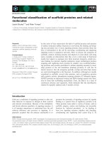
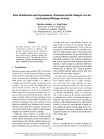
2.4% yield. The purified enzyme showed a single b and o n
SDS/PAGE (Fig. 1A).
Molecular mass and N-terminal amino acid sequence
The molecular m ass was estimated t o be 27 kDa by gel
filtration on a TSK gel G3000SW c olumn. The molecular
mass of the subunit was calculated to be 30 kDa by SDS/
PAGE (Fig. 1A). These r esults indicate t hat the enzyme
is a monomer. The 27 kDa fraction from the TSK gel
G3000SW column showed the e nzyme a ctivity, and the
Table 1. Purification of aspartate racemase from B. bifidum NBRC
14252.
Step
Total
protein
(mg)
Total
activity
(units)
Specific
activity
(units/mg)
Yield
(%)
Cell extract 4390 708 0.161 100
Ammonium
sulphate (25–45%)
2280 432 0.19 61
Heat treatment 194 243 1.25 34.3
Q20 38.4 193 5.03 27.3
Butyl FF 1.11 39.6 35.7 5.59
Sephacryl S200 0.0593 16.9 285 2.38
4800 T. Yamashita et al.(Eur. J. Biochem. 271) Ó FEBS 2004
gel filtration pattern was not changed with or without
50 m
MD
-aspartate in the elution buffer. Thus, the active
component is a monomer. The N-terminal amino acid
sequence of the enzyme was determined to be MRRP-
FFAVLGGMGTLATSYI. The amino a cid sequence o f
the internal peptide, which was isolated from the lysyl
endopeptidase-digest of the enzyme, was ERFHRVGFL-
GTMGSRASGVYRQAVEEAGYTFV.
pH and thermal stabilities
The enzyme was most stable in the pH range of 6.0–7.0
when kept at 30 °Cfor30minin10m
M
BTP buffers
(pH 5.5–9.0) containing 4 m
M
dithiothreitol and 1 m
M
EDTA.
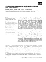
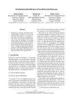
The enzyme was substantially stable up to 30 °Cin0.1
M
Tes buffer (pH 6.5) containing 4 m
M
dithiothreitol and
1m
M
EDTA (Fig. 2A). The coexistence of aspartate ,
however, significantly i ncreased its t hermal stability. The
enzyme was stable up to 60 °C in the presence of 40 m
M
D
-aspartate (Fig. 2 A). We further examined the effects of
the c oncen trations of
L
-and
D
-aspartate on th e e nz yme
stability and found that
D
-aspartate is more effective in the
stabilization than
L
-aspartate (Fig . 2 B).
Under the conditions described above, the enzyme
showed the maximum activity at pH 7.0–7.5 and 45 °C.
Cofactors
The enzyme did not require PLP as a coenzyme and w as not
inhibited by 10 m
M
hydroxylamine and 1 m
M
phenylhydr-
azine. The e nzyme was not affected by 10 m
M
EDTA and
1m
M
FAD, NAD, NADP, ATP, MgCl
2
,andMnCl
2
.
Thus, the enzyme requires no cofactor. The enzyme was
inactivated completely by incubation with 0.1
M
p-chloro-
mercuribenzoate, 0.1
M
HgCl
2
,1m
M
N-ethylmaleimide,
and 1 m
M
5,5¢-dith iobis(2-nitrobenzoate) in 0.1
M
BTP
buffer (pH 7.0) at 30 °C for 30 min. This fact suggests the
essential nature of cysteinyl residue(s) in catalysis.
Substrate specificity
The enzyme exclusively acts on
L
-and
D
-aspartate. Other
amino acids, including both enantiomers of glutamate,
asparagines, glutamine, alanine, serine, lysine, and arginine,
were inactive as substrates. The enzyme was not inhibited
by these nonsubstrate amino acids (10 m
M
), N-methyl-
DL
-aspartate (10 m
M
), and
DL
-threo-b-methylaspartic acid
(10 m
M
). a-Methyl-
DL
-aspartic acid (10 m
M
) slightly inhib-
itedtheenzyme(12%).
Kinetics
The ap parent velocity of the enzymatic aspartate racemiza-
tion was measured against various concentrations of both
enantiomers of the amino acid. As shown in T able 2, the K
m
AB
Fig. 1. SDS/PAGE of aspartate racemase from B. bifidum (A) and the
recombinant enzyme (B). (A) L ane M, the molecular marker proteins;
and lane E, the purified aspartate racemase from B. bifidum (1 lgof
protein). (B) Lane M, the molecular marker proteins; lane 1, the cell
extract of E. coli JM109 (10 lg); lane 2, t he cell extract of the E. coli
JM109/pBASPR c lone (10 lg); and lane 3, the enzyme purified from
E. coli JM109/pBASPR clone cells (5 lg).
Fig. 2. Effects of
D
-aspartate on the enzyme stability. (A) The enzyme was kept at the indicated tem pera ture for 30 min in a test so lutio n [0.1
M
Tes
buffer (pH 6.5) containing 4 m
M
dithiothreitol and 1 m
M
EDTA] in the absence (d) or th e presence o f 40 m
MD
-aspartate ( s). (B) The en zyme was
incubated at 60 °C for 30 min in the test solution with various concentrations of
L
- (white bars) or
D
-aspartate (black bars). The resulting enzyme
fractions that w ere in cubated wi th the
D
-amino acid were s ubjected t o the limit ed filtration w ith Microcon YM -1 0 a nd their enzyme activities we re
assayed according to the methods described in Materials and methods.
Ó FEBS 2004 Bifidobacterial aspartate racemase (Eur. J. Biochem. 271) 4801
and V
max
values for
L
-aspartate were about 14.3 and 13.9
times higher than those for the
D
-enantiomer. However, the
K-value of the reaction was nearly one.
Gene cloning and sequencing
As described above, the gene encoding the monomeric
aspartate racemase w as cloned and sequenced. T he gene
encodes a protein c onsisting of 241 amino a cid r esidues
(Fig. 3 ). The predicted sequence of the first 20 amino acids
was identical with that of the enzyme purified from
B. bifidum . The predicted molecular mass (26 784 Da) was
in good agreement with that of the enzyme isolated from
B. bifidum .
Overproduction of the recombinant aspartate racemase
and its properties
In this study, we s ucceeded in constructing the overproducer
of the monomeric aspartate racemase o f B. bifid um,namely,
E. coli JM109/pBASPR. As shown in F ig. 1B, the r ecom-
binant enzyme was p roduced abundan tly in the E. coli clone
and was found primarily as a s oluble e nzyme. A crude
extract of the recombinant cells (15.1 unitsÆmg
)1
) had about
94-fold higher enzyme activity than that of B. bifidum
NBRC 14252 (0.161 unitsÆmg
)1
). The recombinant enzyme
was puri fied to homogeneity with a 20% yield, without
ammonium sulfate fractionation and heat treatment as
described above. The molecular mass of the recombinant
enzyme was e stimated to be 27 kDa in an intact form ( by gel
filtration o n a TSK gel G3000SW column) and 3 0 kDa
under denatured conditions (by SDS/PAGE analysis)
(Fig. 1 B). The spectrophotometric analysis of the recom-
binant enzyme r evealed an absorption maximum at 278 nm,
and no absorption peak was detected in the region from 300
to 500 nm. The enzymological and kinetic properties of the
recombinant enzyme were almost the same as those of the
enzyme from B. bifidum NBRC 14252.
Discussion
The PLP-independent aspartate racemase has only b een
characterized from the lactic a cid bacterium, S. thermophi-
lus [7,12]. In this study, for the fi rst time, we succeeded in
identifying the aspartate racemase from B. bifidum .Enzy-
mological properties of aspartate racemase purified from
B. bifidum NBRC 14252, such as cofactor independency
and susceptibility to thiol-modifying reagents, are similar to
those of aspartate racemase from S. thermophilus [7,12]. The
B. bifidum enzyme, h owever, is a monomer, w hereas the
enzymes from S. thermophilus [7,12] and Pyrococcus hori-
koshii [18] are dimers.
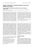
The predicted amino acid sequence of the enzyme from
B. bifidum was similar to tho se of PLP-ind ependent, dimeric
aspartate racemases studied so far [8,17,18], as shown in
Fig. 3. The similarity scores to the racemases from the lactic
acid bacterium S. thermophilus [17], from the sulfur-
dependent hyperthermophilic archaeon Desulfurococcus
strain SY [8], and from the hyperthermophilic archaeon
Pyrococcus horikoshii OT3 [18] were 45, 31, and 32%,
respectively. The whole geno me sequence o f Bifidobacterium
longum NCC2705 was published recently [16]; t his bacter-
ium has a taxonomically close relation with B. bifidum.
Table 2. Kinetic parameters of t he m onomeric as partate ra cemase.
Activity was measured at 30 °C for 10 h.
Parameter Value
L
-Aspartate
K
m
(m
M
) 13.4
V
max
(lmolÆh
)1
Æmg
)1
) 200
V
max
/K
m
14.9
D
-Aspartate
K
m
(m
M
) 0.94
V
max
(lmolÆh
)1
Æmg
)1
) 14.4
V
max
/K
m
15.3
K
eq
(
L
/
D
) 0.974
Fig. 3. Linear alignment of amino acid se-
quences of PLP-independent aspartate race-
mases. Bb, the monomeric aspartate racemase
from B. bifidum; St, the dimeric aspartate
racemase from S. thermophilus [17]; D s, the
dimeric aspartate racemase from Desulfuro-
coccus [8]; and Ph, the dimeric aspartate rac-
emase from P. horikoshii [18]. Asterisks
indicate identical residues among the four se-
quences. Two highly conserved regions that
encompass the catalytic cysteinyl residue are
indicated by bold underlines.
4802 T. Yamashita et al.(Eur. J. Biochem. 271) Ó FEBS 2004
However, unlike B. bifidum, B. longum does not contain
D
-aspartate as the essential component of peptidoglycans
[4], and no orthologous enzyme protein with the PLP-
independent type of aspartate racemase can be found in
B. longum NCC2705. In general, enzymatic aspartate
racemization is thought to proceed via a Ôtwo-baseÕ mech-
anism involving the strictly conserved cysteinyl residue(s)
[12,18]. Yamauchi et al. [12] had concluded that the
dimerization of aspartate racemase is substantial in catalysis
because its composite a ctive site requires a n identical
cysteinyl residue from each subunit as the catalytic acid/
base pair. However, Liu et al.[18]recentlyfoundthetwo
essential cysteinyl residues in each subunit of the dimeric
aspartate racemase and reported that the active sites in the
same dimer are independent of each o ther. Our finding of
the monomeric a spartate racemase strongly supports the
latter hypothesis. Although the bifidobacterial enzyme
contains f our cysteinyl r esidues (Cys85, Cys195, Cys197 ,
and Cys204), its two catalytic residues (Cys85 and Cys204)
and the surrounding regions (CN TAH and G CTE) are
highly conserved (Fig. 3).
Recent research s uggests that the dimerization of
aspartate racemase, which a ssembles through t he disulfide
bonds, may be involved in an increase in its solubility
and thermal stability [18]. On the other hand, we re cently
observed another s trategy for elevating t hermal stability
during the study of the monomeric, bifidobacterial
enzyme (Fig. 2). Kinetic analysis demonstrated that the
monomeric enzyme had a comparatively high affinity for
D
-aspartate (Table 2), suggesting t hat the enzyme bound
tightly with the
D
-amino acid so as to form a confor-
mation exhibiting high stability.
The detailed analysis of the crystal structure of the
dimeric a spartate racemase of P. horikoshii proved that its
active sites are arranged in a pseudo-mirror symmetry [18].
This characteristic of the enzyme is probably c oncerned w ith
the fact that dimeric aspartate racemases generally reveal
little difference in the kinetic parameters for
D
-and
L
-aspa-
rate [7,8,12]. I n contrast, the d ata in T able 2 point to a
significant difference in the kinetic parameters of the
monomeric enzyme for t he different e nantiomers of the
substrate. Therefore, our observations may contribute to
the first identification of nonmirror-symmetric aspartate
racemase. Studies on the tertiary structure of the monomeric
aspartate racemase are now being conducted in order to
understand the general principle in molecular recognition
mechanisms of the mirror-symmetric amino acid enantio-
mers in amino acid racemases.
References
1. Zhang, X.B. & Ohta, Y. (1991) Binding of mutagens by fractions
of the cell wall skeleton of lactic acid bacteria on mutagens.
J. Dairy Sci. 74, 1477–1481.
2. Se kine, K., Ohta, J., Onishi, M., Tatsuki, T., Shimokawa, Y.,
Toida, T., Kawashima, T. & Hashimoto, Y. (1995) Analysis of
antitumor p roperties of effector cells stimulated with a cell wall
preparation (WPG) of Bifidobacterium infantis. Biol. Pharm. Bull.
18, 148–153.
3. Sasaki, T., Samegai, T. & Na mioka, S. (1996) Phagocytosis of
splenetic neutrophils of mice enhanced by orally administered
peptidoglycan from Bifidobacterium thermophilum. J. Vet. Med.
Sci. 58, 85–86.
4. Schleifer, K.H. & K andler, O. (1972) Peptidogl ycan types of
bacterial cell walls and their taxonomic implic ations. Bacteriol.
Rev. 36, 407–477.
5. Yamashita,T.,Ashiuchi,M.,Ohnishi,K.,Kato,S.,Nagata,S.&
Misono, H. (2003) Molecular characterization of alanine racemase
from Bifidobacterium bifidum. J. Mol. Catal. B: Enzym. 23, 213–
222.
6. Ashiuchi, M., Soda, K. & Misono, H. (1999) Characterization of
yrpC gene product of Bacillus subtilis IFO 3336 as glutamate
racemase isozyme. Bios ci. Biotechnol. Biochem. 63, 792–798.
7. Okada,H.,Yohda,M.,Giga-Hama,Y.,Ueno,Y.,Ohdo,S.&
Kumagai, H. (1991) Distribution and purification of aspartate
racemase in lactic acid bacteria. Biochim. Biophys. Acta 1078,
377–382.
8. Yohda,M.,Endo,I.,Abe,Y.,Ohta,T.,Iida,T.,Maruyama,T.&
Kagawa, Y. (1996) Gene for aspartate racemase from the sulfur-
dependent hyperthermophilic archacum, Desulfurococcus strain
SY. J. Biol. Chem. 271, 22017–22021.
9.Matsumoto,M.,Honmma,H.,Long,Z.,Imai,K.,Ida,T.,
Maruyama,T.,Aikawa,Y.,Endo,I.&Yohda,M.(1999)
Occurrence of free
D
-amino acids and aspartate racemases in
hyperthermophilic archaea. J. Bacteriol. 181, 6560–6563.
10. Long, Z., Lee, J A., Okamoto, T., Sekine, M., Nimura, N., Imai,
K., Yohda, M., Maruyama, T., Sumi, M., Kamo, N., Yamagishi,
A., Oshima, T. & Homma, H. (2001) Occurrence of
D
-amino acids
and a pyridoxal 5¢-phosphate-depende nt aspartate racem ase in the
acidothermophilic archaeon, Thermoplasma acidophilum. Bio-
chem. Biophys. Res. Commun. 181, 317–321.
11. Shibata, K., Watanabe, T., Yoshikawa, H., Abe, K., Takahashi,
S., Kera, Y. & Yamada, R. (2003) Purification and characteriza-
tion of aspartate racemase from the bivalve mollusk Scapharaca
broughtonii. Comp.Biochem.Physiol.PartB134, 307–314.
12. Yamauchi,T.,Choi,S Y.,Okada,H.,Yohda,M.,Kumagai,H.,
Esaki, N. & Soda, K. (1992) Properties of aspartate racemase, a
pyridoxal 5¢-p hosphate-inde penden t a mino acid racemase. J. Biol.
Chem. 267, 18361–18364.
13. Hashimoto, A., Nishikawa, T., Oka, T., Takahashi, K. & Haya-
shi, T. (1992) D etermination of free am ino a cid enantiomers in rat
brain and serum by high-performance liquid chromatography
after d er iv atiz at io n w it h N-tert-butyloxycarbonyl-
L
-cysteine and
o-phthaldiald ehyd e. J. Chromatogr. 582, 41–48.
14. Laem mli U .K. ( 1970) C leavage of structural proteins during
the assembly of the head of bacteriophage T4. Nature 227,
680–685.
15. Saito, M. & Miura, K. (1963) P reparation o f tran sforming d eoxy-
ribonucleic acid by phenol treatment. Biochim. Biophys. Acta 72,
619–629.
16. Sc hell, M.A., Karmirantzou, M., Snel, B., Vilanova, D., Berger,
B., Pessi, G., Zwahlen, M.C., Desiere, F., Bork, P ., Delley, M.,
Pridmore, D. & Arigon i, F. (2002) The g en ome sequence of
Bifidobacterium longum reflects its adaptation to the human
gastrointestinal tract. Pr oc. Natl Acad. Sci . USA 99, 1 4422–
14427.
17. Yohda, M., Okada, H. & Kumagai, H. (1991) Molecular cloning
and n ucleot ide sequencing of the aspartate racemase gene f rom
lactic acid bacteria Streptococcus thermophilus. Biochim. Biophys.
Acta 1089, 234–240.
18. Liu,L.,Iwata,K.,Kita,A.,Kawarabayasi,Y.,Yohda,M.&
Miki, K. (2002) Crystal structure of aspartate racemase from
Pyrococcus h o rikoshii OT3 and its implications for molecular
mechanism o f P LP-indep ende nt ra cemiza tion. J. Mol. Biol. 319,
479–489.
Ó FEBS 2004 Bifidobacterial aspartate racemase (Eur. J. Biochem. 271) 4803