báo cáo khoa học: " Paget’s disease of the breast in a male with lymphomatoid papulosis: a case report" doc
Bạn đang xem bản rút gọn của tài liệu. Xem và tải ngay bản đầy đủ của tài liệu tại đây (280.94 KB, 3 trang )
CAS E REP O R T Open Access
Paget’s disease of the breast in a male with
lymphomatoid papulosis: a case report
Dina Fouad
Abstract
Introduction: Paget’s disease is an eczematous skin change of the nipple that is usually associated with an
underlying breast mali gnancy. Male breast cancer represents only 1-3% of all breast malignancies and Paget’s
disease remains very rare.
Case presentation: We present the case of a 67-year-old Caucasian man with lymphomatoid papulosis who was
diagnosed with Paget’s disease of the nipple and who was treated successfully with surgery alone. We discuss the
presentation, investigations, management and pathogenesis of Paget’s disease of the nipple.
Conclusion: The case highlights the need to be vigilant when new skin lesions arise in the context of an
underlying chronic skin disorder.
Introduction
Paget’s disease is an eczematous skin change of the nip-
ple that is usually associated with an underlying breast
malignancy [1]. It may present with erythema, scaling,
ulceration, bleeding or a painful nipple [2,3]. Male
breast cancer accounts for less than 1% of all breast can-
cer with Paget’s disease remaining very rare. Paget ’sdis-
ease of the nipple may be associated with an underlying
invasive cancer, a non-invasive cancer ductal carcinoma
in situ or no underlying cancer. Prognosis is dependent
upon the status of invasion and treatment is tailored
accordingly. Approximately 90% of patients presenting
withapalpablemassorwhohaveavisiblemasson
mammography will have underlying invasive disease.
Notably, invasive cancer can occur with Paget’sdisease
in 38% of patients with no underlying mass [3,4].
Case Presentation
The patient is a 67-year-old Caucasian man who pre-
sented to the Breast Clinic in August 2008 with a six-
month history of a painful right nipple and one episode
of clear nipple discharge. His problem had not resolved
with use of a topical ointment prescribed by his general
practitioner and he was admitted to the Breast ward of
our hospital in September 2008 for further investigations.
The patient’s past medical history includes 30 years of
lymphomatoid papulosis, a chronic papulonodular der-
matological condition, which has been controlled with
long-term methotrexate treatment and folic acid supple-
mentation. There was no report that the control of this
had been parti cularly poor recently, however the patient
had several previous recorded flare ups (1992, 2000,
2004, 2006) requiring clinic appointments and adjust-
ment of medication (mainly methotrexate).The patient
has also suffered from essential hypertension, atrial
fibrillation and atrial flutter since 1990 for which he
takes bendrofluamethiazide and digoxin respectively. In
addition, the patient was diagno sed with mixed cellular-
ity Hodgkin’s lymphoma nine years ago (1999) and suc-
cessfully treated with six cycles of combination
chemotherapy (ABVD: doxorubic in, bleomycin, vincris-
tine and dacarbazine), being in remission to date. The
lymphoma was discovered on palpation of two left sided
inguinal nodes, one right sided inguinal node, palpable
lumps in the left upper thigh, left lower quadrant of the
abdomen and the right hypochondrium. A computed
tom ography (CT) scan revealed retroperitoneal lympha-
denopathy, bilateral inguinal lymphadenoapathy and
nodes present in the both iliac chains. The patient
received no radiotherapy for this disease or for a ny
other reason. Moreover, the patient has been extensively
investigated for ongoing neurological symptoms that
include paraesthesia of hands and left foot and some
Correspondence:
Aberdeen Royal Infirmary, University of Aberdeen, Scotland
Fouad Journal of Medical Case Reports 2011, 5:43
/>JOURNAL OF MEDICAL
CASE REPORTS
© 2011 Fouad; licensee BioMed Central Ltd. This is an Open Access article distributed under the terms of the Creative Commons
Attribution License ( w hich permits unrestricted use, distribution, and reproduction in
any medium, provided the original work is properly cited.
gait imbalance but the aetiology remains unexplained to
date. The only positive family history is of a sister who
died aged 68 from an unknown cancer.
On examination, the right nipple appeared inflamed,
mildly erythematous and thickened with tenderness on
palpation. The erythema, inflammation and thi ckening
did not extend further than the nipple-areolar region.
There was no obvious nipple inversion, masses, ulcera-
tion or active nipple discharge and no axilla ry or supra-
clavicular lymphadenopathy were palpa ble. Notably,
faded scattered, pale pink, papules were visible across
the upper chest, upper back and lower abdomen.
The patient had a mammogram, which was normal,
and he proceeded to have a punch biopsy. The result of
this confirmed Paget’s disease of nipple and the patient
was scheduled for a right mastectomy and sentinel node
biopsy.
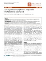
The mastectomy was uneventful and he recovered well
post-operatively (Figure 1). Histopathology confirmed
Paget’sdiseaseoftherightnipplewithnoevidenceof
underlying invasive ductal carcinoma, ductal carcinoma
in situ of the breast tissue or lymph node invasion.
Discussion
Paget’s disease is an eczematous skin change of the nip-
ple that is usually associated with an underlying breast
malignancy [1]. It may present with erythema, scaling,
ulceration, bleeding or a painful nipple [2,3]. The condi-
tion was first described in 1874 by the surgeon, Sir
James Paget, who noted that the chronic eczematous
rash of the nipple preceded an underlying intraductal
carcinoma [1].
Male breast cancer accounts for less than 1% of all
breast cancer and Paget’s disease represents 1-3% of all
breast malignancies, havi ng a higher incide nce in males
(5%) than females (1-4%) [3,4]. Paget’ s disease may pre-
sent concomitantly with an underlying invasive
carcinoma, ductal carcinoma in situ or with no underly-
ing breast cancer. Forty six percent of Paget’scasespre-
sent without a mass and of these, underlying invasive
breast cancer is usually found in only 38% with ductal
carcinoma in situ being found in the majority [4,5].
The patient had no obvious risk factors for breast can-
cer such as testicular abnormalities, infertility, obesity,
cirrhosis or Klinefelter’s syndrome nor was he known to
be positive for any BRCA2 mutations [4]. However, the
patient may have been at increased risk of malignancy
due to long term methotrexate treatment. methotrexate
has anti-folate effects and studies have shown there to
be an increased risk of malignancy in those deficient of
folic acid [6].
Clinical examination of the breast is usually followed
by imaging, either mammography or ultrasound. Ima-
ging may show subareolar microcalcifications, architec-
tural distortion or nipple changes such as thickening [7].
Imaging is followed by fine needle aspiration cytology or
punch biopsy. Histology may reveal hyperkeratosis, para-
keratosis or acanthosis of the epidermis and infiltration
with the classical Paget cell that is large, ovoid, has pale
staining cytoplasm and hyperchromic nuclei [1,2].
The pathogenesis of Paget’s disease is still a subject of
debate with two main hypotheses. The epidermotropic
hypothesis proposes that Paget’s cells originate from
ductal epithel ium, from where they migrate towards the
epidermis. This hypothesis is supported by the associa-
tion between Paget’s and an underlying breast carci-
nomainthemajorityofpatients.Thesecond
hypothesis, the intraepidermal transformation theory,
considers the presence of malignant keratinocytes that
originate from the areolar epidermis. Our case supports
this origin since there was no underlying carcinoma
[8,9].
Treatment is usually a mastectomy plus axillary node
sampling or clearance. Adju vant treatment may be con-
sidered depending on nodal and receptor status [3].
Breast conservation surgery with radiotherapy, or radio-
therapy alone, are not usually considered due to high
recurrence rates [8]. However, studies have shown
breast conserving surgery to be a feasible and safe
option [10-12]. The prognosis of Paget’s depends on the
presence of an invasive cancer and axillary lymph node
spread.Thiscaseisstage0asthereisnounderlying
breast malignancy or lymph node spread and the five-
year survival is 92-94% [9].
Several differential diagnoses should be considered
when Paget’s disease is suspected including malignant
melanoma, pagetoid dyskeratosis, Bowen’ s disease and
inflammatory skin conditions of the nipple e.g. sebor-
rhoeic dermatitis, contact dermatitis, post-rad iation der-
matitis, eczema and psoriasis [5]. This patient has
lymphomatoid papulosis, a condition in which groups of
Figure 1 Patient two weeks post right-sided mastectomy for
Paget’s disease. Medical illustration, University of Aberdeen.
Fouad Journal of Medical Case Reports 2011, 5:43
/>Page 2 of 3
pruritic papules at different stages of development
recurrently arise mainly on the trunk and limbs. It is
conceivable that the papulosis may have masked his
Paget’s nipple lesion and delayed its diagnosis. More-
over, research has shown that lymphomatoid papulosis
and Hodgkin ’ s disease along with cutaneous T-cell lym-
phoma are all connected, being derived from the same
T-cell clone [13]. Cases have been reported of patients
developing lymphomatoid papulosis, followed by Hodg-
kin’s disease and lastly developing cutaneous T-cell lym-
phoma [9]. Therefore this man may be at high risk for
cutaneous T-cell lymphoma, which can present as
erythematous patches resembling eczema. It is essential
that the patient is monitored closely.
Conclusion
The case highlights the need to be vigilant when new
skin lesions present in the context of an underlying
chronic skin disorder.
Consent
Written informed consent was obtained from the patient
for publication of this case report and accompanying
images. A copy of the written consent is available for
review by the journal’s Editor-in-Chief.
Acknowledgements
Professor Emad El-Omar. Professor of Gastroenterology, University of
Aberdeen.
Authors’ contributions
DF performed the literature search, gathered and analysed the relevant test
results and wrote the report. EEO reviewed the manuscript. The author
approved the final manuscript prior to submission.
Competing interests
The authors declare that they have no competing interests.
Received: 28 February 2010 Accepted: 28 January 2011
Published: 28 January 2011
References
1. Serour F, Birkenfeld S, Amsterdam E, Treshchan O, Krispin M: Paget’s
disease of the male breast. Cancer 1988, 62:601-605.
2. Harris JR, Hellman S, Henderson CI, Kinne DW: Breast Diseases. J. B.
Lippincott; 1987.
3. Piekarski J, Kubiak R, Jeziorski A: Clinically silent Paget disease of male
nipple. J Exp Clin Cancer Res 2003, 22:495-496.
4. Ucar AE, Korukluoglu B, Ergul E, Aydin R, Kusdemir A: Bilateral Paget’s
disease of the male nipple: First report. The Breast 2008, 17:317-318.
5. Lloyd J, Flanagan AM: Mammary and extramammary Paget’s disease. J
Clin Pathol 2000, 53:742-749.
6. Blount BC, Mack MM, Wehr CM, MacGregor JT, Hiatt RA, Wang G,
Wickramasinghe SN, Everson RB, Ames BN: Folate deficiency causes uracil
misincorporation into human DNA and chromosome breakage:
implications for cancer and neuronal damage. Proc Natl Acad Sci USA
1997, 94:3290-3295.
7. Kao GF, Garmyn M, Wells M, Farley M, Crawford G, Elston D: Paget Disease,
Mammary 2007 [ />Accessed 20/10/08, 2008 [Au; we can’t find this reference - can you provide
more detail? The URL links to a 404 error page].
8. Brenin DR: 2-6 Paget Disease of the Breast: Changing Patterns of
Incidence, Clinical Presentation, and Treatment in the US. Breast Diseases:
A Year Book Quarterly 2007, 18:142-142.
9. Nedelcu I, Costache DO, Costache RS, Nedelcu D, Berbecar G, Nedelcu LE:
Breast Paget Disease: clinical, histopathological and
immunohistochemical aspects. Balkan Military Medical Review 2006,
9:71-75.
10. Dalberg K, Hellborg H, Warnberg F: Paget’s disease of the nipple in a
population based cohort. Breast Cancer Res Treat 2008, 111:313-319.
11. Caliskan M, Gatti G, Sosnovskikh I, Rotmensz N, Botteri E, Musmeci S, Rosali
dos Santos G, Viale G: Luini A. Paget’s disease of the breast: the
experience of the European Institute of Oncology and review of the
literature. Breast 2008, 112:513-521.
12. Seetharam S, Fentiman IS: Paget’s disease of the nipple. Womens Health
(Lond Engl) 2009, 5:397-402.
13. Davis T, Morton C, Miller-Cassman R, Balk S, Kadin M: Hodgkin
’s disease,
lymphomatoid papulosis, and cutaneous T-cell lymphoma derived from
a common T-cell clone. N Engl J Med 1992, 326:1115-1122.
doi:10.1186/1752-1947-5-43
Cite this article as: Fouad: Paget’s disease of the breast in a male with
lymphomatoid papulosis: a case report. Journal of Medical Case Reports
2011 5:43.
Submit your next manuscript to BioMed Central
and take full advantage of:
• Convenient online submission
• Thorough peer review
• No space constraints or color figure charges
• Immediate publication on acceptance
• Inclusion in PubMed, CAS, Scopus and Google Scholar
• Research which is freely available for redistribution
Submit your manuscript at
www.biomedcentral.com/submit
Fouad Journal of Medical Case Reports 2011, 5:43
/>Page 3 of 3