Báo cáo khoa học: Huntington’s disease: revisiting the aggregation hypothesis in polyglutamine neurodegenerative diseases pptx
Bạn đang xem bản rút gọn của tài liệu. Xem và tải ngay bản đầy đủ của tài liệu tại đây (196.6 KB, 11 trang )
MINIREVIEW
Huntington’s disease: revisiting the aggregation
hypothesis in polyglutamine neurodegenerative diseases
Ray Truant, Randy Singh Atwal, Carly Desmond, Lise Munsie and Thu Tran
Department of Biochemistry and Biomedical Sciences, McMaster University, Hamilton, Canada
The toxic aggregate hypothesis in
polyglutamine diseases
With the identification of expanded CAG repeats of
the X-linked spinal and bulbar muscular atrophy
(SBMA or Kennedy’s disease) gene at the androgen
receptor in 1991 [1], followed by the Huntington’s
disease (HD) gene in 1993 [2], and the cloning of the
spinocerebellar ataxia type 1 gene [3], the expanded
polyglutamine tract as the result of a CAG DNA
expansion became the focus of intense interest to
investigators in these diseases. Two seminal papers
appeared near that time that presented hypotheses
concerning the pathogenic mechanism of polygluta-
mine expansion. One was from Nobel laureate Max
Perutz, demonstrating the concept of polyglutamine
‘polar zipper’ interactions with the side groups of
glutamine residues [4]. Perutz focused on the fact that
the genetics of some (but not all) polyglutamine dis-
eases demonstrated that the minimal length of polyglu-
tamine expansion required for disease was 37 repeats,
and that a repeat length beyond 37 led to earlier dis-
ease onset. That paper demonstrated that polygluta-
mine alone was toxic to Escherichia coli and Chinese
hamster ovary cells, and concluded that polyglutamine
had the ability to adopt a pleated b-sheet structure
that could cause a displacement of water molecules
and hence render the protein insoluble. This theory
was consistent with the genetic gain-of-function seen
with mutant proteins in HD, in the ataxin-1 protein in
spinocerebellar ataxia (SCA) type 1, and other poly-
glutamine diseases. Polar zippers were predicted to
form tighter interactions with increasing polyglutamine
length, thus potentially affecting the severity of disease.
Keywords
huntingtin; Huntington’s disease;
polyglutamine; protein aggregation; protein
misfolding; Spinocerebellar ataxia
Correspondence
R. Truant, Department of Biochemistry and
Biomedical Sciences, McMaster University,
1200 Main Street West, HSC 4H24A,
Hamilton, Ontario L8N3Z5, Canada
Fax: +1 905 522 9033
Tel: +1 905 525 9140 ext. 22450
E-mail:
Website:
(Received 1 March 2008, revised 21 April
2008, accepted 12 May 2008)
doi:10.1111/j.1742-4658.2008.06561.x
After the successful cloning of the first gene for a polyglutamine disease in
1991, the expanded polyglutamine tract in the nine polyglutamine disease
proteins became an obvious therapeutic target. Early hypotheses were that
misfolded, precipitated protein could be a universal pathogenic mechanism.
However, new data are accumulating on Huntington’s disease and other
polyglutamine diseases that appear to contradict the toxic aggregate
hypothesis. Recent data suggest that the toxic species of protein in these
diseases may be soluble mutant conformers, and that the protein context of
expanded polyglutamine is critical to understanding disease specificity.
Here we discuss recent publications that define other important therapeutic
targets for polyglutamine-mediated neurodegeneration related to the con-
text of the expanded polyglutamine tract in the disease protein.
Abbreviations
AR, androgen receptor; FRAP, fluorescence recovery after photobleaching; FRET, fluorescence resonant energy transfer; GFP, green
fluorescent protein; HD, Huntington’s disease; NLS, nuclear localization signal; SCA, spinocerebellar ataxia; SMBA, spinal and bulbar
muscular atrophy; YAC, yeast artificial chromosome.
4252 FEBS Journal 275 (2008) 4252–4262 ª 2008 The Authors Journal compilation ª 2008 FEBS
Consistent with this hypothesis, aggregates of protein
are not seen in proteins expressing polyasparagine, an
amino acid that differs from glutamine by only one
methyl group [5]. Although what exactly polyglutamine
aggregates were doing to trigger toxicity was not
hypothesized by the authors, they did conclude that
this toxic property was universal to all cell types and
species.
The second seminal paper concerning the prediction
of aggregation of polyglutamine disease proteins was
the report on the first HD model mouse using trans-
genic insertion technology [6]. For this study, the
authors expressed the first exon of mutant human hun-
tingtin as a transgene in the mouse, thus expressing the
expanded polyglutamine tract. The resultant ‘R6 ⁄ 2’
mouse lines developed severe disease in as little as
3 weeks, and obvious movement disorders that resem-
bled the chorea seen in HD, as well as some brain mass
loss and total body weight loss. Brain slice imaging
from these mice revealed the abundance of ubiquitin-
rich inclusions of huntingtin fragments in many areas
of the brain, suggesting that these inclusions may be
the toxic trigger of cell death and dysfunction leading
to the HD-like phenotype in these mice.
As a result of these two papers, HD research was
focused on what the gain-of-function was of the poly-
glutamine aggregates. Published work on this small
fragment of huntingtin has implicated its role in seques-
tering important proteins in aggregates [7,8], blocking
cell vesicle trafficking [9], inhibiting proper proteasome
function [10], and toxic titration of chaperones away
from the rest of the cell [11]. The important distinction
of this work is that they define mutant huntingtin
aggregates as static, misfolded, precipitated proteins
that the cell clearance machinery has a problem in deal-
ing with. The central theme is that the toxic nature of
huntingtin depends upon the formation of protein
‘aggregates’. Although these ubiquitin-rich inclusions
are evident in the huntingtin exon 1 mouse models and
other small-fragment HD models [12,13], they can
become cleared in conditional expression models cor-
recting the disease phenotype to normal, for both
huntingtin exon 1 [13] and SCA1 [14] models. The
conditional expression models are the most promising
for treatment of these diseases, implying that even at
the point of severe phenotypic manifestation, the toxic
effects can be reversed by stopping production of the
mutant protein, either by the alleviation of dysfunction
in neurons, or through the brain’s inherent plasticity.
Protein aggregation in neurodegenerative disease is
not unique to polyglutamine diseases, and is a com-
mon theme with other amyloid diseases, including
transmitted spongiform encephalopathies, Parkinson’s
disease, and Alzheimer’s disease [15]. Polyglutamine
diseases have often been historically considered as
amyloid diseases.
New models, new insights
One problem with huntingtin exon 1 mouse models is
that these models express only a fragment of mutant
huntingtin protein that comprises roughly 3% of the
total protein, and controls for observations in this
mouse model are difficult, as a wild-type exon 1 trans-
genic mouse is not typically used, and controls related
to the positional effects of transgene insertion in geno-
mic DNA are difficult to construct. More genetically
accurate huntingtin mouse models now exist that
express the polyglutamine expansion in a full-length
(3144 amino acid) context, with control wild-type
length strains, using a wide variety of technologies,
including: yeast artificial chromosomes (YACs) [16];
human CAG expansion knock-in to the mouse
huntingtin allele [17]; conditional mutant huntingtin
knock-outs [18,19]; and expanded polyglutamine
knock-in to the mouse huntingtin allele [20]. The phe-
notypes of these mice are generally much more attenu-
ated, with little impact on animal longevity at 3 years.
The incidence of visible aggregates is much lower, and
aggregates cannot be detected in the early stages of
disease in the mouse when there are measurable
phenotypic changes as compared to wild-type mice. In
the absence of any early biomarkers for HD to date,
the huntingtin exon 1 model is still the mouse model
in use for drug development, due to the relatively fast
and severe phenotype.
In full-length huntingtin HD genetic mouse models,
aspects of the disease phenotype seem more similar to
the human disease, with the exception of specific stria-
tal cell loss. These models caused a rethinking of
aggregates in polyglutamine disease, raising the possi-
bility that whereas they can be seen in induced disease
models and HD brains, they may not be the patho-
genic trigger of disease. One of the conceptual prob-
lems regarding the pathology of aggregates in exon 1
models is that the pathogenic mechanisms implied do
not explain disease specificity in certain neuronal pop-
ulations. Many of the polyglutamine disease proteins
are expressed in many cell types, even outside the
brain, but pathology is typically restricted to specific
cell loss in a few brain areas. The most striking exam-
ple of this is in SCA17, where the affected protein is
the TATA box-binding protein, which is ubiquitously
expressed and required for RNA polymerase II
transcription initiation at most promoters, but only
manifests as ataxia when expanded beyond 60 repeats
R. Truant et al. Revisiting the aggregation hypothesis
FEBS Journal 275 (2008) 4252–4262 ª 2008 The Authors Journal compilation ª 2008 FEBS 4253
[21]. SCA17 challenges many aspects of the hypotheses
concerning polyglutamine toxicity, as TATA box-bind-
ing protein has normal polymorphic polyglutamine
tract lengths that can exceed 40 repeats with no dis-
ease, and is a normal nuclear protein. The manifesta-
tion of the nine specific human diseases challenges the
concept that expanded polyglutamine expression alone
is toxic to all cells.
Unfortunately, to the nonexpert, understanding the
field of polyglutamine diseases can be hampered by
inconsistent and inaccurate terminology. Huntingtin
exon 1 model system studies often conclude that effects
are observed solely due to polyglutamine, and imply
similar mechanisms in other polyglutamine diseases,
but are rarely actually tested. ‘Polyglutamine’ is often
mislabeled mutant exon 1 huntingtin, and the term
‘aggregates’ can actually refer to any puncta of inclu-
sions of polyglutamine-containing protein, whether
proven to be misfolded or not. This is an important
distinction, given the role of huntingtin in vesicular
interactions [22,23]. Even the term ‘huntingtin’ is often
inaccurately used when only the exon 1 fragment has
been tested, leading to the assumption that all proper-
ties of exon 1 huntingtin can be attributed to full-length
huntingtin in HD. One conceptual milestone that inves-
tigators will have to deal with is whether all the related
pathology in HD can be recapitulated with only the first
exon fragment of this protein, and that the remaining
97% of the protein may not be relevant to this disease.
Polyglutamine and protein context
One of the first groups to design elegant, proof-
of-principle experiments in the mouse to test the uni-
versal toxicity of expanded polyglutamine was the
long-term collaboration of the Orr and Zoghbi labora-
tories on SCA1 mouse models. In both HD and
SCA1, inclusions of polyglutamine-expanded protein
can be seen within nuclei. Orr’s group defined the
nuclear localization signal (NLS) in ataxin-1 protein,
inactivated it by point mutation, and expressed this
NLS mutant (Q84) ataxin-1 in the mouse [24]. The
mice did not develop any disease, despite high expres-
sion of NLS mutant (Q84) ataxin-1 in the cerebellum.
Thus, two important conclusions could be drawn from
this model: that expression of expanded polyglutamine
in the mouse brain was in itself not sufficient for
degeneration; and that the normal function of the
polyglutamine disease protein probably contributed to
the disease pathology. This work was extended further
by the definition of a phosphoserine near the NLS
in ataxin-1 at position 776 that, when mutated to
alanine, also did not lead to disease, but still allowed
nuclear entry of polyglutamine-expanded ataxin-1 [25].
Thus, nuclear localization of polyglutamine is not in
itself sufficient to cause disease, and, perhaps of great-
est interest to the polyglutamine diseases community,
a serine kinase signaling pathway could modulate the
toxicity of SCA1, defining another, potentially better
drug target for a polyglutamine disease outside of the
polyglutamine tract. This single serine mutant also
affected the ability of ataxin-1 to form nuclear inclu-
sions, suggesting that functions in the host protein
could affect the inclusion or aggregation ability of
that protein.
The concept of targeting protein function for a poly-
glutamine disease is best illustrated with SBMA or
Kennedy’s disease and the polyglutamine-expanded
protein androgen receptor (AR) [26]. Males with
SBMA typically exhibit more severe disease than
sibling females, owing to higher levels of circulating
testosterone, leading to increased nuclear signaling of
the AR. Male mice treated with the gonadotropin-
releasing hormone antagonist leuprorelin showed
reduced levels of circulating testosterone and a dra-
matic decrease in the SBMA-like phenotype, a result
that has now directly translated to the clinic with treat-
ment of SBMA patients [27]. Thus, SBMA represents
a success story for the therapeutic development of
treatment that does not target polyglutamine and
aggregation, but targets the well-described known
function of the AR. SCA1 and SBMA are two striking
examples of the importance of the protein context of
polyglutamine mediating its toxic effects.
But what of universal polyglutamine toxicity? A
major aspect of polyglutamine-mediated toxicity that
was not considered in early biochemical work, and in
typical longer-term cell overexpression models in
HEK293, CHO, or Cos7 cell lines, is the level of
huntingtin exon 1 fragment required to see effects,
typically in these cell lines orders of magnitude in molar-
ity above the levels of endogenous huntingtin. This is
particularly evident in biochemical studies in vitro.In
tissue culture cell models with typical very strong cyto-
megalovirus-promoted expression vectors and relatively
large amounts of protein expressed (relative to endoge-
nous huntingtin), quantifiable in vivo by green fluores-
cent protein (GFP) fusions, the incidence of visible
aggregates of mutant huntingtin fragments decreases
dramatically with the increased length of huntingtin
protein the expanded polyglutamine tract is expressed
within. Whereas visible aggregates are very frequent
with huntingtin 1–81 or 1–171 fragment expression,
they do not appear in the context of larger huntingtin
fragments, regardless of expression levels (J. Xia,
McMaster University, unpublished observations).
Revisiting the aggregation hypothesis R. Truant et al.
4254 FEBS Journal 275 (2008) 4252–4262 ª 2008 The Authors Journal compilation ª 2008 FEBS
What are the spots in polyglutamine
diseases?
If polyglutamine-expanded proteins form insoluble,
static and precipitated protein, then quantitative bio-
physical methods such as fluorescence recovery after
photobleaching (FRAP) in living cells could establish
that once polyglutamine-expanded protein enters an
inclusion, it does not exit, consistent with the original
aggregate hypothesis. Three groups, including ours,
have independently used FRAP in the context of
mutant huntingtin exon 1, ataxin-1 and ataxin-3 pro-
teins. Some polyglutamine-expanded proteins in puncta
can exchange back to the soluble phase, others appear
to be static and sequester soluble protein, and some
can move from inclusion to inclusion [7,28,29]. Thus,
the effect of polyglutamine expansion on protein
dynamics is not universal for all proteins. This suggests
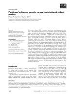
that a third species of soluble, mutant protein can
exist, and that this protein can exist in both the soluble
and insoluble states and move between those two
states (Fig. 1). FRAP studies also confirm that fusions
of GFP to polyglutamine disease proteins are not
misfolded when in inclusions, as they continue to fluo-
resce quantitatively as protein is localized to the inclu-
sions, even when in excess of 5 lm in diameter. In the
case of ataxin-1, normal ataxin-1 function dictates the
formation of nuclear ataxin bodies, which exist even in
the complete absence of the polyglutamine tract [28].
Ataxin-1 inclusion formation is dictated by signaling
and post-translational phosphorylation of a single
serine in ataxin-1 at position 776, regardless of poly-
glutamine tract length [25]. These live cell dynamic
observations and mouse model data obtained with
ataxin-1 are inconsistent with the hypothesis that poly-
glutamine has a universal effect on protein misfolding
and insolubility, rendering all proteins ‘amyloid’.
Another inconsistency with the amyloid hypothesis
for HD is in a YAC mouse model of HD that resulted
from a cloning artefact that was carefully character-
ized. The ‘shortstop’ mouse expressed only 120 amino
acids of huntingtin on a YAC, or roughly 35 amino
acids beyond exon 1 in a polyglutamine-expanded con-
text, and displayed large visible aggregates throughout
the brain, but this mouse had no measurable disease
[30]. The corresponding full-length mutant huntingtin
YAC construct does show a slow, progressive HD-like
phenotype, but without large visible aggregates [16].
These models demonstrate that with HD, as with
SCA1, other sequences within the polyglutamine dis-
ease protein may be able to modulate toxicity, but that
the formation of aggregates is not necessarily corre-
lated with disease.
Correlation between aggregates and
toxicity
The connection between visible protein aggregates and
polyglutamine diseases has been largely circumstantial.
In human brains, the incidence of aggregates is impos-
Ataxin-1 Q82-GFP
30 s 60 s 120 s 480 s240 s
Loss
Gain
A
C
D
E
F
G
Bleach area
B
Fig. 1. Polyglutamine-expanded protein can exist in two reversible states. FRAP experiment with overexpressed ataxin-1–GFP. All of the
protein is bleached except for one mutant ataxin-1 body in the nucleus. Gain of fluorescence is first seen in the same inclusions bleached
prior to recovery closest to the unbleached inclusion; the corresponding loss of fluorescence over time is seen in the unbleached inclusion.
Thus, polyglutamine-expanded mutant ataxin-1 can move from one inclusion of highly concentrated protein to another through a soluble
phase.
R. Truant et al. Revisiting the aggregation hypothesis
FEBS Journal 275 (2008) 4252–4262 ª 2008 The Authors Journal compilation ª 2008 FEBS 4255
sible to follow with disease in any one individual,
although increased aggregates are noted in more severe
stages or grades of HD [31,32]. In order to directly
follow the fate of individual neurons expressing a
small fragment of polyglutamine-expanded huntingtin,
Arrasate and colleagues transfected huntingtin exon 1–
GFP expression plasmids in primary neuronal cultures,
and used robotic 4D fluorescent microscopy to track
the fate of single cultured neurons over time, imaging
them repeatedly [33]. From this work, they observed
an inverse correlation between huntingtin exon 1 frag-
ment inclusion size and cell death; that is, the larger
the aggregate, the more likely the neuron was to
survive longer than a neuron expressing mutant
huntingtin without any visible aggregates. This work
took advantage of recent technology and trends in cell
biology towards quantitative measurement of effects.
This data thus indicated that large aggregates of hun-
tingtin fragments may constitute a cellular protective
mechanism to localize the toxic soluble mutant protein
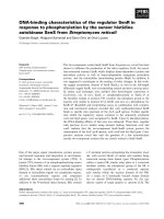
to insoluble and inactive protein reservoirs (Fig. 2).
This localization to large inclusions may also contrib-
ute to the loss-of-function seen in HD [34], whereas
the soluble mutant protein can participate in normal
protein functions with an additional gain-of-function.
We know that mutant huntingtin protein can assume
the functions of wild-type protein, as it can lead to
normal development in mutant homozygous mice and
humans [17].
The concept of neuroprotection of aggregates of
polyglutamine disease proteins is not limited to HD.
In SCA7, two groups independently showed an inverse
correlation of aggregate formation of ataxin-7 with
toxicity, both in cultured neurons and in a mouse
model [35,36]. Ataxin-7 has a known role as a compo-
nent of the transcription mediator complex known as
STAGA, and when polyglutamine-expanded, can
affect the proper recruitment and composition of this
complex [37]. Therefore, with ataxin-7, it is likely that
the toxic version of the protein is not that found in
>36 Repeats
Long repeats
<36 Repeats
No structure
No structure
Gain of structure
Gain of toxicity
Highly stable structure
highly toxic protein
Biological or
chemical
modulators
Biological or
chemical
modulators
Toxic
Verytoxic
Inert?
Loss-of-function?
Inert?
Fig. 2. Polyglutamine expansion lengths may disrupt the equilibria between toxic and healthy protein and between toxic soluble species and
inert insoluble species. Polyglutamine lengths beyond 37 repeats in HD are predicted to form a structure leading to gain of toxic function.
Mutant protein can exist in three states: soluble and without structure (healthy); soluble with a structure leading to gain of toxic function;
and insoluble with a structure leading to loss of normal function. Longer expansion lengths can skew this equilibrium to essentially two con-
formers, either loss-of-function or gain-of-function, both contributing to the manifestation of disease. Biological or chemical modulators are
able to skew equilibria in vivo, suggesting that the optimal modulator may be a molecule that can push all mutant protein into the insoluble,
unstructured and hence inert state. This modulator may not necessarily need to interact with polyglutamine, and may be different for
different protein contexts related to biological functions.
Revisiting the aggregation hypothesis R. Truant et al.
4256 FEBS Journal 275 (2008) 4252–4262 ª 2008 The Authors Journal compilation ª 2008 FEBS
aggregates, but rather the soluble mutant protein that
is able to participate in complexes with STAGA to
exert dominant effects over wild-type protein. In
SCA2, although aggregates can be seen in a small
number of neurons, they are not seen within the
nucleus, as they are in HD or SCA1 [38]. In
SCA3 ⁄ Machado–Joseph disease, as in HD and SCA7,
an inverse correlation is seen between nuclear inclu-
sions of ataxin-3 protein and cell death, both by exam-
ination of brain slices [39,40] and in tissue culture
models [41].
These newer data can therefore allow us to revisit
the early brain pathology data obtained with HD
patients from another perspective. One of the hall-
marks of HD in humans, but not as much in mouse
models, is the striking loss of the striatum, and up to
30% of total brain mass, prior to death [31]. In the
neurons that remain to be seen post mortem, aggre-
gates of huntingtin N-terminal fragments can be seen
[32]. One hypothesis was that these aggregates may be
the cause of cell death, and when they were visualized,
they were in neurons en route to death. However, from
a revisionist perspective, one can also hypothesize that
these neurons may have survived longer than the
missing striatal neurons, due to the presence of the
aggregates. The consideration of aggregates in HD fol-
lows many of the conundrums seen with polyglutamine
diseases and the struggle to understand what is cause
and what is effect in these diseases.
Hunting the elusive toxic
polyglutamine conformer
A thorough search of crystallographic databases
reveals that polyglutamine tracts seen in a variety of
normal cellular proteins are either annotated as
‘unstructured’ or have to be removed to facilitate crys-
tallization. Obtaining structural information on poly-
glutamine in proteins is technically difficult, as even
wild-type polyglutamine lengths can tend to be insolu-
ble at the high concentrations required for crystallo-
graphic or NMR studies. Wetzel’s group has focused
on the identification of the toxic structure of poly-
glutamine. Led by the antiparallel b-sheet model
originally proposed by Perutz [4], they inserted
proline–glycine substitutions in pure polyglutamine
tracts to induce a b-strand structure, and found that
even short lengths of polyglutamine could form aggre-
gates similar to pure Q45 lengths when b-strands and
b-turns were induced [42]. The group of Ross then
showed that these structured constructs were similarly
toxic in primary cultured neurons and tissue culture
models [43]. This work led to the concept that the
genetic gain-of-function of polyglutamine could be
tied to a gain of structure [44], but that this structural
gain did not necessarily have to exert toxicity by the
formation of aggregates. Recently, Onodera’s group
confirmed the parallel b-sheet model or cylindrical
b-sheet of polyglutamine in atrophin-1 by the use of
fluorescence resonant energy transfer (FRET) studies
in vivo. This FRET-based ‘spectroscopic ruler’ tool
allowed the investigators to distinguish between solu-
ble expanded polyglutamine oligomers, soluble mono-
mer and inclusion bodies in live cells. In neuronal cell
culture toxicity assays, they demonstrated that the
toxic species appeared to be soluble oligomers, and
not the protein in aggregates [45]. The caveat of this
work is that the authors assume that polyglutamine in
the context of atrophin-1 fragments has the same
structure in all polyglutamine disease proteins, but
given the importance of flanking sequences to polyglu-
tamine structure, this model needs to be tested in
other polyglutamine disease contexts. Biophotonic
methods such as FRET and fluorescence correlation
spectroscopy have led, and will probably continue to
lead, to major biochemical insights into polyglutamine
folding in vivo [46].
With small huntingtin fragments, many groups,
including ours, have independently reported the impor-
tance of flanking sequences next to the polyglutamine
tract in huntingtin exon 1 as modulators of toxicity. In
the yeast toxicity model, the positioning of flag-tags on
the expression constructs modulated toxicity and the
nature of aggregated protein, with tight, compact
aggregates being benign, but amorphous aggregates
being much more toxic [47]. Another group observed
modulation of polyglutamine aggregation by the use of
structured chimeras with the cellular retinoic-acid
binding protein in E. coli [48]. Again revisiting the
seminal Perutz paper [4], investigators have shown that
the glutathione S-transferase fusion to polyglutamine
does affect the aggregation dynamics, and may not be
an innocuous purification tag, as it was once cons-
idered to be. Aggregation may occur through forma-
tion of a reservoir of soluble intermediates whose
populations and stabilities increase with polyglutamine
length [49]. However, these sequences were exogenous
to huntingtin exon 1, and toxicity was not assayed in
mammalian cells. Deletion of the proline-rich region in
huntingtin exon 1 greatly increases the toxicity of
exon 1 fragments in yeast, which are otherwise inno-
cuous [50]. Therefore, the proline-rich region appears
to be protective against the effects of expanded
polyglutamine. The effects of polyproline in cis, in vitro
can be seen to affect the structure of expanded
polyglutamine [51].
R. Truant et al. Revisiting the aggregation hypothesis
FEBS Journal 275 (2008) 4252–4262 ª 2008 The Authors Journal compilation ª 2008 FEBS 4257
The first 17 amino acids of huntingtin, prior to the
polyglutamine tract, are highly conserved (100% simi-
larity) in all vertebrate species, and were originally
annotated as unstructured [4]. However, by exhaustive
mutational analysis in vivo and CD spectroscopy
in vitro with peptides, our group has determined the
first 17 amino acids to be an amphipathic a-helix, with
membrane-associating properties with regard to the
endoplasmic reticulum [23]. Like the proline-rich tract,
this region of huntingtin, present in all mouse models
of HD, was shown to modulate the toxicity of Q138
huntingtin 1–171 in a structure-dependent manner. A
single point mutant in the middle of the helix, shown
to disrupt the a-helical structure, resulted in three
surprising phenotypes: constitutive nuclear entry of
full-length huntingtin, or any huntingtin small N-ter-
minal fragments; the complete abrogation of any
visible aggregates of polyglutamine-expanded hun-
tingtin 1–171, even in the context of 250 repeats; and a
corresponding increase of toxicity of this fragment of
huntingtin in a polyglutamine-dependent manner of
close to four-fold over Q138 huntingtin 1–171. Thus,
loss of structure in regions adjoining the polyglutamine
tract on either side of the tract can lead to increased
huntingtin toxicity, with an inverse correlation with
aggregation. These results predict that regions on
either side of the polyglutamine tract in huntingtin
may interact with each other, with a critical compo-
nent of normal interaction being the flexible region of
at least four glutamine residues seen in all vertebrate
huntingtin proteins. Huntingtin 1–17 and the proline-
rich region adjacent to the polyglutamine tracts are
both involved in targeting vesicular populations
[23,52]. In HD, the gain-of-structure may perturb
huntingtin functions in vesicular trafficking by a ‘rusty
hinge’ model, where important on–off interactions may
be stuck on or off by the structure gained as a result
of polyglutamine expansion (Fig. 3). Similar models
may apply to other polyglutamine disease proteins,
with different consequences.
Basic residues in the ataxin-3 protein form an inter-
action motif with VCP ⁄ p97 protein, and this inter-
action can modulate ataxin-3 aggregation and toxicity
in Drosophila models [53]. Serine mutations in the
N-terminus of the AR can modulate polyglutamine-
expanded AR’s ability to aggregate, with increased
aggregation but less toxicity being seen in a Drosophila
model [54]. Thus, many different sequences flanking
polyglutamine tracts can affect polyglutamine tract-
mediated toxicity and the potential to form aggregates.
The importance of the structure on either side of an
expanded polyglutamine tract may be due to imp-
rinting of structure on polyglutamine by adjoining
sequences that interact with the flexible polyglutamine
tract in cis. This is consistent with peptides or small
molecules in trans that are able to mediate the aggrega-
tion potential of polyglutamine tracts and skew the
equilibrium distribution of polyglutamine-expanded
protein towards soluble or insoluble. Some of the
factors that may be able to affect this equilibrium may
include normal interacting proteins, such as chaper-
ones, or the HYPK protein interaction with hunting-
tin’s N-terminus modulating its ability to form
aggregates [55,56].
Modifiers of polyglutamine structure
and toxicity
Even if large visible ‘aggregates’ are not the actual
targets of therapeutic development in HD and other
polyglutamine diseases, proteins, small molecules or
other factors that affect polyglutamine-dependent
aggregation may have important effects on the toxic
soluble species of polyglutamine-expanded proteins.
Early high-throughput (biochemical) assays used filter-
trapped aggregates as the readout for screening
of small molecules. Benzothiazole compounds were
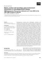
Fig. 3. The ‘rusty hinge’ hypothesis of gain of structure leading to
toxic function in HD. We speculate that there is an overall superhe-
lical structure of huntingtin, owing to the large number of HEAT
repeats throughout the entire 3144 amino acid protein. The normal
polyglutamine tract, present in all vertebrates with least four gluta-
mines, provides an important flexible region in the huntingtin scaf-
fold for factors that can interact with the first 17 amino acids and
downstream regions. With increasing polyglutamine lengths, the
pool of total mutant protein is skewed towards b-sheet structured
polyglutamine, leading to a loss of flexibility and the ability of hun-
tingtin 1–17 to interact with the rest of huntingtin via factors or
complexes. Normal interactions that should switch on or off will
then be stuck in either the on or off position or pools of either posi-
tion, both of which may be toxic. Normal interaction between the
proline-rich region and huntingtin 1–17 influences the structure of
expanded polyglutamine in cis, leading to increased toxicity if the
normal structures of these regions are disrupted.
Revisiting the aggregation hypothesis R. Truant et al.
4258 FEBS Journal 275 (2008) 4252–4262 ª 2008 The Authors Journal compilation ª 2008 FEBS
identified as being able to prevent aggregation or solu-
bilize aggregates [57]. The small molecule C2–8 was
identified from high-content screening (cell biological)
as an inhibitor of polyglutamine aggregate growth [58],
but its efficacy in mouse models was modest, despite it
crossing the blood–brain barrier effectively [59]. One
surprising finding from a FRET-based high-content
screen of a kinase inhibitor library is that the Rho
kinase inhibitor, Y-27632, could prevent huntingtin
exon 1 fragment-mediated aggregation [60]. What is
not known is what the mechanism of this inhibition is,
but Rho kinase inhibition suggests that other functions
of huntingtin exon 1 fragment, perhaps in actin associ-
ation, may be necessary for the formation of aggre-
gates. These classes of small molecules that affect
huntingtin aggregation now allow cell biologists to use
these molecules as tools of ‘chemical biology’ in the
study of huntingtin function and mutant huntingtin
pathology.
One of the strongest lines of evidence for a soluble
oligomeric or misfolded toxic species of polyglutamine,
and effects of peptides in trans, comes from the studies
of the polyglutamine-binding trytophan-rich peptide
QBP1 (WKWWPGIF). Although it was originally
described as a suppressor of polyglutamine-mediated
toxicity through the suppression of aggregation [61,62],
more detailed studies have shown that this peptide can
inhibit the transition of polyglutamine from an
unstructured state to the toxic soluble b-sheet mono-
mer structure [63], consistent with independent work
on the b-sheet structure of polyglutamine from many
other groups.
Another look at the amyloid hypothesis
In the past, it has been tempting to place polygluta-
mine diseases into the category of amyloid diseases,
a family of neurodegenerative disorders caused by
misfolded proteins leading to large protein ultrastruc-
tures within or outside affected neurons. However,
recent research evidence from Alzheimer’s and Par-
kinson’s diseases is starting to cast doubt on the uni-
versality of the amyloid hypothesis in those diseases
as well. In Alzheimer’s disease, genetic mutations in
familial Alzheimer’s disease reveal that Alzheimer’s
disease in those cases may be caused by a redox
imbalance, leading to the effect of amyloid plaques
[64]. In Parkinson’s disease, a-synuclein accumula-
tion, like mutant huntingtin aggregation, can be seen
to be neuroprotective [65]. Small molecules that
encourage aggregation appear to be effective in toxic-
ity assays for many amyloid diseases and HD
[66,67]. Although it now appears that understanding
polyglutamine disease probably cannot be achieved
without the disease protein context, important lessons
have been learned from huntingtin small-fragment
models and studies focusing on the toxic species of
polyglutamine in different disease contexts. Proof-of-
concept successes with SCA1 pointing to serine
kinase inhibition as a therapeutic strategy, and clini-
cal success with the treatment of SBMA by leupro-
relin, underscore the importance of analysis of
huntingtin toxicity in the full protein context and the
importance of elucidating the normal biological func-
tion of huntingtin. From that milestone, HD
researchers can then have a new vantage point from
which to consider alternative or coincident therapeu-
tic strategies related to huntingtin function along
with antiaggregation compounds. The hallmark of
any good drug is selective toxicity for its target, and
thus expanded polyglutamine remains a valid target
in polyglutamine diseases, with the appeal that drug
toxicity will be specific to the mutant, and not wild-
type, protein.
Acknowledgements
The Truant laboratory is supported by current and
past grants from the Hereditary Disease Foundation,
(HDF) USA, the Cure Huntington’s Disease Initiative
(CHDI) USA, the Huntington’s disease Society of
America (HDSA), the Huntington’s Society of Canada
and the Canadian Institutes of Health Research
(CIHR), Genetics and Mental Health and Addiction
Institutes. R. Truant is Chair of the Huntington’s
disease Society of Canada (HSC) scientific advisory
board.
References
1 La Spada AR, Wilson EM, Lubahn DB, Harding AE
& Fischbeck KH (1991) Androgen receptor gene muta-
tions in X-linked spinal and bulbar muscular atrophy.
Nature 352, 77–79.
2 The Huntington’s Disease Collaborative Research
Group (1993) A novel gene containing a trinucleotide
repeat that is expanded and unstable on Huntington’s
disease chromosomes. Cell 72, 971–983.
3 Orr HT, Chung MY, Banfi S, Kwiatkowski TJ
Jr, Servadio A, Beaudet AL, McCall AE, Duvick
LA, Ranum LP & Zoghbi HY (1993) Expansion of an
unstable trinucleotide CAG repeat in
spinocerebellar ataxia type 1. Nat Genet 4, 221–226.
4 Perutz MF (1996) Glutamine repeats and inherited
neurodegenerative diseases: molecular aspects. Curr
Opin Struct Biol 6, 848–858.
R. Truant et al. Revisiting the aggregation hypothesis
FEBS Journal 275 (2008) 4252–4262 ª 2008 The Authors Journal compilation ª 2008 FEBS 4259
5 Oma Y, Kino Y, Sasagawa N & Ishiura S (2004) Intra-
cellular localization of homopolymeric amino acid-con-
taining proteins expressed in mammalian cells. J Biol
Chem 279, 21217–21222.
6 Mangiarini L, Sathasivam K, Seller M, Cozens B, Har-
per A, Hetherington C, Lawton M, Trottier Y, Lehrach
H, Davies SW et al. (1996) Exon 1 of the HD gene with
an expanded CAG repeat is sufficient to cause a pro-
gressive neurological phenotype in transgenic mice. Cell
87, 493–506.
7 Chai Y, Shao J, Miller VM, Williams A & Paulson HL
(2002) Live-cell imaging reveals divergent intracellular
dynamics of polyglutamine disease proteins and sup-
ports a sequestration model of pathogenesis. Proc Natl
Acad Sci USA 99, 9310–9315.
8 Preisinger E, Jordan BM, Kazantsev A & Housman D
(1999) Evidence for a recruitment and sequestration
mechanism in Huntington’s disease. Phil Trans R Soc
Lond B Biol Sci 354, 1029–1034.
9 Gunawardena S, Her LS, Brusch RG, Laymon RA,
Niesman IR, Gordesky-Gold B, Sintasath L, Bonini
NM & Goldstein LS (2003) Disruption of axonal trans-
port by loss of huntingtin or expression of pathogenic
polyQ proteins in Drosophila. Neuron 40, 25–40.
10 Bence NF, Sampat RM & Kopito RR (2001) Impair-
ment of the ubiquitin–proteasome system by protein
aggregation. Science 292, 1552–1555.
11 Satyal SH, Schmidt E, Kitagawa K, Sondheimer N,
Lindquist S, Kramer JM & Morimoto RI (2000) Poly-
glutamine aggregates alter protein folding homeostasis
in Caenorhabditis elegans. Proc Natl Acad Sci USA 97,
5750–5755.
12 Schilling G, Becher MW, Sharp AH, Jinnah HA, Duan
K, Kotzuk JA, Slunt HH, Ratovitski T, Cooper JK,
Jenkins NA et al. (1999) Intranuclear inclusions and
neuritic aggregates in transgenic mice expressing a
mutant N-terminal fragment of huntingtin. Hum Mol
Genet 8, 397–407.
13 Yamamoto A, Lucas JJ & Hen R (2000) Reversal of
neuropathology and motor dysfunction in a conditional
model of Huntington’s disease. Cell 101, 57–66.
14 Zu T, Duvick LA, Kaytor MD, Berlinger MS, Zoghbi
HY, Clark HB & Orr HT (2004) Recovery from poly-
glutamine-induced neurodegeneration in conditional
SCA1 transgenic mice. J Neurosci 24, 8853–8861.
15 Koo EH, Lansbury PT Jr & Kelly JW (1999) Amyloid
diseases: abnormal protein aggregation in neurodegener-
ation. Proc Natl Acad Sci USA 96, 9989–9990.
16 Hodgson JG, Agopyan N, Gutekunst CA, Leavitt BR,
LePiane F, Singaraja R, Smith DJ, Bissada N,
McCutcheon K, Nasir J et al. (1999) A YAC mouse
model for Huntington’s disease with full-length mutant
huntingtin, cytoplasmic toxicity, and selective striatal
neurodegeneration. Neuron 23, 181–192.
17 Fossale E, Wheeler VC, Vrbanac V, Lebel LA, Teed A,
Mysore JS, Gusella JF, MacDonald ME & Persichetti
F (2002) Identification of a presymptomatic molecular
phenotype in Hdh CAG knock-in mice. Hum Mol Genet
11, 2233–2241.
18 Clabough EB & Zeitlin SO (2006) Deletion of the trip-
let repeat encoding polyglutamine within the mouse
Huntington’s disease gene results in subtle behav-
ioral ⁄ motor phenotypes in vivo and elevated levels of
ATP with cellular senescence in vitro. Hum Mol Genet
15, 607–623.
19 Reiner A, Dragatsis I, Zeitlin S & Goldowitz D (2003)
Wild-type huntingtin plays a role in brain development
and neuronal survival. Mol Neurobiol 28, 259–276.
20 Menalled LB, Sison JD, Dragatsis I, Zeitlin S & Chess-
elet MF (2003) Time course of early motor and neuro-
pathological anomalies in a knock-in mouse model of
Huntington’s disease with 140 CAG repeats. J Comp
Neurol 465, 11–26.
21 Nakamura K, Jeong SY, Uchihara T, Anno M, Naga-
shima K, Nagashima T, Ikeda S, Tsuji S & Kanazawa I
(2001) SCA17, a novel autosomal dominant cerebellar
ataxia caused by an expanded polyglutamine in TATA-
binding protein. Hum Mol Genet 10, 1441–1448.
22 Pal A, Severin F, Lommer B, Shevchenko A & Zerial
M (2006) Huntingtin–HAP40 complex is a novel Rab5
effector that regulates early endosome motility and is
up-regulated in Huntington’s disease. J Cell Biol 172,
605–618.
23 Atwal RS, Xia J, Pinchev D, Taylor J, Epand RM &
Truant R (2007) Huntingtin has a membrane associa-
tion signal that can modulate huntingtin aggregation,
nuclear entry and toxicity. Hum Mol Genet 16, 2600–
2615.
24 Klement IA, Skinner PJ, Kaytor MD, Yi H, Hersch
SM, Clark HB, Zoghbi HY & Orr HT (1998) Ataxin-1
nuclear localization and aggregation: role in polygluta-
mine-induced disease in SCA1 transgenic mice. Cell 95,
41–53.
25 Emamian ES, Kaytor MD, Duvick LA, Zu T, Tousey
SK, Zoghbi HY, Clark HB & Orr HT (2003) Serine 776
of ataxin-1 is critical for polyglutamine-induced disease
in SCA1 transgenic mice. Neuron 38, 375–387.
26 Sobue G (1995) X-linked recessive bulbospinal neuron-
opathy (SBMA). Nagoya J Med Sci 58, 95–106.
27 Katsuno M, Adachi H, Waza M, Banno H, Suzuki
K, Tanaka F, Doyu M & Sobue G (2006) Pathogene-
sis, animal models and therapeutics in spinal and
bulbar muscular atrophy (SBMA). Exp Neurol 200,
8–18.
28 Irwin S, Vandelft M, Pinchev D, Howell JL, Graczyk J,
Orr HT & Truant R (2005) RNA association and nucle-
ocytoplasmic shuttling by ataxin-1. J Cell Sci 118, 233–
242.
Revisiting the aggregation hypothesis R. Truant et al.
4260 FEBS Journal 275 (2008) 4252–4262 ª 2008 The Authors Journal compilation ª 2008 FEBS
29 Stenoien DL, Mielke M & Mancini MA (2002) Intranu-
clear ataxin1 inclusions contain both fast- and slow-
exchanging components. Nat Cell Biol 4, 806–810.
30 Slow EJ, Graham RK, Osmand AP, Devon RS, Lu G,
Deng Y, Pearson J, Vaid K, Bissada N, Wetzel R et al.
(2005) Absence of behavioral abnormalities and neu-
rodegeneration in vivo despite widespread neuronal
huntingtin inclusions. Proc Natl Acad Sci USA 102,
11402–11407.
31 Vonsattel JP, Myers RH, Stevens TJ, Ferrante RJ, Bird
ED, Richardson EP Jr (1985) Neuropathological classi-
fication of Huntington’s disease. J Neuropathol Exp
Neurol 44, 559–577.
32 DiFiglia M, Sapp E, Chase KO, Davies SW, Bates GP,
Vonsattel JP & Aronin N (1997) Aggregation of hun-
tingtin in neuronal intranuclear inclusions and dystro-
phic neurites in brain. Science 277, 1990–1993.
33 Arrasate M, Mitra S, Schweitzer ES, Segal MR &
Finkbeiner S (2004) Inclusion body formation reduces
levels of mutant huntingtin and the risk of neuronal
death. Nature 431, 805–810.
34 Zhang Y, Li M, Drozda M, Chen M, Ren S, Mejia
Sanchez RO, Leavitt BR, Cattaneo E, Ferrante RJ,
Hayden MR et al. (2003) Depletion of wild-type
huntingtin in mouse models of neurologic diseases.
J Neurochem 87, 101–106.
35 Taylor J, Grote SK, Xia J, Vandelft M, Graczyk J, Ell-
erby LM, La Spada AR & Truant R (2006) Ataxin-7
can export from the nucleus via a conserved exportin-
dependent signal. J Biol Chem 281, 2730–2739.
36 Bowman AB, Yoo SY, Dantuma NP & Zoghbi HY
(2005) Neuronal dysfunction in a polyglutamine disease
model occurs in the absence of ubiquitin–proteasome
system impairment and inversely correlates with the
degree of nuclear inclusion formation. Hum Mol Genet
14, 679–691.
37 Helmlinger D, Hardy S, bou-Sleymane G, Eberlin A,
Bowman AB, Gansmuller A, Picaud S, Zoghbi HY,
Trottier Y, Tora L et al. (2006) Glutamine-expanded
ataxin-7 alters TFTC ⁄ STAGA recruitment and chroma-
tin structure leading to photoreceptor dysfunction.
PLoS Biol 4, e67, 0432–0445.
38 Huynh DP, Figueroa K, Hoang N & Pulst SM (2000)
Nuclear localization or inclusion body formation of
ataxin-2 are not necessary for SCA2 pathogenesis in
mouse or human. Nat Genet 26, 44–50.
39 Evert BO, Schelhaas J, Fleischer H, de Vos RA, Brunt
ER, Stenzel W, Klockgether T & Wullner U (2006)
Neuronal intranuclear inclusions, dysregulation of cyto-
kine expression and cell death in spinocerebellar ataxia
type 3. Clin Neuropathol 25, 272–281.
40 Rub U, de Vos RA, Brunt ER, Sebesteny T, Schols L,
Auburger G, Bohl J, Ghebremedhin E, Gierga K, Seidel
K et al. (2006) Spinocerebellar ataxia type 3 (SCA3):
thalamic neurodegeneration occurs independently from
thalamic ataxin-3 immunopositive neuronal intranuclear
inclusions. Brain Pathol 16 , 218–227.
41 Yoshizawa T, Yoshida H & Shoji S (2001) Differen-
tial susceptibility of cultured cell lines to aggregate
formation and cell death produced by the truncated
Machado–Joseph disease gene product with an
expanded polyglutamine stretch. Brain Res Bull 56,
349–352.
42 Thakur AK & Wetzel R (2002) Mutational analysis of
the structural organization of polyglutamine aggregates.
Proc Natl Acad Sci USA 99, 17014–17019.
43 Poirier MA, Jiang H & Ross CA (2005) A
structure-based analysis of huntingtin mutant polyglu-
tamine aggregation and toxicity: evidence for a
compact beta-sheet structure. Hum Mol Genet 14,
765–774.
44 Ross CA & Poirier MA (2005) Opinion: what is the role
of protein aggregation in neurodegeneration?
Nat Rev
Mol Cell Biol 6, 891–898.
45 Takahashi T, Kikuchi S, Katada S, Nagai Y, Nishizawa
M & Onodera O (2008) Soluble polyglutamine oligo-
mers formed prior to inclusion body formation are
cytotoxic. Hum Mol Genet 17, 345–356.
46 Takahashi Y, Okamoto Y, Popiel HA, Fujikake N,
Toda T, Kinjo M & Nagai Y (2007) Detection of poly-
glutamine protein oligomers in cells by fluorescence cor-
relation spectroscopy. J Biol Chem 282, 24039–24048.
47 Duennwald ML, Jagadish S, Muchowski PJ & Lind-
quist S (2006) Flanking sequences profoundly alter
polyglutamine toxicity in yeast. Proc Natl Acad Sci
USA 103, 11045–11050.
48 Ignatova Z, Thakur AK, Wetzel R & Gierasch LM
(2007) In-cell aggregation of a polyglutamine-containing
chimera is a multistep process initiated by the flanking
sequence. J Biol Chem 282, 36736–36743.
49 Bulone D, Masino L, Thomas DJ, San Biagio PL &
Pastore A (2006) The interplay between polyQ and
protein context delays aggregation by forming a reser-
voir of protofibrils. PLoS ONE 1, e111.
50 Dehay B & Bertolotti A (2006) Critical role of the
proline-rich region in Huntingtin for aggregation and
cytotoxicity in yeast. J Biol Chem 281, 35608–35615.
51 Bhattacharyya A, Thakur AK, Chellgren VM, Thiaga-
rajan G, Williams AD, Chellgren BW, Creamer TP &
Wetzel R (2006) Oligoproline effects on polyglutamine
conformation and aggregation. J Mol Biol 355, 524–
535.
52 Qin ZH, Wang Y, Sapp E, Cuiffo B, Wanker E, Hay-
den MR, Kegel KB, Aronin N & DiFiglia M (2004)
Huntingtin bodies sequester vesicle-associated proteins
by a polyproline-dependent interaction. J Neurosci 24,
269–281.
53 Boeddrich A, Gaumer S, Haacke A, Tzvetkov N,
Albrecht M, Evert BO, Muller EC, Lurz R, Breuer P,
Schugardt N et al. (2006) An arginine ⁄ lysine-rich motif
R. Truant et al. Revisiting the aggregation hypothesis
FEBS Journal 275 (2008) 4252–4262 ª 2008 The Authors Journal compilation ª 2008 FEBS 4261
is crucial for VCP ⁄ p97-mediated modulation of ataxin-3
fibrillogenesis. EMBO J 25, 1547–1558.
54 Funderburk SF, Shatkina L, Mink S, Weis Q, Weg-
Remers S & Cato AC (2008) Specific N-terminal
mutations in the human androgen receptor induce
cytotoxicity. Neurobiol Aging, doi:10.1016/j.neurobiola-
ging.2007.12.023.
55 Raychaudhuri S, Sinha M, Mukhopadhyay D & Bhat-
tacharyya NP (2008) HYPK, a Huntingtin interacting
protein, reduces aggregates and apoptosis induced by
N-terminal huntingtin with 40 glutamines in Neuro2a
cells and exhibits chaperone-like activity. Hum Mol
Genet 17, 240–255.
56 Raychaudhuri S, Majumder P, Sarkar S, Giri K,
Mukhopadhyay D & Bhattacharyya NP (2008)
Huntingtin interacting protein HYPK is intrinsically
unstructured. Proteins 71, 4, 1686–1698.
57 Heiser V, Engemann S, Brocker W, Dunkel I, Boedd-
rich A, Waelter S, Nordhoff E, Lurz R, Schugardt N,
Rautenberg S et al. (2002) Identification of benzothiaz-
oles as potential polyglutamine aggregation inhibitors
of Huntington’s disease by using an automated filter
retardation assay. Proc Natl Acad Sci USA 99(Suppl.
4), 16400–16406.
58 Zhang X, Smith DL, Meriin AB, Engemann S, Russel
DE, Roark M, Washington SL, Maxwell MM, Marsh
JL, Thompson LM et al. (2005) A potent small mole-
cule inhibits polyglutamine aggregation in Huntington’s
disease neurons and suppresses neurodegeneration
in vivo. Proc Natl Acad Sci USA 102, 892–897.
59 Chopra V, Fox JH, Lieberman G, Dorsey K, Matson W,
Waldmeier P, Housman DE, Kazantsev A, Young AB &
Hersch S (2007) A small-molecule therapeutic lead for
Huntington’s disease: preclinical pharmacology and effi-
cacy of C2-8 in the R6 ⁄ 2 transgenic mouse. Proc Natl
Acad Sci USA 104, 16685–16689.
60 Pollitt SK, Pallos J, Shao J, Desai UA, Ma AA,
Thompson LM, Marsh JL & Diamond MI (2003)
A rapid cellular FRET assay of polyglutamine
aggregation identifies a novel inhibitor. Neuron 40,
685–694.
61 Nagai Y, Tucker T, Ren H, Kenan DJ, Henderson BS,
Keene JD, Strittmatter WJ & Burke JR (2000) Inhibi-
tion of polyglutamine protein aggregation and cell
death by novel peptides identified by phage display
screening. J Biol Chem 275, 10437–10442.
62 Ren H, Nagai Y, Tucker T, Strittmatter WJ & Burke
JR (2001) Amino acid sequence requirements of
peptides that inhibit polyglutamine–protein aggre-
gation and cell death. Biochem Biophys Res Commun
288, 703–710.
63 Nagai Y, Inui T, Popiel HA, Fujikake N, Hasegawa K,
Urade Y, Goto Y, Naiki H & Toda T (2007) A toxic
monomeric conformer of the polyglutamine protein.
Nat Struct Mol Biol 14, 332–340.
64 Lee HG, Zhu X, Takeda A, Perry G & Smith MA
(2006) Emerging evidence for the neuroprotective role
of alpha-synuclein. Exp Neurol 200, 1–7.
65 Lee HG, Zhu X, Nunomura A, Perry G & Smith MA
(2006) Amyloid beta: the alternate hypothesis. Curr
Alzheimer Res 3, 75–80.
66 Bodner RA, Housman DE & Kazantsev AG (2006)
New directions for neurodegenerative disease therapy:
using chemical compounds to boost the formation
of mutant protein inclusions. Cell Cycle 5, 1477–1480.
67 Bodner RA, Outeiro TF, Altmann S, Maxwell MM,
Cho SH, Hyman BT, McLean PJ, Young AB, Hous-
man DE & Kazantsev AG (2006) Pharmacological
promotion of inclusion formation: a therapeutic
approach for Huntington’s and Parkinson’s diseases.
Proc Natl Acad Sci USA 103, 4246–4251.
Revisiting the aggregation hypothesis R. Truant et al.
4262 FEBS Journal 275 (2008) 4252–4262 ª 2008 The Authors Journal compilation ª 2008 FEBS