báo cáo khoa học: "Long-term follow-up after en bloc resection and reconstruction of a solitary paraganglioma metastasis in the first lumbar vertebral body: a case report" ppt
Bạn đang xem bản rút gọn của tài liệu. Xem và tải ngay bản đầy đủ của tài liệu tại đây (1.51 MB, 6 trang )
CAS E REP O R T Open Access
Long-term follow-up after en bloc resection and
reconstruction of a solitary paraganglioma
metastasis in the first lumbar vertebral body:
a case report
Alexander Richter
1*
, Henry F Halm
2
, Thomas Lerner
3
, Ulf R Liljenqvist
3
, Markus Quante
2
Abstract
Introduction: Paragangliomas are rare tumors that originate from the autonomic nervous system-associated
paraganglia. They metastasize infrequently. Malignancy can only be demonstrated by the presence of chromaffin
tissue at sites where it usually is not present, such as bone, lung or liver, or local recurrence after total resection of
a primary mass. Paragangliomas within the central nervous system are usually intradural near the conus medullaris.
The metastatic spread of a retroperitoneal paraganglioma to a vertebral body is extremely rare, and there are only
a few cases reported in the literature.
Case presentation: We report the case of a 16-year-old Caucasian girl who had undergone resection of a
retroperitoneal paraganglioma that measure d 15 × 11.5 × 9.5 cm. After further staging, a solitary metastatic
paraganglioma was detected in the first lumbar vertebral body. After initial chemotherapy, marginal en bloc
resection and reconstruction were performed followed by radiotherapy. Histologic examination of the specimen
revealed that the tumor cells did not show any response to preoperative chemotherapy, which is in line with a
few other reports in the literature. Ten years after operative treatment, the patient is free of complaints, very
satisfied with the result and without signs of local recurrence or distant metastases.
Conclusion: We recommend en bloc spondylectomy and local radiotherapy in the treatment of solitary spinal
metastatic paragangliomas.
Introduction
Paraganglioma is a rare tumor that o riginates from the
autonomic nervous system-associated paraganglia.
Approximately 90% of paragangliomas arise from the
adrenal medulla, carotid body and glomus jugulare [1-3].
These metastasize infrequently. Within the central ner-
vous system, the majority of paragangliomas arise intradu-
rally in the area of the cauda equina [2]. For extra-adrenal
retroperitoneal paragangliomas, a 50% rate of metastasis
has been described [4,5]. Extra-adrenal paragangliomas are
divided on the basis of their anatomic distribution into
cervical, t horacic and intraabdomina l tumors [6]. About
15% to 20% of childhood paragangliomas are extra-adrenal
[7]. Metast atic spine involvement is uncommon, and if it
occurs, it is generally intradural at the level of the cauda
equina, very rarely within the vertebral bodies [1,3,8-17].
The individual behavior of paragangliomas i s unpredict-
able because the fundamental characteristics of malignant
neoplasms such as vascular invasion and extensive loc al
invasion are of limited value in assessing neuroendocrine
tumors [17].
We present one rare case of a solitary L1 metastatic
paraganglioma, which was detected after removal of an
intraabdominal paraganglioma. Preoper ative chemother-
apy, en bloc spondylectomy and postoperative radiother-
apy were performed.
Case presentation
An otherwise healthy 16-year-old Caucasian girl pre-
sented with a sudden onset of cramp like pain in the
* Correspondence:
1
Spine Center Hamburg, Asklepios Klinik St. Georg, Lohmühlenstrasse 5,
20099 Hamburg, Germany
Full list of author information is available at the end of the article
Richter et al. Journal of Medical Case Reports 2011, 5:45
/>JOURNAL OF MEDICAL
CASE REPORTS
© 2011 Richter et al; licensee BioMed Central Ltd. This is an Open Access article distributed under the terms of the Creative Commons
Attribution License ( which permits unrestricted use, distr ibution, and reproduction in
any medium, provided the original work is properly cited.
right lower abdomen. After e xamination, her gynecolo-
gist performed laparoscopy and found extreme varicosis
of th e internal genital tract but without further p atholo-
gic findings. To exclude thrombosis and consecutive
collateral circulation, postoperative phlebography was
done and showed excessi ve displacement of the inferior
vena cava. A retroperitoneal tumor was suspected, and
magnetic resonance imaging (MRI) revealed a tumor
measuring14×10×14cmintherightabdomenwith
a craniodorsal shift of the kidney. Laboratory para-
meters, includ ing tumor markers (24-hour urinary cate-
cholamines and metabolites, dopamine, serum and
plasma a-fetoprotein, neuron-specific enolase (NSE),
b-human chorionic gonadotropin) we re within normal
ranges.
Explorative laparotomy was performed, and the retro-
peritoneal tumor was resected. The tumor weighed 817
g, and macroscopic examination demonstrated a thinly
encapsulated neoplasm. The diagnosis of a paragan-
glioma was confirmed by histologic and immunohistolo-
gic examinations. Because vascular invasion and focal
infiltration of the fibrous capsule could be shown, it was
an R1 marginal resection.
The postoperative course was uneventful, but becaus e
of the potential malignant behavior of extra-adrenal
paragangliomas, Tc-99-MDP (Tc-99m-methylene dipho-
sphonate) and I-123-MIBG (123 I-metaiodobenzylguani-
dine) scintigraphy was performed 10 and 21 days
postoperatively. An increased uptake in the first lumbar
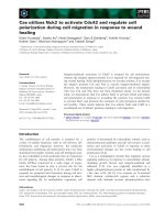
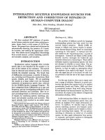
ver tebra was noted and MRI showed a l esion in the left
dorsal third of the L1 vertebral body (Figure 1). The
supposed metastatic paraganglioma was confirmed by
computed tomography- (CT-) guided needle biopsy.
Chemotherapy was applied using a n euroblastoma pro-
tocol (NB 90 of the German Society of Paediatric
Oncology and Haematology).
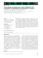
Five months later, combined posteroanterior en bloc
resection of the L1 vertebra was performed. Because of
partial infiltration of the left pedicle, it was left en bloc
with the vertebral body (Figure 2). Reconstruction was
performed with posterior transpedicular screw instrumen-
tation and anterior reconstruction us ing a modular cage
filled with autologous morselized rib grafts (Figure 3).
Macroscopically, the cut surface of the vertebral body
showed a reddish tumor in the left dorsolateral part of
the vertebral body (Figure 4). Histologic morphologic fea-
tures similar to the primary tumor were found, and
because of the penetration of the posterior cortex with
intact tumor capsule (but microscopic focal infiltration),
the resection was considered marginal as well. The
tumor cells did not show any response to preoperative
chemotherapy as found in the macroscopy and micro-
scopy pathology. The postoperative course was again
completely uneventful.
Because of the marginal resection and the poor
response to preoperative chemotherapy, postoperative
radiation therapy was added with a dose of 50 Gy. Ten
years postoperatively, the now 26-year-old female
patient is in excellent general condition without signs of
local recurrence or further distant metastasis. Concern-
ing instrumented fusion, no signs of lysis around the
pedicle screws or signs of cage dislocation have been
detected (Figure 5). CT has revealed that the autologous
bone within the cage is mineralized and has most p rob-
ably fused with the adjacent endplates o f the T12 and
L2 vertebral bodies, as far as thi s can be evaluated with
this or any other imaging technique.
Discussion
Paraganglia (or glomus bodies) are extra-adrenal rests of
neural crest-derived cells that are closely associated with
the autonomic nervous system. They are found in dispa-
rate areas of the body, including the head, neck, thorax,
abdomen and retroperitoneal space. Paragangliomas
arising from carotid bodies appear to have the highest
propensity for metastatic spread to the spine [1]. The
retroperitoneal extra-adrenal paraganglioma is the most
aggressive one w ith malignant behavior in up to 50% of
the cases [4,5]. So far no publications have come to our
attention that predict clinical outcome of patients with
paraganglioma by conventional histology. Therefore,
malignancy can only be demonstrated by the presence
of chromaffin tissue at sites where it is usually not pre-
sent, such as bone, lung or liver, or local recurrence
aft er total resectio n of a primary mass. In this case, sta-
ging after resection of the primary tumor revealed a
solitary metastasis in the vertebral body of L1. This is
unusual because metastases have been reported to occur
usually intradurally when t he spine is involved
[3,11,13,18,1 9]. Isolated me tastatic involvement of ver-
tebral bodies is extremely rare, and o nly isolated case
reports have been published. Brodke y et al [1] presented
the case of a 54-year-old man with a metastatic lesion
in the body of C2, which was resected. They did not
mention whether the procedure was intralesional or
marginal. Over a 30-month period, the patient’s myelo-
pathy resolved, and there had b een no progression of
the disease.
Razakaboay et al [20] reported on three patients who
developed bone metastasis of a retroperitoneal paragan-
glioma occurring up to 17 years after resection of the
primary tumor. T he treatment of choice was surgery
and radiotherapy.
A third case was published by Hamilton and Tait [21],
who described metastatic retroperitone al paraganglioma
associated with spinal cord compression in two young
men. One was metastatic at presentation, and the other
became metastatic 19 years after surgical resection of
Richter et al. Journal of Medical Case Reports 2011, 5:45
/>Page 2 of 6
the primary tumor. Both men died because of wide-
spread metastatic disease.
The latest rep ort was publis hed by Lehmen et al [22],
who described the case of metastatic lesio n in a cervical
vertebra treated by surgery and adjuvant radiation.
In our case, the superficial intraosseous extension of
the tumor within the vertebral body, which occupied
zones 4 to 9, according to the staging system of Boriani
et al [23], (Weinstein Boriani Biagini (WBB) staging sys-
tem) made en bloc resection possible. However, because
of the destruction of the posterior wall of the vertebral
body, only a marginal resection could be obtained. The
pseudocapsule was examined and considered intact.
With posterior bisegmental transpedicular screw instru-
mentation using a rigid internal fixator and anterior
strut g rafting using a modular cage filled with autolo-
gous morselized rib graft, a primary stable load-sharing
situation c ould be obtained, and the patient was mobi-
lized without additional external support. Ten ye ars
after surgery, the instrumented spine seems to be fused
and is absolutely stable.
Chemotherapy was applied before en bloc resection of
L1, according to the recommendation of our pediatric
oncologists, but histologic microscopy e xamination of
Figure 1 Magnetic resonance image showing the metastatic lesion within the vertebral body with destruction of the posterior cortex,
encroachment of the spinal canal and invasion of the left pedicle.
Figure 2 En bloc resected vertebral body with the affected left
pedicle left en bloc.
Richter et al. Journal of Medical Case Reports 2011, 5:45
/>Page 3 of 6
Figure 3 Postoperative anteroposterior and lateral plane radiograph showing reconstruction with modular tumor cage and a pedicle-
screw instrumentation.
Figure 4 Horizontal cut through the resected vertebral body. Complete d estruction of the posterior cortical lamellae with intact
pseudocapsule. Metastatic lesion in zones 4 to 9 and layer B (intraosseous superficial), according to Boriani et al [23].
Richter et al. Journal of Medical Case Reports 2011, 5:45
/>Page 4 of 6
the s pecimen did not show any response of the tumor
cells to the preoperative chemotherapy. This finding is
in line with a numb er of disappointing reports on che-
motherapy for this type of tumor [21,24,25], and it must
be emphasized that preoperative neoadjuvant che-
motherapy seems to be of no value in the treatment of
patients with metastatic retroperitoneal paragangliomas.
In a 1992 review, Schild et al [13] showed that radio-
therapy is beneficial in the treatment of paragangliomas.
Later, postoperative radiotherapy was recommended by
several authors [1,2,21].
Therefore, we deci ded to apply radiotherapy with
50 Gy postoperatively. Ten years after surgery, the
patient is without signs of local recurrence or distant
metastasis, completely asymptomatic and very satisfied
with the result of the operation.
Conclusion
En bloc resection of a solitary metastatic parag anglioma
combined with postoperative radiotherapy seems to be
the ideal and only curative therapeutic modality, which
is in line with other report on the treatment of specific
solitary metastasis as well as primary tumors of the
spine [2,23,26-30]. Chemotherapy is without any value,
acco rding to the literat ure and our own experience, and
therefore should not be recommended. With posterior
short segmented transpedicular screw instrumentation
and anterior strut grafting using a modular cage filled
with morselized autologous bone grafts, primary and
long-term stable instrumented fusion can be obtained.
Patient outc ome in th is case with a disease-fr ee interval
of now 10 years at present strongly justifies e n bloc
spondylectomy and instrumented reconstruction in a
solitary paraganglioma metastasis of a vertebral body.
Because of descriptions of recurrence up to 19 years
[20,21] after primary tumor resection, further surveil-
lance screening (including 24-hour urinary fractionated
metanephrines and catecholamines) is recommended.
Consent
Written informed consent was obtained from the patient
for publication of this case report and a ny accompany-
ing images. A copy o f the written consent is available
for review by the Editor-in-Chief of this journal.
Figure 5 Anteroposterior and lateral plane radiograph at 10-year follow-up showing no signs of lysis or cage dislocation.
Richter et al. Journal of Medical Case Reports 2011, 5:45
/>Page 5 of 6
Author details
1
Spine Center Hamburg, Asklepios Klinik St. Georg, Lohmühlenstrasse 5,
20099 Hamburg, Germany.
2
Department of Spine Surgery and Scoliosis
Center, Klinikum Neustadt, 23730 Neustadt i. H., Germany.
3
Department of
Spine Surgery, St. Franziskus Hospital, 48145 Münster, Germany.
Authors’ contributions
AR and HFH contributed to this case report’s conception and design. They
also performed the literature research, prepared the manuscript and
reviewed it for publication. URL, TL and MQ were involved in the literature
review and helped draft parts of the manuscript. MQ supervised the writing
of the manuscript. URL and HFH performed the operation. HFH, URL and TL
supervised the general management and follow -up of the patient. All
authors have read and approved the final manuscript.
Competing interests
The authors declare that they have no competing interests.
Received: 11 February 2010 Accepted: 1 February 2011
Published: 1 February 2011
References
1. Brodkey JA, Brodkey JS, Watridge CB: Metastatic paraganglioma causing
spinal cord compression. Spine 1995, 20:367-372.
2. Laufer I, Edgar MA, Hartl R: Primary intraosseous paraganglioma of the
sacrum: a case report. Spine J 2007, 7:733-738.
3. Sonneland PR, Scheithauer BW, LeChago J, Crawford BG, Onofrio BM:
Paraganglioma of the cauda equina region. Clinicopathologic study of
31 cases with special reference to immunocytology and ultrastructure.
Cancer 1986, 58:1720-1735.
4. Sclafani LM, Woodruff JM, Brennan MF: Extraadrenal retroperitoneal
paragangliomas: natural history and response to treatment. Surgery 1990,
108:1124-1129.
5. Singh NG, Sarkar C, Sharma MC, Garg A, Gaikwad SB, Kale SS, Mehta VS:
Paraganglioma of cauda equina: report of seven cases. Brain Tumor
Pathol 2005, 22:15-20.
6. Fries JG, Chamberlin JA: Extra adrenal phaeochromocytoma. Surgery 2009,
13:268-279.
7. Lack EE: Extra-adrenal paragangliomas. Pathology of Adrenal and Extra-
adrenal Paraganglia. Major Problems in Pathology Philadelphia: WB Saunders;
1994, 273-292.
8. Abe H, Maeda M, Koshimoto Y, Baba H, Noriki S, Takeuchi H, Kubota T,
Ishii Y: Paraganglioma of the cauda equina: MR findings. Radiat Med
1999, 17:235-237.
9. Asdourian PL: Metastatic disease of the spine. In The Textbook of Spinal
Surgery. Edited by: Bridwell K, DeWald R. Philadelphia: Lippincott Raven;
1997:1983-2006.
10. Ashkenazi E, Onesti ST, Kader A, Llena JF: Paraganglioma of the filum
terminale: case report and literature review. J Spinal Disord 1998,
11:540-542.
11. Boker DK, Wassmann H, Solymosi L: Paragangliomas of the spinal canal.
Surg Neurol 1983, 19:461-468.
12. Cybulski GR, Nijensohn E, Brody BA, Meyer PR Jr, Cohen B: Spinal cord
compression from a thoracic paraganglioma: case report. Neurosurgery
1991, 28:306-309.
13. Schild SE, Foote RL, Buskirk SJ, Robinow JS, Bock FF, Cupps RE, Earle JD:
Results of radiotherapy for chemodectomas. Mayo Clin Proc 1992,
67:537-540.
14. Falavigna A, Righesso O, Volquind D, Salgado KB, Teles AR: Intraosseous
sacral paraganglioma with extradural extension: case report. Acta
Neurochir (Wien) 2010, 152:475-480.
15. Persu A, Amyere M, Gutierrez-Roelens I, Rustin P, Sempoux C, Lecouvet FE,
Van Beers BE, Horsmans Y, Plaen JF, Vikkula M: Rare presentation of
familial paraganglioma without evidence of mutation in the SDH, RET
and VHL genes: towards further genetic heterogeneity. J Hypertens 2009,
27:76-82.
16. Kwan RB, Erasmus AM, Hunn AW, Dubey A, Waites P, Jessup PJ, Burgess JR,
Beasley A: Pre-operative embolisation of metastatic paraganglioma of
the thoracic spine. J Clin Neurosci 2010, 17:394-396.
17. Falkmer S, Hansson G: Phaeochromocytomas and paragangliomas. In
Diagnostic Histopathology of Neuroendocrine Tumors. Edited by: Polak JM.
New York: Churchill Livingstone; 1993:203-226.
18. Jindel R, Gupta AK, Mahapatra AK, Bal CS, Singhal RM: Extradural
paraganglioma with multiple skeletal metastases. Br J Radiol 1992,
65:938-940.
19. Sundgren P, Annertz M, Englund E, Stromblad LG, Holtas S:
Paragangliomas of the spinal canal. Neuroradiology 1999, 41:788-794.
20. Razakaboay M, Maillefert JF, Wendling D, Juvin R, Toussirot E, Tavernier C,
Phelip X: Bone metastases from a paraganglioma. A review of five cases.
Rev Rhum Engl Ed 1999, 66:86-91.
21. Hamilton MA, Tait D: Metastatic paraganglioma causing spinal cord
compression. Br J Radiol 2000, 73:901-904.
22. Lehmen JA, Babbel DM, Mikhitarian K, Choma TJ: Paraganglioma
presenting as metastatic lesion in a cervical vertebra: a case report and
review of the literature. Spine (Phila Pa 1976) 2010, 35:E152-E154.
23. Boriani S, Weinstein JN, Biagini R: Primary bone tumors of the spine.
Terminology and surgical staging. Spine 1997, 22:1036-1044.
24. Konowitz PM, Lawson W, Som PM, Urken ML, Breakstone BA, Biller HF:
Laryngeal paraganglioma: update on diagnosis and treatment.
Laryngoscope 1988, 98:40-49.
25. Majumdar S, Friedrich CA, Koch CA, Megason GC, Fratkin JD, Moll GW:
Compound heterozygous mutation with a novel splice donor region
DNA sequence variant in the succinate dehydrogenase subunit B gene
in malignant paraganglioma. Pediatr Blood Cancer 2010, 54:473-475.
26. Kawahara N, Tomita K, Matsumoto T, Fujita T: Total en bloc
spondylectomy for primary malignant vertebral tumors. Chir Organi Mov
1998, 83:73-86.
27. Kostuik JP, Errico TJ, Gleason TF, Errico CC: Spinal stabilization of vertebral
column tumors. Spine 1988, 13:250-256.
28. Levine AM, Crandall DG: Treatment of primary tumors of the spine and
sacrum. In The Textbook of Spinal Surgery. Edited by: Bridwell K, DeWald R.
Philadelphia: Lippincott Raven Publishers; 1997:1983-2006.
29. Roy Camille R, Mazell C: Vertebrectomy through a enlarged posterior for
tumor and malunion. In The Textbook of Spinal Surgery. Edited by: Bridwell
K, DeWald R. Philadelphia: JB Lippincott; 1991.
30. Tomita K, Kawahara N, Baba H, Tsuchiya H, Nagata S, Toribatake Y: Total en
bloc spondylectomy for solitary spinal metastases. Int Orthop
1994,
18:291-298.
doi:10.1186/1752-1947-5-45
Cite this article as: Richter et al.: Long-term follow-up after en bloc
resection and reconstruction of a solitary paraganglioma metastasis in
the first lumbar vertebral body: a case report. Journal of Medical Case
Reports 2011 5:45.
Submit your next manuscript to BioMed Central
and take full advantage of:
• Convenient online submission
• Thorough peer review
• No space constraints or color figure charges
• Immediate publication on acceptance
• Inclusion in PubMed, CAS, Scopus and Google Scholar
• Research which is freely available for redistribution
Submit your manuscript at
www.biomedcentral.com/submit
Richter et al. Journal of Medical Case Reports 2011, 5:45
/>Page 6 of 6