Báo cáo y học: " Brain microischemic phenomena in a woman receiving bevacizumab treatment: a case report" ppt
Bạn đang xem bản rút gọn của tài liệu. Xem và tải ngay bản đầy đủ của tài liệu tại đây (413.21 KB, 5 trang )
CAS E REP O R T Open Access
Brain microischemic phenomena in a woman
receiving bevacizumab treatment: a case report
Carlo C Quattrocchi
1*
, Andrea M Alexandre
2
, Giuseppe Tonini
3
, Yuri Errante
1
, Rosario F Grasso
1
,
Bruno Beomonte Zobel
1
Abstract
Introduction: Several adverse events have been associated with the use of bevacizumab during the treatment of
neoplasms such as colorectal cancer, breast cancer, non-small cell lung cancer, pancreatic cancer and renal cell
carcinoma. The present case demonstrates how focal neurological symptoms lead to the magnetic resonance
imaging-based differential diagnosis between focal parenchymal metastases and microischemic phenomena, with
crucial implications for patient management.
Case presentation: We describe the case of a 37-year-old Italian Caucasian woman with metastatic colon cancer
who developed focal neurological symptoms during a chemotherapy regimen involving the use of bevacizumab.
Brain magnetic resonance imaging examination revealed millimetric lesions with restricted diffusion without
perilesional edema or contrast enhancement after gadodiamide intravenous injection, suggestive of acute
microischemic phenomena. This complication is very rare but c linically significant.
Conclusion: The differential diagnosis in patients with cancer undergoing beva cizumab treatment should include
microischemic phenomena.
Introduction
We describe the case of a patient w ith metastatic colon
cancer who developed focal neurolo gical symptoms dur-
ing a chemotherapy regimen including bevacizumab.
Some millimetric lesions were detected by a first mag-
netic resonance imaging (MRI) examination and were
not detectable on the MRI examination performed six
months later. In patients with cancer undergoing bevaci-
zumab treatment, the occurrence of neurological f ocal
symptoms leads to an MRI differential diagnosi s
between focal parenchymal metastases and microis-
chemic phenomena, with crucial decisions that must be
made for patient management.
Case presentation
A 37-year-old Italian Caucasian woman underwent a
colonscopy that revealed a polypoid forma tion 28cm
from the external anal margin. The biopsy showed areas
of adenocarcinoma in the context of tubulovillous and
villous adenoma with mild to severe dysplasia. Com-
puted tomography (CT) staging was negative fo r regio-
nal or distant metastases. Surgical removal was
performed by partial colectomy. The tumor histology
confirmed the diagnosis of adenocarcinoma with infiltra-
tion of the serosa and pathological TNM staging of
pT4pN1M0.
A follow-up CT examination three months later
revealed eight focal hepatic lesions distributed through-
out both lobes. Chemotherapy treatment with the folinic
acid, fluorouracil and oxaliplatin (FOLFOX) scheme was
started, and the patient showed a partial response after
the fourth course of treatment. She underwent surgical
resection of metastases localized at hepatic segments IV
and V. CT examination showed disease progression in
the lung and liver parenchyma six months later. Several
lines of trea tment were started, including XELO X (cape-
citabine plus oxaliplatin), FOLFIRI (folinic acid, fluorour-
acil and irinotec an) and radiofrequency thermoablation,
with no response. CT showed a partial hepatic response
after 12 courses of cetuximab and irinotecan therapy, but
hepatic progression was observed after 24 courses.
Therefore, chemoimmunotherapy with bevacizumab
* Correspondence:
1
Interdisciplinary Center for Biomedical Research, Department of Radiology,
University Campus Bio-Medico of Rome, via Longoni, 47 I-00155 Rome, Italy
Full list of author information is available at the end of the article
Quattrocchi et al. Journal of Medical Case Reports 2011, 5:84
/>JOURNAL OF MEDICAL
CASE REPORTS
© 2011 Quattrocchi et al; licensee BioMed Central Ltd. This is an Open Access article distributed under the terms of the Creative
Commons Attribution License ( which permits unrestricted use, distribution, and
reproduction in any medium, provided the original work is properly cited.
(Avastin; Genentech, South San Francisco, CA, USA) and
FOLFOX was started, but it was suspended after nine
cycles as the patient developed left hemiparesis, hemifa-
cial left anesthesia and right-hand paresthesia.
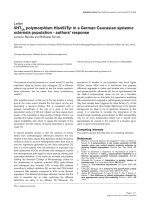
A brain MRI scan showed three millimetric lesions
located in the right temporooccipital lobe (Figure 1A),
the left pontine region (Figure 2B) and the right parietal
lobe (Figure 2C) with restricted diffusion (Figures 1A to
1C) and no enhancement after gadodiamide injection.
These findings, that is, hyperintensity on f luid attenu-
ated inversion recovery (FLAIR) images, slight enhance-
ment on postgadoliniu m T1-we ighted images, restricted
Figure 1 (A) Right temporooccipital l esion can be easily detected as a hyperintense spot in T1-weighted sequences. (B) The lesion
shows restricted diffusion, an absence of perilesional edema. (C) No enhancement is observed after gadodiamide injection. These findings are
suggestive of areas of microischemic phenomena.
Quattrocchi et al. Journal of Medical Case Reports 2011, 5:84
/>Page 2 of 5
diffusio n and no contrast enhancement, were suggestive
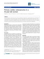
of areas of acute microischemic strokes. Although unu-
sual in the context of stroke, the subcortical lesio n at
the level of the right tempo rooccipital white matter was
confirmed to be unchanged with regard to size and
signal intensity on FLAIR images obtained at the one-
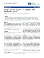
year follow-up examination (Figure 3). Moreover, other
bilateral centrum semiovale lesions not detected on dif-
fusion-weighted images and not showing contrast
enhancement were hypointense with a hyperintense
Figure 2 Other lesions suggestive of areas of acute embolic strokes are (A) the right parietal lobe, (B) the left pontine regio n and (C)
the right parietal lobe (alternate view). (D) Another bilateral centrum semiovale lesion not detected on diffusion-weighted images, without
contrast enhancement, is hypointense in this fluid attenuated inversion recovery (FLAIR) image with a hyperintense gliotic peripheral ring,
suggestive of small vascular ischemic microlacunae.
Quattrocchi et al. Journal of Medical Case Reports 2011, 5:84
/>Page 3 of 5
gliotic peripheral ring visualized on FLAIR images,
suggestive of areas of chronic microischemic origin
(Figure 2D). These lesions appeared as millimetric spots
of hyperintensity on T2-weighted images obtained at
the one-year follow-up examination. No other risk fac-
tors for thromboembolic events were recognized: the
patient’s clinical history was negative for hypertension,
hypercholesterolemia, hyper triglyceridemia, diabetes,
obesity, smoking, atrial fibrillation, heart d isease, atrial
or ventricular septal defects and previous episodes of
thrombosis or symptoms correlated to thrombosis. Her
pharmacological history was negative for anticoagulant
or procoagulant drugs. Her platelets were 161 × 10
3
/μL
(normal range, 150 to 450), her International Normal-
ized Ratio was 1.08 (normal range, 0.85 to 1.16) and her
activated partial thromboplastin time ratio w as 0.84
(normal range, 0.82 to 1.20).
An ultrasound Doppler study was obtained, which
showed normal morphology of supraaortic vessels. In
addition, the patient’s electrocardiogram and echocar-
diogram were normal.
Low-molecular-weight heparin (6,000 IU twice daily),
edema therapy (8 mg of dexamethasone twice daily) and
antiplatelet therapy (200 mg/day aspirin) were adminis-
tered, resulting in complete resolution of the pat ient’ s
neurological symptoms.
Discussion
Bevacizumab , a humanized antibody directed against the
vascular endothelial growth factor (VEGF) that is used as
an angiogenesis inhibitor, has been examined in combi-
nation with chemotherapeutic agents in several clinical
trials in patients with advance d colorectal cancer [1],
even as a first-line treatment [2]. The addition of bevaci-
zumab increased the overall response rate and extended
median survival. In the past four years, bevacizumab has
been used with increasing frequency for the treatment of
other neoplasms, such as breast cancer, non-small cell
lung cancer, pancreatic cancer and renal cell carcinoma.
Several adverse events have been associated with the
use of bevacizumab: h ypertension (the most common
side effect), gastrointestinal pe rforatio n, wound-healing
complications, hemorrhage, arterial thomboembolic
events, proteinuria, congestive h eart failure, leukopenia
and diarrhea [3]. Arterial thromboembolic events have
been observed in 0.9% of the cases in the BEATrial [3]
and in 2.1% of the cases in the BRiTE Registry [4]. The
mech anism of co ncurrent thrombosis and bleeding dur-
ing bevacizumab treat ment is not clear, being relat ed to
the role of VEGF in maintaining a healthy endothelium.
Conclusion
Vascular events involving the central nervous system
have been reported as reversible posterior leukoence-
phalopathy syndrome following a bevacizumab or FOL-
FIRI treatment regimen for metastatic colon cancer,
which are likely related to high systolic blood pressure
levels [5]. Furthermore, as thromboembolic events and
microischemic phenomena are a well-known complica-
tion of bevacizumab chemotherapeutic treatment [6],
the o ccurrence of neurological focal symptoms leads to
an MRI-based differential diagnosis between focal par-
enchymal metastases and microischemic phenomena,
which lead to crucial decisions for patient management.
Consent
Written informed consent was obtained from the patient
for publication o f this case report and accompanying
images. A copy of the written consent is available for
review by the Editor-in-Chief of this journal.
Author details
1
Interdisciplinary Center for Biomedical Research, Department of Radiology,
University Campus Bio-Medico of Rome, via Longoni, 47 I-00155 Rome, Italy.
2
Department of Bio-imaging and Radiological Sciences, Catholic University of
Figure 3 FLAIR image obtained at the patient’ s one-year
follow-up magnetic resonance imaging (MRI) examination
demonstrating the subcortical lesion at the level of the right
temporooccipital white matter. The lesion was unchanged with
regard to size and signal intensity compared with the previous MRI
(see Figure 1A).
Quattrocchi et al. Journal of Medical Case Reports 2011, 5:84
/>Page 4 of 5
Sacred Heart, Policlinico A. Gemelli. Largo F. Vito, I-00100 Rome, Italy.
3
Interdisciplinary Center for Biomedical Research, Oncology, University
Campus Bio-Medico of Rome, via Longoni, 47 I-00155 Rome, Italy.
Authors’ contributions
GT and YE analyzed and interpreted the patient data regarding the primary
disease (colon cancer) and decided on the therapeutic strategy. CCQ, RFG
and BBZ performed the brain magnetic resonance imaging. CCQ and AMA
were the major contributors in writing the manuscript. All authors read and
approved the final manuscript.
Competing interests
The authors declare that they have no competing interests.
Received: 1 April 2010 Accepted: 27 February 2011
Published: 27 February 2011
References
1. Meyerhardt JA, Mayer RJ: Systemic therapy for colorectal cancer. N Engl J
Med 2005, 352:476-487.
2. Saif MW, Merritt J, Robbins J, Stewart J, Schupp : Phase III multicenter
randomized clinical trial to evaluate the safety and efficacy of CoFactor/
5-fluorouracil/bevacizumab versus leucovorin/5-fluorouracil/bevacizumab
as initial treatment for metastatic colorectal carcinoma. Clin Colorectal
Cancer 2006, 6:229-234.
3. Van Cutsem E, Michael M, Berry S, Dibartolomeo M, Rivera F, Kretzschmar A,
Mazier M, Lutiger B, Cunningham D: Preliminary safety and efficacy of
bevacizumab with first-line FOLFOX, XELOX, FOLFIRI, and capecitabine
for mCRC: First BEATrial. Presented at the 2007 Gastrointestinal Cancers
Symposium, Orlando, FL, USA;, [Abstract 346].
4. Kozloff M, Hainsworth J, Badarinath S, Cohn A, Flynn P, Steis R, Dong W,
Suzuki S, Sugrue M, Grothey A: Efficacy of bevacizumab plus
chemotherapy as first-line treatment of patients with metastatic
colorectal cancer: updated results from a large observational registry in
the US (BRiTE). Presented at the 2006 ASCO annual meeting
proceedings, Part I (Abstract 3537). J Clin Oncol 2006, 24(18S).
5. Glusker P, Recht L, Lane B: Reversible posterior leukoencephalopathy
syndrome and bevacizumab. N Engl J Med 2006, 354:980-981.
6. Friedman HS, Prados MD, Wen PY, Mikkelsen T, Schiff D, Abrey LE,
Yung WK, Paleologos N, Nicholas MK, Jensen R, Vredenburgh J, Huang J,
Zheng M, Cloughesy T: Bevacizumab alone and in combination with
irinotecan in recurrent glioblastoma. J Clin Oncol 2009, 27:4733-4740.
doi:10.1186/1752-1947-5-84
Cite this article as: Quattrocchi et al.: Brain microischemic phenomena
in a woman receiving bevacizumab treatment: a case report. Journal of
Medical Case Reports 2011 5:84.
Submit your next manuscript to BioMed Central
and take full advantage of:
• Convenient online submission
• Thorough peer review
• No space constraints or color figure charges
• Immediate publication on acceptance
• Inclusion in PubMed, CAS, Scopus and Google Scholar
• Research which is freely available for redistribution
Submit your manuscript at
www.biomedcentral.com/submit
Quattrocchi et al. Journal of Medical Case Reports 2011, 5:84
/>Page 5 of 5