báo cáo khoa học: "The use of some nanoemulsions based on aqueous propolis and lycopene extract in the skin’s protective mechanisms against UVA radiation" pps
Bạn đang xem bản rút gọn của tài liệu. Xem và tải ngay bản đầy đủ của tài liệu tại đây (613.97 KB, 9 trang )
RESEARCH Open Access
The use of some nanoemulsions based on
aqueous propolis and lycopene extract in the
skin’s protective mechanisms against UVA
radiation
Monica V Butnariu
1*†
, Camelia V Giuchici
2†
Abstract
Background: The use of natural prod ucts based on aqueous extract of propolis and lycopene in the skin’s
protective mechanisms against UVA radiation was evaluated by means of experimental acute inflammation on rat
paw edema. The aim of the present study was to evaluate the harmlessness of propolis - lycopene system through
evaluation of skin level changes and anti-inflammatory action. The regenerative and protective effect of the
aqueous propolis and lycopene extract is based on its richness in biologically active substances such as:
tocopherols, flavonoids, amino acids, polyunsaturated fatty acids, the chlorophyll pigment, all substances with
strong antioxidant activity, that modify the oxidative stress, main ly by reducing the prooxidant processes and
enhancing the antioxidant ones. These substances participate in the synthesis of prostaglandins and phospholipids
components of cell membrane thus enhancing skin protection mechanisms.
Results: The experimental systems offered a sustained release of the drug, in vitro, for aim eight hours. The
prepared formulations aim did not reveal a deteriorating effect on tissues. They proved a better therapeutic
efficiency Compared to standard suspension, they provided a better therapeutic efficiency coupled with extended
time interval of tested parameters (24 hours). Preliminary examination of tissues showed that the experimental
formulations did not irritate. Local application of propolis and lycopene aqueous extract nanoemulsi on has a high
potential both regarding its efficiency (the analgesic effect) and therapeutic safety.
Conclusions: This study demonstrates that propolis and lycopene extract nanoemulsions, preparations contains
active substances, can confer better therapeutic effects than those of the conventional formulations, based on local
control-release of dozed form, for a longer period of time, which probably improve its efficiency and skin acceptance,
meaning a better compliance. The information obtained in the present study suggests that administration of propolis
and lycopene aqueous extract nanoemulsion is safe. The preparation can be useful for further preclinical studies
lycopene embedded in aqueous propolis extract to be used in pharmaceuticals (targeted medical therapy).
Background
In recent years, it has been noticed that the incidence of
skin cancer has increased alarmingly. Exposure to UV
irradiation has instantaneous effects (erythema and pig-
mentation) and delayed effects (premature skin ageing
and differe nt forms of cancer) [1]. UVB radiation has a
stronger energy compared to UVA radiation and is
absorbed directly by a series of cellular constituents,
such as nucleic acids, proteins and urocanic acid. UVB
radiation has also mutational effect [2]. UVA radiation
penetrates easily throu gh epidermis and acts on its basal
proliferative layer and even on blood components of the
dermis [3,4]. It acts indirectly on the cellular constitu-
ents, through oxidative mechanisms that forma reactive
oxygen species [5,6]. Reactive oxygen species have a
relative short lifespan, nevertheless are highly reactive
* Correspondence:
† Contributed equally
1
Exact Sciences Department, Banat’s University of Agricultural Sciences and
Veterinary Medicine from Timisoara, Calea Aradului no.119, 300645 Timisoara,
Romania
Full list of author information is available at the end of the article
Butnariu and Giuchici Journal of Nanobiotechnology 2011, 9:3
/>© 2011 Butnariu and Giuchici; licensee BioMed Central Ltd. This is an Open Access art icle distributed under the terms of the Creative
Commons Attribution License ( g/licenses/by/2.0), which permits unrestricted use, distribution, and
reproduction in any medium, provi ded the original work is properly cited.
with the vast majority of cellular components: nucleic
acids, proteins, lipids, polysaccharides. Frequently their
action induces irreversible modifications [7,8]. UVA
radiation acts upon biological environments through
oxidative mechanisms, correlated with the formation of
reactive oxygen species: singlet oxygen, hydroxyl radi-
cals, superoxide anions, hydrogen peroxide [9]. Nucleic
acids and proteins adsorb poorly radiation however but
the initial event triggering biological effects is made up
of absorption of UVA photons by different chromo-
phores in the cellular environment such as: quinones,
steroids, porphyrins, proteins with flavin coenzymes and
heme group (cytochrome, peroxidase, catalase) [10].
Many cellular components are targed by reactive oxygen
species generated by UVA irradiation [11,12].
Hydroxyl radic als react with almost all cell molecules
types: carbohydrates, phospholipids, nucleotides, organic
acids and amino acids. On enzymes, the effect of reac-
tive oxygen species results in catalytic capacity reduc-
tion, often determined by sulphhydryl oxidation and
modification of amino groups by malonylation [13].
Organisms are protected against reactive oxygen species
attack in several ways: cellular compartmentalization,
protection afforded by antioxidant compounds and
enzyme systems, their ability to develop adaptive
responses inducible under oxidative stress conditions.
Repair and turnover processes help to minimize these
[14,15]. Under normal circumstances there is a balance
between antioxidant systems and reactive oxygen gen-
erative systems. Lack of balance in favor of prooxidant
systems causes the apparition of oxidative s tress, with
pathological implications [16]. Skin is the organ most
exposed to solar radiation [17]. Skin presents a ser ies of
structures with a protective role, such as stratum cor-
neum and melanin. Superficial corneum layer functions
as optical barrier by reflection, scattering and absorption
of incident radiation. Larger part of UVA radiation
penetrates deeply into the skin, to dermis [18]. UVA
radiation can be absorbe d by diff erent components of
the blood, at the level of blood vessels. UVA radiation
acts as inducer of enzymes responsible for polyamine
synthesis. An additional mechanism of epidermis protec-
tion is to stimulate skin pigmentation with melanin [19].
The protection mechanisms are established and induci-
ble protections at the skin level. Inducible defence
mechanisms were not identified at epidermis [20], but
was identified in dermis, w here increased heme oxyge-
nase, which are correlated with an increase in ferritin
levels [21]. Pharmaceutical and cosmetics industries
have launched a wide range of substances that act as fil-
ters capable of absorbing UV photons [22]. Photoprotec-
tion products are characterized by the protection factor.
An accurate assessment of the effectiveness of photopro-
tection products should be based on their ability to
inhibit the isomerisation reaction of urocanic acid and
prevent accumulation of the protein [23]. “Quantum
dot” nanostructures have been used (nanopartic les wit h
quantum properties and ability to change size according
to light emission). Another reason for the use o f these
products is their ability to “connect” many substances,
thanks to a large su rface area, and easy transport due to
their small sizes (10 to 100 nanometers). These sub-
stances can also remain and accumulate preferential ly at
skin level, facilitated by surface drainage [24]. Currently,
nanomedicine is seen not only as a possible and promis-
ing path to an early and effective treatment, but also a
possible way to prevent certain types of diseases [25].
Results
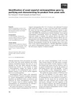
Characterization of lycopene extract
Lycopene (F igure 1) belongs to the class of natural pig-
ments, called carotenoids, with a role in pro tecting the
body from the destructive effects of oxidants. Lycopene
neutralizes the negative effects of free radic als. Further-
more, recent studies have shown that lycopene has a
significant potential to counter free radicals in compari-
son with b-Carotene. Validation was confirmed by appli-
cation of standard techniques lycopene determination.
Aqueous extract of propolis has a high concentration of
polyphenols and is standardized in polyphenol car-
boxylic acids (caffeic acid), responsible, among other
active substances, for its healing and anti-inflammatory
action upon tegument affected by dandr uff and sebor-
rheic dermatitis. Thus, the antimicrobial and anti-
inflammatory action of lycopene is enhanced and
regeneration of skin affected by f unga l and/or microbial
infections is stimulated. The product formulation also
considered physiological aspects of the skin, resulting in
a product with low allergenic potential and high
degreasing capacity. Because of t he nanoemulsion phar-
maceutical form, it has several advantages over other
topical products with similar action. Thus, in contact
with skin it quickly releases active substances due to its
good adherence to skin and close contact it has a high
therapeutic efficiency, easy administration low all ergenic
potential, good local tolerance and an increased viscos-
ity, allowing the required concentrat ion in bioactive
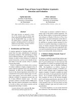
compounds. Figure 2 clearly shows the absorption spec-
trum of the extracted lycopene solution. The absorption
spectrum very closely coincided with the three peaks
characteristic of trans-lycopene (l = 446, 472, 505 nm).
Figure 1 Molecule of lycopene (chemical structure).
Butnariu and Giuchici Journal of Nanobiotechnology 2011, 9:3
/>Page 2 of 9
Any analytical method (bio analytical, in particular), in
order to be valid ated, must demonstrate first that it is
specified in relation to existing endogenous substances
in the biological matrix, to metabolism products and
reagents used in the sample preparation. For that, the
specific ity of this method was verified using six different
sources of blank solutions. We aimed to see if there was
any endogenous interfe rence at the retention times of
the experimental analytes. The linearity of the method
was verified by the method of smallest squares, on the
0-3.0 mg/L lycopene domain, using internal standard
calibration as calibration model. Lycopene area and
standard area ratio were calculated for seven leve ls of
concentration in the selected domain and used for cali-
bration curves. For each calibration point we examined
the distribution, the relative percentage deviation of
recalculated concentration from calibration curve equa-
tion. The calibration model was considered correct if
residuals were within boundaries of ± 20% at lower limit
and ± 15% at other concentrations and did not have a
trend of increase or decrease along with concentration.
Correlation was considered linear at a value of the
determination coefficient greater than 0.99, as seen in
Figure 3. Accuracy, expressed as relative pe rcentage
deviation of measured concentration in relation to the a
obtained concentration, and precision, expressed as
standard relative percentage deviation or coefficient of
variation CV%, were determined at thre e concentration
levels. Both, precision and accuracy were de termined on
the same day, based on five measurements on five dif-
ferent samples at each concentration, and accuracy and
precision on different days based on the analysis on di f-
ferent days of five standard samples at each of the three
levels of tested concentrat ion. The lowest limit of quan-
tification was considered the lowest concentration on the
calibration line with an accuracy and pre cision within
± 20%. Retrieval was assessed at four c oncentrations,
including the lower limit of quantification, comparing the
response obtained after application of UV radiation with
that obtained with a standard solution of the same con-
centration in water and similarly processed as biological
samples.
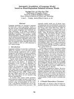
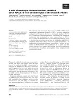
FTIR analysis
All FTIR analysis is considered technically “non-destruc-
tive” therefore further analysis can be performed. Com-
position analysis by FTIR spectroscopy (Fourier
Transform Infrared) allows quantitative estimates and
the study of links nature that appear during the nanoe-
mulsions process. Following FTIR spectra analysis, no
significant differences were apparent between the pro-
ducts. As shown in Figure 4, differences were found in
FTIR spectra in the region 900-500 nm regarding
experimental conditions and the resulting products. IR
spectra highlight the presence of -CH
2
- groups illu-
strated by the characteristic bands at 1450 cm
-1
and
1460 cm
-1
respectively. The nanoemulsion ester group
causes the appear ance of characteristi c frequency bands
at 1700 cm
-1
(-C = O stretching) given by a larger
amount of propolis and lycopene. Increase in propolis
content (the 30% option) determines the appearance of
new frequency bands characteristic of the carbonyl
group (1900-1600 cm
-1
-C = O stretching). Growth
leads (in propolis) to the disappearance of intense bands
at 750-1280 cm
-1
, assigned to ring vibrations: 1235-1280
cm
-1
, 810-905 cm
-1
and 805-875 cm
-1
and can be attrib-
uted to characteristic methyl bands shielding or to the
dissolution of certain groups during the process of
obtaining the nanoemulsion. In the case of the bands
group in the range of 115 0-1250 cm
-1
, characteristic of
ester groups (-C-O stretching), an increase in the inten-
sity of the bands is observed, which is reflected in the
decrease of its transmittance from 96% to 56%. This is
also explained by the increasing of the quantity of pro-
polis from 27% v/v to 35% v/v in the nanoemulsion.
High lycopene content of 35% has the effect of increased
intensity of certain bands at 1450 cm
-1
(-CH
3
and CH
2
= strain) refle cted in t he decrease of the transmittance
Figure 2 UV-VIS spectrum of lycopene extracted from
tomatoes and of standard of lycopene.
Figure 3 The calibration curve of lycopene standard.
Butnariu and Giuchici Journal of Nanobiotechnology 2011, 9:3
/>Page 3 of 9
from 92% to 70.846%. An intensity increase was observed
at 1730 cm
-1
(-C = O stretching) a characteristic of unsa-
turated esters. This growth is highlighted by the decrease
of transmittance from 97.95% to 47.126% and is
explained by 35% propolis content. The intensity signifi-
cant increase of nanoemulsion bands characteristic is
attributed to the increase of distance interactions
between atoms and molecules, through altered angles
between the links. In the case of experimental formulated
nanoemulsions, for second formulation spectral bands
intensity was reduced, which is consistent with the
reported kinetic data (Table 1). Table 1 shows correlation
coefficients of data regarding the release of formulated
nanoemulsions, obtained from the Higuchi model, zero
order kinetic and release exponent values (n) obtained
from the equation Mt/Mo = ktn regarding the prepared
nanoemulsions based on propolis lycopene (n = 3). Dur-
ing these experiments no changes were observed in the
organoleptic properties of experimental formulations and
pH value of the two nanoemulsions remained within the
limits of ± 0.3 pH units. No significant changes were
noticed in terms of particle size (p < 0.05).
The properties of the two experimental formulations
Micrometric properties of the two experimental formul a-
tions are shown in Table 2. This experiment highlighted
the improved performances of the se emulsions without
side effects and adverse reactions by replacing synthetic
chemicals with natural products. It has been documen-
ted, that some UV radiation absorbers can be partially
degraded therefore causing skin alterations while exposed
Figure 4 IR spectrum of nanoemulsions in KBr disc, obtained from experimental analysis.
Table 1 Data correlation coefficients regarding the
release of active constituents of propolis lycopene
systems
Sample “n” R Higuchi Model Zero-order kinetics
k(%h
-1/2
)R k(%h
-1
)R
formulation with 20% lycopene, 27% propolis, 53% water vol./vol
(nanoemulsion 1)
Newly prepared 4.45 0.997 0.62 0.994
After one week 0.80 0.971 3.18 0.998 0.70 0.998
After two weeks 4.21 0.982 0.78 0.998
After three weeks 4.16 0.987 0.77 0.999
formulation with 35% lycopene, 35% propolis, 30% water vol./vol
(nanoemulsion 2)
Newly prepared 3.47 0.983 0.71 0.998
After one week 0.98 0.980 3.87 0.996 0.67 0.995
After two weeks 3.68 0.999 0.62 0.999
After three weeks 3.48 0.989 0.68 0.990
Table 2 Micrometric properties of the two formulations
tested
No. Micrometric property Nanoemulsion
1
Nanoemulsion
2
1 Response angle 35.60 ± 1.33 51.94 ± 1.68
2 Density of the nanoemulsion
(merged)
0.38 ± 0.03 0.41 ± 0.02
3 Density of the nanoemulsion 0.53 ± 0.07 0.42 ± 0.02
4 % Porosity 42 ± 1.3 68 ± 2.4
Each value represents the average (± SD) of three independent
determinations.
Butnariu and Giuchici Journal of Nanobiotechnology 2011, 9:3
/>Page 4 of 9
to UV influencing the effectiveness of sunscreen protec-
tion. For information on UV absorbers, the nanoemul-
sions SPF in vitro parameters were investigated. UVA
radiation, the UVA/UVB ratio and in vitro SPF were
measured using the Diffey and Robson method. The effi-
ciency of these products is measured by a coefficient,
index or protective factor, noted SPF (sun protection fac-
tor) or PI (protection index), which is the ratio of the
minimum dose of solar radiation t hat causes lowest skin
redness, after and before skin application of these
preparations.
The higher the SPF values, the more efficient the
photoprotection
This experiment was conducted to determine the photo-
protection capacity of natural substances in nanoemul-
sions. In both nanoemulsions we also introduced a
UVR-absorber (Saliform), accepted by European stan-
dards, to observe its influence on the analyzed samples.
In Table 3 presents the absorbance’s of two nanoemul-
sions measured by UVA, UVA/UVB ratio and SPF. The
obtained data show that most absorbance in the U V
domain in the relative parameter 5 is present in nanoe-
mulsion 2+ absorber-UVR (Saliform) 1:1 (v/v). Regard-
ing UVA/UVB ratio with the relative parameter 0.82,
most absorbance is found in the same nanoemulsion
and in the case of SPF with absolute parameter 10.9, in
nanoemulsion 2. As shown by these results, highest
absorbance parameters may be assigned to nanoemul-
sion 2. Experimental formulations showed a pseudo-
plastic rheology, under the influence of shear stress
(Figure 5). The viscosity is directly dependent on the
formulations content of propolis. Microscopic observa-
tions of the experimental nanoemulsions confirmed for-
mation of spherical particles, as shown in Figure 5.
Differences in droplet size of these formulations were
not statistically significant (droplet size was found to be
45 μ m). Nanoemulsions were kept for three weeks at
4°C in order to provide a stable formulation for local
application. Nanoemulsion stability is shown in Figure 6,
7 and 8. This study suggests that the nanoemulsions
made of aqueous extract of propolis and lycopene and
prepared with active substances may confer better
therapeutic eff ects than conventional formulations, as a
result of dose controlled local release longer period of
time, which could lead to a greater efficiency and to a
greater acceptance b y the skin (i.e. better compliance).
These results are supported by presented data in Table
4. Collagenase (inactive form of pre-collagenase, acti-
vated by trypsin) is an enzyme that cleaves the peptide
bonds of fibrillar collagen types I and III. Experimental
results showed that both nanoemulsions induced a
reduction in collagenase activity. The intensity of anti-
inflammatory effect varies with time interval covered by
the assessment of therapeutic response according to
data in Table 4. The activity of propolis and lycopene
nanoemulsions (Table 5) was emphasized by measuring
the induced inflammation. Produced inflammation
decreased by 75% for nanoemulsion 1 and 100% for
nanoemulsion 2. The maximal inflammation reduction
effect occurred after 8 hours from the initial application.
Preliminary tissue examination showed that these for-
mulations did not produce irritation. Local application
of the experimental nanoemulsion (propolis and lyco-
pene) has g reat potential , both in te rms of effi cacy
Table 3 UVA radiation, the UVA/UVB ratio and
nanoemulsion SPF with and without UVR-absorber
No. Samples UVA UVA/UVB SPF
1 Nanoemulsion 1+absorber-UVR
(Saliform)1:1 (v/v)
4.0 0.71 5.9
2 Nanoemulsion 2 +absorber-UVR
(Saliform)1:1 (v/v)
5 0.82 6.9
3 Nanoemulsion 1 3.1 0.24 9.1
4 Nanoemulsion 2 3.2 0.815 10.9
Figure 5 Dependence between the absorb ance parameters of
the two formulations.
Figure 6 Nanoemulsi on 2 layer (aqueous extract of p ropolis
and lycopene) after one week.
Butnariu and Giuchici Journal of Nanobiotechnology 2011, 9:3
/>Page 5 of 9
(analgesia) and therapeutic safety. Nanoemulsions have
proved a better therapeutic efficacy compared to stan-
dard suspension, was observed improving monitored
parameters for a longer period of time (24 hours). These
systems can be seen as a viable alternative to conven-
tional creams, due to their ability to improve residence
time and thereby, bioavailability.
Discussion
Emulsions are a mixture of molecules in a combination
of two liquids that keep their properties unaltered. This
feature has been used in the delivery of poorly soluble
drugs [26]. N anoemulsions have a greater capacity for
micellar solubilisation compared to simple solutions and
offer advantages in thermodynamic stability to unstable
dispersions (suspensions), as can be produced with less
energy input and have a greater shelf life [27]. The
nanoemulsions are systems with droplet sizes of
approximately 45 μm, having surfactant ratios of 47/53
and respectively 70/30 of aqueous extract of propolis-
lycopene. UVA absorbance with relative parameter 5 is
manifested by nanoemulsion 2 +absorber-UVR (Sali-
form) 1:1 (v/v), in the case of the UVA/UVB ratio with
relative parameter 0.82 by the same nanoemulsion and
in the case of SPF with absolute parameter 10.9 by the
nanoemulsion 2. Highest absorbance parameters can be
assigned to nano emulsion 2. Prepared formulations
showed a pseudo-plastic rheology, under the influence
of shear stress. It appears that viscosity is directly
dependent on the propolis content of the formulation.
Lycopene is insoluble in water; it can be dissolved only
in organic solvents and oils [28-30]. Researchers have
correlated the antioxidant function of lycopene (ability
to protect cells and other body structures caused by oxi-
dative damage) with the protection of DNA (ou r genetic
material) inside the white blood cells [31].
White blood cells (WBC) are mediators of inflamma-
tion and the immune response. Unlike other foo d phy-
tonutrients, whose effects have only been studied in
animals, lycopene from tomatoes has been repeatedly
studied in humans, where research has shown additional
protection against many types of diseases [32,33].
Conclusions
The experimental nanoemulsions in a high kinetic stabi-
lity, and reduction in collagenase activity by 37.14% for
a 70/30 surfactant ratio and respectively 26.81% for a
47/53 ratio. These nanoemulsions provided a sustained
drug release in vitro for a period of 8 hours. Lycopene
antioxidant as a nanoemulsion component beside its
moisturizer characteristicimprovestheabilityofthe
skin to defend against sunlight.
Methods
Aqueous extract of propolis
Aqueous extract of propolis was obtained by refluxing
100 g of propolis powder and 250 ml of double distilled
water. It was concentrated in a water bath then filtered
resulting in 1.5 cm
3
of extract with 95% of dry substance.
Determination of lycopene
Lycopene was obtained from ripe tomatoes (Lycopersi-
con esculentum) by solvent extraction. Samples were
homogenized in a laboratory homogenizer. 5ml 0.05%
BHT in acetone, 5 ml of ethanol and 10 ml of hexane
were added to 0.6 g homogenated sample. The supple-
mented homoge nate was kept on ice and stirred with a
magnetic stirrer for 15 minutes. Then 3 ml of deionized
Figure 7 Nanoemulsi on 2 layer (aqueous extract of p ropolis
and lycopene) after two week.
Figure 8 Nanoemulsi on 2 layer (aqueous extract of p ropolis
and lycopene) after three weeks.
Table 4 Enzymatic activity of collagenase in the presence
of nanoemulsions
Substance Concentration
(μg/ml)
Enzymatic activity
(Units/mg of
protein)
%
Inhibition
Nanoemulsion 1 40 1.205 ± 0.001 26.81
Nanoemulsion 2 40 1.033 ± 0.003 37.14
Without
nanoemulsion
- 1.648 ± 0.007 -
Reaction conditions: T = 25°C, pH 7.5, l = 345 nm, t = 5 min.
Butnariu and Giuchici Journal of Nanobiotechnology 2011, 9:3
/>Page 6 of 9
water were added and samples were mixed for addi-
tional 5 minutes. Samples w ere then left at room tem-
perature for 5 minutes to allow phase sep aration. The
absorption of the hexane layer (upper layer) was mea-
sured in a 1cm quartz cuvette at a wavelength of 503
nm, against hexane as blank. Lycopene was measured
quantitatively by UV-VIS spectrophotometer T60U, PG
Instruments Limited, UV WIN
®
version 5.05; detection
was performed at 503 nm and calculated using the fol-
lowing formula:
Absorbance at 503 nm (A
503
)=
ε (M
-1
·cm
-1
)·b(cm)
[Lycopene concentration (M)]
The measuring conditions were: scan speed 90 nm/
min and an interval of 1 nm. After extraction, was hex-
ane evaporated to dryness in a vacuum evaporator,
under a nitrogen stream [34]. All substances were pur-
chased from Sigma Chemical.
Preparation of nanoemulsions
Nanoemulsions were prepared by adding lycopene to
aqueous solution of propolis (50 mg propolis in 10 ml D.
W.), using a magnetic stirrer at ~ 2000 rpm . The mixture
was introduced for in an ultrasonic bath at 20 kHz 20
minutes. The nanoparticles that are formed have a lyco-
pene-propolis loaded shape. After ultrasonic treatment
the solution is brought at room temperature (22°C).
The excess organic solvent in excess was evaporated
using a rotary evaporator and samples were kept for
further analysis by lyophilisation. Independent of pre-
paration temperature, samples were kept at 25°C [35].
Fourier transforms infrared spectrometry (FTIR)
For spectral characterization (FTIR spectrometry), sam-
ples were prepared as follows: compounds obtained
after heat treatment were mixed at a temperature of
1300°C with potassium bromide powder, previously
dried for 24 hours at a temperature of 120°C, at a mass
ratio of 0.04:1. After a vigorous mix to obtain uniformi-
sation, pills with a thickness of 0.5-0.75 mm and 13 mm
in diameter at a pressure of 0.3 GPa in normal atmo-
spher e were prepared. The pills were analyzed using the
JASCO 660 PLUS spectrophotometer, which recorded
the IR absorption spectra in the area 4000 cm
-1
100-
400 cm
-1
.Forin vitro characterization of the experimental
nanoemulsions, permeability studies and rheological mea-
surements were carried out [36].
Permeability studies
Membrane permeability of the experimental nanoemul-
sions was investigated by filling the donor compartment
of a diffusion cell with a 2 g test mixture. All other
experimental conditions identical to those described for
permeability studies of solid systems [37].
Viscosity measurement
Apparent viscosity of the experimental nanoemulsions
was determined using a viscometer Brookfield Rheos-
tress DV-III + Rheometer. Measurements were per-
formed 3 times at 25°C using SC4 spindle. To
determine the influence of shear stress applied to the
microstructure of t he prepared nanoemulsions, mea-
surementsweremadeatarotationspeedof1and10
rpm.
Apparent viscosity of the controlled product (2%
HPMC dispersion i n water), were examined under simi-
lar conditions [38].
Enzyme activity measurements
Enzyme activit y is determined by a c ontinuous spectro-
photometric method, using as substrate 2-L-leuc ylglycyl
furanacryloyl-L-prolyl-L-alanine (FALGPA, a specific
collagenase substrate), as it is preferentially hydrolyzed
much faster than other synthetic substrates.
Measurement of the substrate absorbance decrease
was done at 345 nm.
Pharmacological evaluation of formulations
Identification and quantification are done according to
the methods described in the European Directorate for
the Quality of Medicines [39].
Animal testing
Preparation: adult, young, he althy animals of the species
of guinea pigs, breed albino were used. The animals
were acclimatized to laboratory conditions for at least
five days before test. Animals were divided randomly
into treatment and control groups before test. Their
skin was cleaned by clipping, shaving or, if possible, by
chemical depila tion without excoriation (cleaning
Table 5 Activity of aqueous extract of propolis and lycopene assessed on induced mouse paw edema
Compound Mean ± SE difference in right and left paw volumes (ml) Reduction of edema (%)
4h 6h 8h 24h 4h 6h 8h 24h
Control 0.147 ± 0.02 0.186 ± 0.11 0.246 ± 0.03 0.165 ± 0.01
Regular sunscreen 0.039 ± 0.03 0.026 ± 0.10 0.020 ± 0.14 0.015 ± 0.004 69 67 72 73
Nanoemulsion 1 0.019 ± 0.03 0.014 ± 0.01 0.011 ± 0.02 0.007 ± 0.005 97 85 78 75
Nanoemulsion 2 0.013 ± 0.04 0.008 ± 0.02 0.003 ± 0.02 0.001 ± 0.00 99 99 99 100
Butnariu and Giuchici Journal of Nanobiotechnology 2011, 9:3
/>Page 7 of 9
method is based on the test method used). Animals have
been weighed before and after test.
Experimental procedure
Superficial skin burns were i nduced using a UV radia-
tion lamp.
Testing the anti-inflammatory action
Assessment of anti-inflammatory action was achieved by
evaluating the inhibition of rat paw edema i nduced by a
2% solution of carrageenan.
The percentage inhibition of edema was calculated
using the following equation:
Inhibition (%) =
Mean paw diameter (control) - Mean paw diameter (treated)
MMean paw diameter (control)
⋅ 100
The edema inhibition rate of each group was calcu-
lated as follows:
Inhibition (%) =
Mean number of writhing (control)-Mean of writhing (test)
MMean number of writhing (control)
⋅ 100
Monitoring and staging: approximately 2 1 hours after
the patch removal, hair is cleared off the surface exposed
to challenge concentration. After 3 hours (about 30 hours
after the challenge patch application) skin reactions were
observed and recorded. After an additional time of 24
hours (54 hours) skin reactions were observed and
recorded again. “Blind” reading is recommended for tested
and control animals. All skin reactions and any unusual
results, including syst emic reactions caused by induction
and challenge procedures were observed and recorded in
accordance with the Magnusson/Kligman staging. If any
of the reactions are difficult to interpret, other procedures
can be taken into account, for instance hi stopathological
examination or measurements of the skin fold.
Staging Magnusson/Kligman scale for assessing the
post-challenge responses: 0 = no visible change, 1 =
erythema or discrete form of spot, 2 = moderate and con-
fluent erythema, 3 = intense erythema and swelling [40]
Determination of in vitro sun protection factor (SPF)
Determination of SPF (sun protection factor) was per-
formed using a spectrophotometer, equipped with an
integrating sphere, with appropriate software and a
TRANSPOR 3 TM support, with a composition similar
to that of natural skin, on which the amount of 2 mg/
cm
2
of nanoemulsion was applied [40].
Statistical analysis
Values were expressed as mean ± S.D. Statistical signifi-
cance was ev aluated by Stude nts-„t‟ test at 5 % level of
significance (p < 0.05).
Acknowledgements
The authors would like to thank the European regional development fund
(ERDF) to finance project “Environment-Biochemical Cooperation for
prognosis of natural water and soil pollution in the Hungarian and
Romanian cross-border region to Shun Catastrophe” acronym “R & D SZTE,
BAÁE, no. HURO/0801/038”.
Author details
1
Exact Sciences Department, Banat’s University of Agricultural Sciences and
Veterinary Medicine from Timisoara, Calea Aradului no.119, 300645 Timisoara,
Romania.
2
Inspectorate for quality of seed and planting materials,
Delamarina Victor Vlad no. 3, 300077 Timisoara, Romania.
Authors’ contributions
CG participated in the design of the study and performed the statistical
analysis. MB conceived of the study, and participated in its design and
coordination. All authors read and approved the final manuscript.
Competing interests
The authors declare that they have no competing interests. The opinions
expressed in this article are those of the authors and do not necessarily
represent any agency determination or policy.
Received: 23 November 2010 Accepted: 4 February 2011
Published: 4 February 2011
References
1. Kumar A, Bagewadi A, Keluskar V: Efficacy of lycopene in the
management of oral submucous fibrosis. Oral Surg Oral Med Oral Pathol
Oral Radiol Endod 2007, 103:207-213.
2. Applegate LA, Frenk E: Cellular defense mechanisms of the skin against
oxidant stress and in particular UVA radiation. Eur.J.Dermatol 1995,
5:97-103.
3. Peak JG, Pilas B, Dudek EJ, Peak MJ: DNA breaks caused bz
monochromatic 365 nm ultraviolet-A-radiation and their repair in
human epithelioid and xeroderma pigmentosum cell. Photochem.
Photobiol 1991, 54:197-203.
4. Dean RT, Fu S, Stocker R, Davies MJ: Biochemistry and pathology of
radical-mediated protein oxidation. Biochem.J 1997, 324:1-18.
5. Shore RE: Radiation-induced skin cancer in humans. Med.Pediatr.Oncol
2001, 36:549-554.
6. Chandra RV, Prabhuji ML, Roopa DA: Efficacy of lycopene in the treatment
of gingivitis: a randomised, placebo-controlled clinical trial. Oral Health
Prev Dent 2007, 5:327-336.
7. Shao A, Hathcock JN: Risk assessment for the carotenoids lutein and
lycopene. Regul Toxicol Pharmacol 2006, 45:289-298.
8. Sesso HD, Buring JE, Norkus EP: Plasma lycopene, other carotenoids, and
retinol and the risk of cardiovascular disease in men. Am J Clin Nutr
2005, 81:990-997.
9. Halliday GM, Bestak R, Yuen KS, Cavanagh LL: Barnetson R.S. UVA-induced
immunosuppression. Mutat.Res 1998, 422:139-45.
10. Khachik F, Carvalho L, Bernstein PS: Chemistry, distribution, and
metabolism of tomato carotenoids and their impact on human health.
Exp Biol Med 2002, 227:845-51.
11. Balasubramanian D: Ultraviolet radiation and cataract. J.Ocul.Pharmacol.
Ther 2000, 16:285-297.
12. Hasegawa T, Kaneko F, Niwa Y: Changes in lipid peroxide levels and
activity of reactive oxygen scavenging enzymes in skin, serum and liver
following UVB irradiation in mice. Life Sci 1992, 50:1893-1903.
13. Chapple ILC: Reactive oxygen species and antioxidants in inflammatory
diseases. J.Clin.Paradontol 1997, 24:287-296.
14. Kuusilehto A: Transmission of UVA radiation through epithelium of oral
mucosa and skin in rat and man. Photodermatol.Photoimmunol.Photomed
2000, 16:189-191.
15. Cole C:
Sunscreen protection in the ultraviolet A region: how to
measure
the effectiveness. Photodermatol.Photoimmunol.Photomed 2001,
17:2-10.
16. Morliere P, Moysan A, Tirache I: Action spectrum for UV-induced lipid
peroxidation in cultured human skin fibroblasts. Free Rad.Biol.Med 1995,
19:365-371.
Butnariu and Giuchici Journal of Nanobiotechnology 2011, 9:3
/>Page 8 of 9
17. Cunningham ML, Johnson JS, Giovanazzi SM, Peak MJ: Photosenzitized
production of superoxide anion by monochromatic (290-405 nm)
ultraviolet irradiation of NADH and NADPH coenzyms. Photochem.
Photobiol 1985, 42:125-128.
18. Peak MJ, Peak JG, Carnes BA: Induction of direct and indirect single-
strand breaks in human cell DNA by far-and near-ultraviolet radiations:
Action spectrum and mechanisms. Photochem.Photobiol 1987, 45:381-387.
19. Stoker R: Induction of heme oxygenase as a defence against oxidative
stress. Free Rad.Res.Comms 1990, 9:101-112.
20. Wang SQ, Setlow R, Berwick M, Polsky D, Marghoob AA, Kopf AW, Bart RS:
Ultraviolet A and melanoma: a review. J.Am.Acad.Dermatol 2001, 44:837-846.
21. Dudek EJ, Peak JG, Roth RM, Peak MJ: Isolation of V79 fibroblast cell lines
containing elevated metallothionein levels that have increased
resistance to the cytotoxic effects of ultraviolet-A radiation. Photochem.
Photobiol 1993, 58:836-840.
22. Zigman S, McDaniel T, Schultz JB, Reddan J, Meydani M: Damage to
cultured lens epithelial cells of squirrels and rabbit by UV-A (99.9%) plus
UV-B (0.1%) radiation and alpha tocopherol protection. Mol.Cell Biochem
1995, 143:35-46.
23. European Directorate for the Quality of Medicines (EDQM), Council of
Europe. Strasbourg, Cedex 1, France;, 5 2005.
24. Goel RK, Singh A, Mahajan MP, Kulkarni SK: Evaluation of anti
inflammatory and anti hyperalgesic activity of some novel monocyclic
β-lactam compounds in rats. Indian J. Pharm. Sci 2004, 66:87-91.
25. Loftsson T, Masson M: Cyclodextrin in topical drug formulations: theory
and practice. Int. J. Pharm 2001, 225:15-30.
26. Roco Mihail C: Nanotechnology: convergence with modern biology and
medicine. CurrentOpinioninBiotechnology 2003, 14:337-346.
27. Blonska M, Bronikowska J, Pretsz G, Czuba ZP, Scheller S, Krol W: Effects of
ethanol extract of propolis (EEP) and flavones on inducible gene
expression in J774a, 1 makrophages. J. Ethnopharmacol 2004, 91:25-30.
28. Borrelli F, Maffia P, Pinto L, Ianaro A, Russo A, Capasso F, Lalenti A:
Phytochemical compounds involved in the antiinflammatory effectof
propolis extract. Fitoterapia 2002, 73:353-363.
29. Denli M, Cankaya S, Silici S, Okan F, Uluocak AN: Effect-of dietary addition
of Turkish propolis on the growth performance, carcass characteristics
and serum variables of quail (Coturnix Coturnix Japanica). J. Anim. Sci
2005, 18:5.
30. Basuny MA, Mostafa MD, Azouz A: Supplementation of polyunsaturated
oils with lycopene as natural antioxidant and antipolymerization during
heating process. Minia J. Agric. Res. Develop 2006, 26(3):449-469.
31. Chasse GA, Mak ML, Deretey E: An ab initio computational study on
selected lycopene isomers. J. Mol. Struc. (Theochem) 2001, 571:27-37.
32. Riso P, Visioli F, Grande S, Guarnieri S, Gardana C, Simonetti P: Effect of a
tomato-based drink on markers of inflammation, immunomodulation
and oxidative stress. J. Agric. Food Chem 2006, 54:2563-2566.
33. Omoni AO, Aluko RE: The anticarcinogenic and anti-atherogenic effects
of lycopene: a review. Food Sci. Technol 2005, 16:344-350.
34. Kaur D, Ali Abas Wani Oberoi DPS, Sogi DS: Effect of extraction conditions
on lycopene extractions from tomato processing waste skin using
response surface methodology. Food Chem 2008, 108:711-718.
35. Kaur D, Wani AA, Sogi DS, Shivhare US: Sorption isotherms and drying
characteristics of tomato skin isolated from tomato pomace. Drying
Technol 2006, 24:1-6.
36. Loftsson T, Masson M: The effects of water-soluble polymers on
cyclodextrins and cyclodextrins solubilisation of the drugs. J. Drug Del.
Sci. Tech 2004, 14:3-20.
37. WHO, Regional Office for Europe, Largely preventable chronic diseases cause
86% of deaths in Europe: 53 WHO European Member States map a strategy
to curb the epidemic. Press Release EURO/05/06, Copenhagen; 2006.
38. European Technology Platform on NanoMedicine. Nanotechnology for
Health-Vision Paper and Basis for a Strategic Research Agenda for
NanoMedicine 2005.
39. European Medical Research Councils (EMRC): Nanomedicine, an ESF-
European Medical Research Councils (EMRC) Forward Lookreport; 2005.
40. Singh Mritunjai , Singh Shinjini , Prasada S, Gambhir IS: Nanotechnology in
medicine and antibacterial effect of silver nanoparticles. DigestJournal of
Nanomaterials and Biostructures 2008, 3:115-122.
doi:10.1186/1477-3155-9-3
Cite this article as: Butnariu and Giuchici: The use of some
nanoemulsions based on aqueous propolis and lycopene extract in the
skin’s protective mechanisms against UVA radiation. Journal of
Nanobiotechnology 2011 9:3.
Submit your next manuscript to BioMed Central
and take full advantage of:
• Convenient online submission
• Thorough peer review
• No space constraints or color figure charges
• Immediate publication on acceptance
• Inclusion in PubMed, CAS, Scopus and Google Scholar
• Research which is freely available for redistribution
Submit your manuscript at
www.biomedcentral.com/submit
Butnariu and Giuchici Journal of Nanobiotechnology 2011, 9:3
/>Page 9 of 9