Báo cáo y học: "A giant adrenal pseudocyst presenting with right hypochondralgia and fever: a case repor" pptx
Bạn đang xem bản rút gọn của tài liệu. Xem và tải ngay bản đầy đủ của tài liệu tại đây (827.17 KB, 5 trang )
CAS E REP O R T Open Access
A giant adrenal pseudocyst presenting with right
hypochondralgia and fever: a case report
Masashi Momiyama
1*
, Kenichi Matsuo
1
, Kenichi Yoshida
1
, Kuniya Tanaka
1
, Hirotoshi Akiyama
1
, Shoji Yamanaka
2
and Itaru Endo
1
Abstract
Introduction: Adrenal pseudocysts are rare cystic masses that arise from the adrenal gland and which are usually
non-functional and asymptomatic. Adrenal pseudocysts consist of a fibrous wall without an epithelial or endothelial
lining. We report the case of a patient with a gian t adrenal pseudocyst presenting with right hypochondralgia and
high fever.
Case presentation: A 52-year-old Japanese man was admitted with right hypochondralgia and a chill. Abdominal
computed tomography revealed a well-defined cystic mass measuring 19 cm which was located in the right
adrenal region and the contents of which were not enhanced with contrast medium. Abdominal ultrasonography
revealed a heterogeneously hypo -echoic lesion with a peripheral high-echoic rim. Serum hormonal levels were
almost normal. Despite treatment with antibiotics, the high fever persisted. Based on these findings, we made a
preoperative diagnosis of a right adrenal cyst with infection. However, the possibility of malignancy still remained.
The pa tient underwent laparotomy and right adrenal cyst excision with partial hepatectomy in order to relieve the
symptoms and to confirm an accurate diagnosis. Histological examination revealed an adrenal pseudocyst with
infection. His condition improved soon after the operation.
Conclusion: We report a case of a giant adrenal pseudocyst with infection. Surgery is required for sy mptomatic
cases in order to relie ve the symptoms and in cases of uncertain diagnosis.
Introduction
In 1903, Doran attributed the first case of adrenal cyst
to Greiselius [1]. In 1670, he described a 45-year-old
man whose death resulted from a rupture of the cyst.
There were only seven cases o f adrenal cyst reported by
1906. Wahl questioned the rarity of adrenal cysts in
1951 and found an autopsy incidence of 1 in 1555 [2].
The paucity of reports in the literature was a manifesta-
tion of clinical silence rather than true rarity. In 1966,
Foster described 220 cases of adrenal cyst in the world’s
literature [3], while in 1979 Incze et al.reported250
cases [4].
Cystic lesions of the adrenal gland are uncommon and
demonstrate a spectrum of histological changes and may
vary from pseudocysts to malignant cystic neoplasms.
An adrenal pseudocyst is a fibrous-surrounded cyst
within the adrenal gland devo id of a recognizable layer
of lining cells. The incidence of adrenal pseudocyst with
infection is very rare. Only a few cases have been found
in the MEDLINE database search with ‘adrenal pseudo-
cyst’ and ‘ infection’ so far. We report a case of giant
adrenal pseudocyst presenting with a right hypochon-
dralgia and high fever, which was diagno sed as an adre-
nal pseudocyst with infection measuring about 19 cm in
largest diameter.
Case presentation
A 52-year-old Japanese man, who had an intra-abdom-
inal cystic mass, was followed up every year in another
hospital. His previous ultrasonography (US) which was
performed seven years ago, reveal ed a unilobulated cyst,
measuring 14 cm in diameter , adjacent to the liver. The
internal structure of the cyst was homogeneous and
there was no septation. Upon this find ing his lesion was
misdiagnosed as a liver cyst and it was suggested that it
be monitored. He was admit ted to our hospital with a
* Correspondence:
1
Department of Gastroenterological Surgery, Yokohama City University,
Yokohama, Japan
Full list of author information is available at the end of the article
Momiyama et al. Journal of Medical Case Reports 2011, 5:135
/>JOURNAL OF MEDICAL
CASE REPORTS
© 2011 Momiyama et al; licensee BioMed Central Ltd. This is an Open Access article distributed under the terms of the Creativ e
Commons Attribution License ( g/licenses/by/2.0), which permits unrestricted use , distribution, and
reproduction in any medium, provided the origi nal work is properly cited.
two-month history of right hypochondralgia and hig h
fever. On clinical examination, he was febrile with tem-
perature of 38.0°C. His blood pressure was 116/76 mm
Hg without orthostatic changes or tachycardia. A clearly
defined mass occupied the right hypochondrium and
was tender. Laboratory investigation showed: a total
leukocyte count of 12,200/mm
3
; C-reactive protein of
23.7 mg/dL; alkaline phosphatase of 634 U/L; and
gamma glutamyl transpeptidase of 183 U/L. The hormo-
nal examination, serum catecholamine s, cortisol and
aldosterone were all within normal limits. Fasting blood
sugar, renal functions and liver function were within
normal limits. Tumor marker (carcinoembryonic anti-
gen, carbohydrate antigen 19-9 and alpha-fetoprot ein)
levels were within normal limits; urine and stool exami-
nation, chest X-ray, gastrointestinal endoscopy and colo-
noscopy did not reveal any abnormalities. He unde rwent
several imaging investigations. An abdominal US
revealed an 18 × 18 cm heterogeneously hypoechoic
lesion in the right adrenal area with a peripheral highe-
choic rim (Figure 1). An enhanced computed tomogra-
phy (CT) of the abdomen revealed a giant homogeneous
low density mass lesion in the right adrenal region
indenting over the inferior aspect of the right lobe of
the liver, displacing the inferior vena cava with no
abdominal lymphadenopathy (Figure 2a). The content of
the lesion was not enhanced with contrast medium. His
right kidney was also ventrally displaced (Figure 2b).
Clear margins between the mass and the liver could not
be defined in the CT. Magnetic resonance imaging
(MRI) revealed a well-defi ned high intensity mass which
appeared homogeneous intense in the T1-weighted
image (Figure 3a) but heterogeneous intense in the
T2-weighted image (Figure 3b). Based on these findings,
the patient was diagnosed as having an adrenal cyst with
infection.
After admission, t reatment with antibiotics (imipe-
nem/cilastatin sodium) was started. Desp ite treatment
with antibiotics, the high fever persisted. He had blood
culture taken three times during a fever episode and all
of the results were negative. Percutaneous aspiration
was not performed because of the possibility of its
malignancy due to the large size of the mass. The
patient u nderwent reverse-L-type la paroto my and exc i-
sion of the right adrenal cyst. At lap arotom y, the cyst
was found to be densely adhered to the posterior
abdominal wall, the liver, the inferior vena cava and the
right kidney. It was resected concomitant with partial
hepatectomy. The cyst was unilocular, measured 19 ×
18 × 19 cm and weighed 1525 g. It contained a reddish
brown fluid and a culture of the fluid showed Staphylo-
coccus captis. The histological examination showed that
the cystic wall was 0.6 cm to 1.1 cm thick and consisted
of dense fibrous tissue, without an epithelial lining
(Figure 4). There were areas of abscess and chronic
inflammation within the fibrous tissue. A rim of the
normal adrenal tissue was found to be compressed
within the cystic capsule and a diagnosis of an adrenal
pseudocyst was made. The patient’s postoperative course
was uneventful and he was discharged 10 days after the
operation. The right hypochondralgia and high fever
resolved after the removal of the pseudocyst.
Discussion
Adrenal cysts are rare and the documented incidence
varies between 0.064% and 0.18% in autopsy series [5].
However, the rate of detection of adrenal cysts has risen
dramatically due to the more frequent use of CT and
MRI imaging studies in recent years, which account for
approximately 5 % of incidentally discovered adrenal
lesions [6]. Adrenal cysts may occur at any age but most
are found in the 3rd to 5th decades [3]. In some series,
a female preponderance of about 3:1 has been noted for
unknown reasons [7].
Histologically, cystic formations of the adrenals are
divided into four groups: parasitic; epithelial (true cysts);
endothelial (vascular cysts with an endothelial lining);
and pseudocysts [8]. There are also other more infre-
quent subtypes such as lymphangiomas, mesothelial
cysts, dermoid cysts or cystic adrenal carcinomas. Adre-
nal pseudocysts represent approximately 80% of cystic
adrenal masses [9,10]. Adrenal pseudocysts are devoid
of an epithelial or endothelial lining, arise within the
adrenal gland and are surrounded by a fibrous tissue
wall.
The true origin of adrenal pseudocyst remains a mys-
tery. One theory suggests that these lesions result from
an intra-adrenal hemorrhage caused by trauma, a sepsis
Figure 1 Abdominal ultrasonography revealed an 18 × 18 cm,
heterogeneously hypo-echoic lesion in the right adrenal area
with peripheral high-echoic rim.
Momiyama et al. Journal of Medical Case Reports 2011, 5:135
/>Page 2 of 5
event or some other form of shock [11]. The initial
injury leads to the development of a cavity with a
scarred, fibrous lining that slowly enlarges over time.
Another theory suggests that these lesions are true cysts
that have lost their cellular lining because of the inflam-
mation and bleeding within the cyst. The etiology of our
patient’s pseudocyst seemed to be similar to latter the-
ory. The patient’s lesion was diagnosed as a true cyst at
first because of its homogeneity on the US finding. The
internal structure of the cyst changed into heteroge-
neous and, finally, the cyst was diagnosed as pseudocyst.
Most adrenal cysts are asymptomatic because of their
small size [3]. In the case of large cysts, symptoms
occur in relation to their compression of adjacent
organs. This seems to be a common feature in most
pseudocysts (either they arise from the adrenals or from
the pancreas) and seems to be related to the chronically
increased intra-abdominal pressure that these cyst intro-
duce [12]. The three most prominent clinical fe atures
are: a dull pain in the adrenal area; gastrointestinal
symptoms; and a palpable mass. They seldom cause
adrenal hypofunction, Cushing’s syndrome or pheochro-
mocytoma [8]. Acute abdomen or a tender mass may
occasionally be found, when intracystic hemorrhage,
rupture or infection occurs [8]. Our patient had a right
hypochondralgia with tenderness and high fever due to
infection, with no gastrointestinal complaints. He had
no hypertension during the follow-up period.
AB
AB
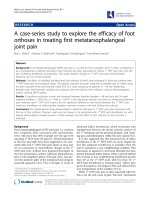
Figure 2 An enhanced compu ted tomography of the abdomen revealed a giant homogeneous low density mass lesion in the right
adrenal region. (A) A giant mass lesion was indented over the inferior aspect of the right lobe of the liver and a displaced inferior vena cava
(arrowhead) with no abdominal lymphadenopathy. (B) The right kidney was also ventrally displaced.
AB
AB
Figure 3 Abdominal magnetic resonance imaging revealed a well-defined high intensity mass which appeared homogeneous intense
in the T 1-weighted image (A) but heterogeneously intense in the T 2-weighted image (B).
Momiyama et al. Journal of Medical Case Reports 2011, 5:135
/>Page 3 of 5
Due to the wide use of the diagnostic imaging modal-
ities, the detection rate of adrenal cystic lesions is
increasing. However, a preoperative confirmatory diagno-
sis of a large adrenal cyst can be very difficult because of
the indistinct boundary with surrounding organs and
adhesion to neighboring organs. Furthermore, even with
integrated fluorine- 18 fluorodeoxyglucose positron emis-
sion tomography (PET), adrenal lesions may be identified
as false-positive at PET, including adrenal adenomas,
adrenal endothelial cysts and inflammatory and infectious
lesions [13].
The differential diagnosis of adrenal pseudocysts
includes splenic, hepatic and renal cysts, as well as
mesenteric or retroperitoneal cysts, urachal cysts and
solid adrenal tumors. An exact diagnosis is clinically
important in large lesion because adrenal incidentalomas
larger than 5 cm [14] carry an increased risk of adrenal
malignancy. The reported incidence of malignancy in
adrenal cystic lesions is approximately 7% [14].
On CT, most pseudocysts demonstrate well-demar-
cated round or oval masses with fluid density but the
CT features of pseudocysts are more complicated than
simple cysts due to the complicated components such
as septa, blood and soft-tissue components. The cysts
wall shows occasional calcifica tion. MRI is the best
modality for visualizing the complicated intracystic com-
ponents. Moreover, MRI is particularly sensitive for
detecting intracystic hemorrhage, which shows hyperin-
tense on both T1- and T2-weighted images.
Treatment of adrenal cysts is determined by size and
the symptoms related to the mass. Surgical excision is
indicated by the presence of symptoms, a suspicion of
malignancy a nd an increase in size, or the detection of,
a functioning adrenal cyst. Surgical treatment may not
be necessary for small asymptomatic lesions as most
cysts are benign [14]. If the adrenal lesion is diagnosed
as a simple nonfunctioning cyst, the patient may be
treated conservatively with aspiration alone. In large
abscesses, where the probability of rupture is increased,
transcutaneous drainage should be avoided as it may
increase the risk of microbial load dissemination [15].
Conclusion
An adrenal pseudocyst is an uncommon clinical finding
and is even rarer when it is giant-sized and infected.
Surgery is required for symptomatic cases in order to
relieve the symptoms and in cases of uncertain diagno-
sis. Radiological and clinical features of the tumor are
nonspecific, thus, histopathological examination is
essential in order to establish a definitive diagnosis.
Consent
Written informed consent was obtained from the patient
for publicatio n of this case report and any accompany-
ing images. A copy of the written consent is available
for review by the Editor-in-Chief of this journal.
Abbreviations
CT: computed tomography; MRI: magnetic resonance imaging; PET: positron
emission tomography; US: ultrasound.
Author details
1
Department of Gastroenterological Surgery, Yokohama City University,
Yokohama, Japan.
2
Division of Anatomic and Surgical Pathology, Yokohama
City University Hospital, Yokohama, Japan.
Authors’ contributions
MM, KM and KY were involved in drafting the manuscript. KT, HA, SY and IE
revised the manuscript. All authors have read and approved the final
manuscript.
Competing interests
The authors declare that they have no competing interests.
Received: 3 July 2010 Accepted: 4 April 2011 Published: 4 April 2011
References
1. Doran AHG: Cystic tumor of the supra-renal body successfully removed
by operation. BMJ 1908, 1558-1563.
2. Wahl HR: Adrenal cysts. Am J Pathol 1951, 27:758.
3. Foster D: Adrenal cysts: Review of literature and report of case. Arch Surg
1966, 92:131-143.
4. Incze JS, Lui PS, Merrian JC, Austin G, Widrich WC, Gerzof SG: Morphology
and pathogenesis of adrenal cysts. Am J Pathol 1979, 95:423-432.
5. Rozenblit A, Morehouse HT, Amis ES Jr: Cystic adrenal lesions: CT features.
Radiology 1996, 201(Suppl 2):541-548.
6. Masumori N, Adachi H, Noda Y, Tsukamoto T: Detection of adrenal and
retroperitoneal masses in a general health examination system. Urology
1998, 52(Suppl 4):572-576.
7. Abeshouse GA, Goldstein RB, Abeshouse BS: Adrenal cysts: Review of the
literature and report of three cases. J Urol 1959, 81(Suppl 6):711-719.
8. Gaffey MJ, Mills SE, Fechner RE, Bertholf MF, Allen MS Jr: Vascular adrenal
cysts: a clinicopathologic and immunohistochemical study of endothelial
and hemorrhagic (pseudocystic) variants. Am J Surg Pathol 1989,
13(Suppl 9):740-747.
Figure 4 Histological examination of the cyst showed an
adrenal pseudocyst. The cystic wall consisted of dense fibrous
tissue without an epithelial lining (arrow). A rim of normal adrenal
tissue was found compressed within the cystic capsule (arrow
head). (hematoxylin and eosin stain, Scale bar = 1 mm).
Momiyama et al. Journal of Medical Case Reports 2011, 5:135
/>Page 4 of 5
9. Erickson LA, Lloyd RV, Hartman R, Thompson G: Cystic adrenal neoplasms.
Cancer 2004, 101:1537-1544.
10. Bellantone R, Ferrante A, Raffaelli M, Boscherini M, Lombardi CP, Crucitti F:
Adrenal cystic lesions: report of 12 surgically treated cases and review
of the literature. J Endocrinol Invest 1998, 21:109-114.
11. Foroughi E: Calcified simple parenchymal cyst of the adrenal gland.
J Urol 1965, 94(Suppl 5):504-510.
12. Papavramidis TS, Duros V, Michalopoulos A, Papadopoulos VN,
Paramythiotis D, Harlaftis N: Intra-abdominal pressure alterations after
large pseudocyst transcutaneous drainage. BMC Gastroenterol 2009,
9:42-46.
13. Chong Semin, Lee Soo Kyung, Kim Young Ha, Kim Kyung Yoon, Kim Byung-
Tae, Chung Jin Myung, Chin AYi, Kwon Young Ghee: Integrated PET-CT for
the characterization of adrenal gland lesions in cancer patients:
diagnostic efficacy and interpretation pitfalls. Radiographics 2006,
26(Suppl 6):1811-24, discussion 1824-1826.
14. Kasperlik-Załuska AA, Otto M, Cichocki A, Rosłonowska E, Słowinska-
Srzednicka J, Zgliczyński W, Jeske W, Papierska L, Tołłoczko T, Polański J,
Słapa R: 1,161 patients with adrenal incidentalomas: indications for
surgery. Langenbecks Arch Surg 2008, 393(Suppl 2):121-126.
15. Papavramidis TS, Sapalidis K, Pappas D, Karagianopoulou G, Trikoupi A,
Souleimanis CH, Papavramidis ST: Gigantic hepatic amebic abscess
presenting as acute abdomen: a case report. J Med Case Reports 2008,
2:325.
doi:10.1186/1752-1947-5-135
Cite this article as: Momiyama et al.: A giant adrenal pseudocyst
presenting with right hypochondralgia and fever: a case report. Journal
of Medical Case Reports 2011 5:135.
Submit your next manuscript to BioMed Central
and take full advantage of:
• Convenient online submission
• Thorough peer review
• No space constraints or color figure charges
• Immediate publication on acceptance
• Inclusion in PubMed, CAS, Scopus and Google Scholar
• Research which is freely available for redistribution
Submit your manuscript at
www.biomedcentral.com/submit
Momiyama et al. Journal of Medical Case Reports 2011, 5:135
/>Page 5 of 5