Advanced therapy in thoracic surgery - part 5 pdf
Bạn đang xem bản rút gọn của tài liệu. Xem và tải ngay bản đầy đủ của tài liệu tại đây (1.22 MB, 40 trang )
steriods. Tracheostomies may be useful in some patients
as the only way to secure an airway. When possible, they
should be placed through the stenosis, preserving the
uninvolved trachea for future reconstruction.
Anesthesia
An experienced anesthesiology team working in close
cooperation with the surgical team is essential.
Replacement of spontaneous breathing with positive
pressure ventilation can convert a partially obstructing
lesion into a complete obstruction. When maintenance of
the airway is a concern, a breathe down with an inhala-
tion agent is employed and paralytics given once the
airway is secured.
20
Anesthesia is maintained with total
intravenous anesthesia (TIVA) using short-acting agents
such as remifentanil and propofol. This allows immediate
extubation at the completion of the procedure and main-
tains continuous anesthesia during periods when inhala-
tional agents are interrupted by the procedure. When a
thoracotomy incision is used, epidural anesthesia signifi-
cantly decreases thoracotomy pain. For lower tracheal
and carinal resections, endotracheal intubation is accom-
plished with an extra-long, armored endotracheal tube.
Its flexibility allows bronchoscopic placement into one of
the main stem bronchi. After transecting the airway, the
orotracheal tube is pulled back into the trachea and
intermittent ventilation is performed with sterile cross-
field equipment. The orotracheal tube is again advanced
once the anastomosis is completed. The anesthesiology
team should be familiar with the techniques of high-
frequency “jet” ventilation. Cardiopulmonary bypass is
not helpful and only introduces unnecessary risks.
Simple Tracheal Resection
This section describes our technique for uncomplicated
resections of the middle and upper trachea. Rigid bron-
choscopy with dilation is performed at the time of
planned resection, and if the lesion appears amenable to
surgery, the patient is intubated, positioned, and pre-
pared for incision.
For most relatively short lesions, the patient is placed
supine with an inflatable airbag beneath the shoulders
with the neck extended. The inflatable bag is important
in alleviating tension since it can be deflated to facilitate
neck flexion just prior to tying down the anastomosis.
The head and neck are supported in a foam “doughnut.”
The arms are tucked at the sides and only the neck and
upper sternum are prepared and draped.
A low collar incision is adequate for most tracheal
resections involving the upper trachea. Occasionally,
vertical extension with a partial sternal split is required
for middle to lower tracheal lesions (Figure 17-8A).
Dissection is carried through the platysma, and sub-
platysmal flaps are elevated superiorly to the level of the
cricoid and inferiorly to the level of the sternal notch.
The strap muscles are separated in the midline, and a
plane of dissection is established very close to the
tracheal wall to avoid injury to the recurrent laryngeal
nerves (Figure 17-8B). The pretracheal plane is dissected
to the level of the carina. The investing fascia of the
innominate artery and the adjacent mediastinal fat is left
intact to guard against postoperative tracheoinnominate
fistulization. The location and extent of the lesion may
sometimes be identified by observation of changes in the
tracheal wall as seen in the operative field. Often,
however, these changes are subtle, and the limits of the
resection must be delineated by the surgeon transillumi-
nating the trachea above and below the lesion with a flex-
ible bronchoscope while the assistant watches the field
and marks the limits of the lesion with fine sutures.
The trachea is sharply dissected circumferentially at
the most distal extent of the lesion, with the dissection
plane maintained on the tracheal wall. Sterile ventilating
tubing is then positioned under the ether screen and
fastened to the drapes. The endotracheal tube is with-
drawn into the upper trachea, the trachea divided at the
most distal extent of the lesion, and bilateral 2-0 Vicryl
traction sutures placed such that they are anchored
around a tracheal ring about 1 cm below the distal tran-
section site. A cuffed, armored Tovell tube is promptly
passed into the distal tracheal segment and attached to
the sterile connecting tubing, and crosstable ventilation
commenced (Figure 17-8C). The diseased segment of
trachea is sharply dissected from the esophagus and tran-
sected at the most proximal extent of the lesion and
passed out of the field.
The patient’s neck is then flexed and the anastomosis
tested for tension. Using the traction sutures, the proxi-
mal and distal segments can be brought towards one
another. When they come together without tension, the
anastomosis can be created. If the limits of flexion and
safe dissection have been reached and anastomotic
tension still exists, then one proceeds with release proce-
dures (see below). It is simplest to anticipate the need for
release procedures and perform them prior to dividing
the trachea, but this is not always possible.
When the surgeon is satisfied that the anastomosis
will not be under tension, interrupted 4-0 Vicryl anasto-
motic sutures are placed (but not tied) such that the
knots will be on the outside, beginning posteriorly in the
midline and proceeding around either side to the front
(Figure 17-8D). The sutures are placed 5 to 6 mm from
the cut edge of the trachea and 4 mm apart. They should
encircle a tracheal ring on either side of the anastomosis
to help prevent dehiscence. Frequently, the Tovell tube
220
/ Advanced Therapy in Thoracic Surgery
strap muscles. In situations where postoperative intuba-
tion is thought to be necessary, a small uncuffed endotra-
cheal tube is left in place initially and a stitch placed on
the trachea to mark the site for tracheostomy should it
become necessary. This allows limited dissection and
accurate placement in a reoperative field. It is best to wait
a few days before placing a tracheostomy to allow skin
flaps and other tissue layers to seal before exposing them
to airway secretions. This also allows for postsurgical
airway edema to resolve before committing to a
tracheostomy tube.
For tumors, the approach is modified in a number of
ways. Considerable experience is required to make the
judgment of whether a tumor can be safely resected with
sufficient tissue to provide a clear margin and yet allow
successful primary reconstruction of the airway. This can
be particularly difficult in patients with adenoid cystic
carcinoma in whom frozen sections may show micro-
scopic tumor at grossly clear resection margins. When
extension of resection to the more distal trachea is
required, an upper sternal split may be extended into the
right fourth interspace. The plane of dissection in tumor
cases must be kept away from the involved portion of
trachea in order to ensure an adequate radial margin.
This endangers the recurrent laryngeal nerves more than
in resections for benign disease. If a recurrent nerve is
involved by tumor, the nerve is sacrificed. Paratracheal
lymph nodes are removed en bloc with the specimen
when possible, but extensive lymph node dissection
cannot be done for fear of destroying the blood supply to
the remaining trachea. Postoperative radiation therapy is
recommended in all cases of bronchogenic or adenoid
cystic carcinoma, unless contraindicated by performance
status or anastomotic complications.
11
Laryngotracheal Resection
In cases where an upper tracheal lesion involves the
cricoid, occuring most commonly in idiopathic laryngo-
tracheal stenosis or tumor, a laryngotracheal resection
will be necessary. In idiopathic laryngotracheal stenosis
the lesion typically involves the cricoid on its anterior
and lateral luminal surface. The operative procedure
must be tailored to address the particular anatomical
involvement encountered (Figure 17-9). The recurrent
laryngeal nerves are protected by bevelling off the cricoid
anteriorly and laterally while preserving the posterior
plate.
21,22
The extent of anterior cricoid resection ranges
from complete, with a line of transection through the
cricothyroid membrane to none at all, depending on the
extent of involvement. Tracheal resection depends on the
distal extent of lesion (Figure 17-10A and B). The trachea
is appropriately tailored so that the proximal trachea
coapts well with the cut edge of the larynx
(Figure 17-10C and D). 2-0 Vicryl “traction sutures” are
placed in the midlateral position both proximally and
distally. Interrupted 4-0 Vicryl sutures were used to fash-
ion the anastomosis. The midline of the thyroid cartilage
is approximated to the midline of tracheal “prow.” 2-0
Vicryl traction sutures are tied followed by individual 4-0
Vicryl anastomotic sutures (Figure 17-10E and F).
This operation is modified in patients in whom the
stenosis affects the mucosa overlying the cricoid plate.
Sparing the posterior cricoid plate preserves the recurrent
laryngeal nerves. The line of mucosal division is
performed high on the posterior cricoid plate to excise
involved mucosa and submucosa (Figure 17-11). Mucosal
resection stops short of the superior border of the cricoid
plate, immediately below the arytenoid cartilages. The
rostrum or “prow” of the proximal tracheal cartilage is
shaped as described above, but posteriorly a broad-based
flap of membranous wall is fashioned, which is advanced
to resurface the denuded posterior criciod plate. The
posterior portion of the anastomosis is made with inter-
rupted 4-0 Vicryl sutures placed only through the full
thickness of mucosa and submucosa of the posterior wall
of the larynx, and then through the full thickness of the
membranous wall of the trachea (Figure 17-12), inverted
so that the suture knots lay external to the lumen. Four
sutures are placed through the cartilaginous portion of
the inferior margin of the cricoid plate and the outer
portion of the membranous wall of the trachea below the
proximal edge of the flap in order to fix the membranous
wall to the inferior edge of the cricoid plate. When the
lesion extends proximally toward the conus elasticus, it is
necessary to accept some residual narrowing because of
the height of the anastomosis.
Lower Tracheal and Carinal Resections
While isolated benign strictures of the lower trachea and
carina are seen, the most common lesions requiring
surgery are tumors. Therefore, the principles of surgical
oncology must be strictly applied to most of these resec-
tions. Patients with bronchogenic carcinoma and N2
disease should be considered to have unresectable
disease, and surgery should only be performed in a
protocol setting.
23–26
Mediastinoscopy is performed on the
day of proposed surgery not only to assess nodal status
and resectability, but to facilitate the resection and recon-
struction by mobilizing the pretracheal plane while visu-
alizing the recurrent laryngeal nerve. Scarring of the
pretracheal plane from prior mediastinoscopy limits
airway mobility, complicates reconstruction, and
increases the likelihood of injury to the left recurrent
222
/ Advanced Therapy in Thoracic Surgery
generous caudal displacement of the trachea. For this
reason, end-to-end anastomosis of trachea to the left
main stem with reimplantation of the right into the
trachea is more commonly employed. A right hilar
release maneuver facilitates this procedure. More exten-
sive resections require end-to-end anastomosis of trachea
to right main stem with reimplantation of left into the
bronchus intermedius. This obviates the need for exten-
sive left main stem mobility. When there is extensive
endobronchial involvement, excessive lung destruction,
or invasion of hilar vessels, then carinal (sleeve) pneu-
monectomy is necessary. Experienced intraoperative
judgment is required to determine the ideal approach.
The anastamosis is fashioned with interrupted simple
4-0 Vicryl sutures placed with knots tied outside the
lumen. Once reconstructed, the anastomoses are tested
for air tightness to 40 cm of water. All suture lines are
circumferentially wrapped with pedicled flaps of pericar-
dial fat or a broad-based pleural flap. In high-risk
patients, especially those who have undergone prior
radiotherapy, an intercostal flap stripped of all perios-
teum or an omentum pedicle is used. These flaps not
only buttress the anastomoses, but more importantly,
separate them from the hilar vessels, helping to prevent
bronchovascular fistulas.
Release Procedures
When extensive resections are required the standard
methods of mobilization by dissection in the pretracheal
plane and flexion of the neck often do not allow a
tension-free anastomosis. In these instances, further
mobilization with “release” procedures is required. In our
experience, this has been necessary in 8.3% of patients
undergoing resections for postintubation stenosis and
15% of patients undergoing resections for tumors.
18
Certain release maneuvers are more effective for achiev-
ing additional mobility of the cervical trachea, whereas
others are more effective for freeing the intrathoracic
trachea.
In resection of the upper trachea, additional length
may be gained by releasing the larynx with a
Montgomery suprahyoid release.
27
This consists of divid-
ing the muscles that insert on the superior aspect of the
central part of the hyoid bone. The hyoid itself is then
divided just medial to its lesser cornua on either side, and
the stylohyoid tendons are divided (Figure 17-13). This
provides an additional approximately 1.5 cm of length. It
is important to realize that laryngeal release maneuvers
may predispose patients to postoperative aspiration,
especially of liquids. In time, however, this problem has
resolved in virtually all patients.
For intrathoracic tracheal or carinal resections, addi-
tional length is best achieved by hilar release.
28
Mobilization of the right hilum should be done first,
along with division of the inferior pulmonary ligament.
Then, a U-shaped incision is made in the pericardium
below the inferior pulmonary vein. If required, the peri-
cardium can be incised 360° around the hilus for maxi-
mal mobility. In this event, the vascular and lymphatic
pedicle to the main stem bronchus is left preserved
behind the pericardium. The left hilum may be similarly
mobilized (Figure 17-14) in the unusual case where
unilateral mobilization is insufficient. However, left-sided
hilar release can only be accomplished easily through a
median sternotomy by opening the pericardium anteri-
orly, bilateral thoracotomies, or an extended clamshell
incision. As with most airway surgery, neck flexion is
helpful. Laryngeal release has not been shown to produce
meaningful mobility at the level of the carina.
29
Tracheal Resection in an
Irradiated Field
In patients who have received radiation therapy prior to
coming to surgical resection, the risk of anastomotic
dehiscence is increased. The detrimental effects of irradi-
ation on tissue and, more specifically, tracheal healing
have been amply demonstrated in animals. The early
Massachusetts General Hospital experience with tracheal
resection in patients who had received high doses of radi-
ation, particularly when this occurred remotely in time,
confirmed these findings.
30
In these patients there was a
markedly increased incidence of anastomotic failure.
When a patient has received either high-dose irradia-
tion (more than 4,500 cGy) or who has undergone irra-
Techniques of Tracheal Resection and Reconstruction
/
225
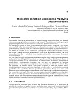
FIGURE 17-13. The dotted lines indicate the point where the hyoid
bone is divided, separating its body from the greater horn on each
side. Reprinted with permission from Montgomery ww.
27
A collar incision is performed which circumscribes
the stoma (Figure 17-16). Dissection identical to that
described above for simple tracheal resection is
performed up to the point of division of the trachea
below the fistula. As the posterior wall of the trachea is
dissected from inferior to superior, the fistulous connec-
tion is isolated circumferentially. It is detached from the
esophagus with a small rim of normal esophageal tissue
and kept attached to the tracheal segment with which it
will be removed (Figure 17-17). After removal of the
specimen, the esophagus is closed longitudinally with
two layers of 4-0 silk (Figure 17-18A and B). The ster-
nohyoid or sternothyroid muscle is sutured into place to
buttress the esophageal closure and interpose healthy
tissue between the esophageal and tracheal suture lines
(Figure 17-19). The end-to-end tracheal anastomosis is
then performed as described previously. If the fistulous
opening is long and the tracheal wall is not circumferen-
tially damaged as far down as the fistula extends, the
margin of the tracheal opening may be excised as a V and
repaired with a vertical suture line prior to creating the
end-to-end tracheal anastomosis. In the rare case where
there is no significant damage to the trachea associated
with the fistula, tracheal resection is unnecessary, and
simple esophageal and tracheal repair with muscle
buttress is performed.
Postoperative Issues
The patient’s postoperative course is largely determined
by intraoperative technique. The goals of both intraoper-
ative and postoperative care are the maintenance of good
pulmonary toilet and the promotion of anastomotic
Techniques of Tracheal Resection and Reconstruction
/
227
FIGURE 17-16. Exposure for most tracheoesophageal fistulas is through a low collar incision. Occasionally, a partial upper sternotomy is required
for more distal exposure of the trachea. Reprinted with permission from Mathisen DJ et al.
31
FIGURE 17-15. Endoscopic view of tracheoesophageal fistula.
dure. Postoperatively, these include minimizing fluids,
elevating the head of the bed and administering racemic
epinephrine to help prevent laryngeal edema. Rarely, an
especially high laryngotracheal resection will cause
enough laryngeal edema to necessitate one or two doses
of steriods to avoid impending re-intubation and/or
tracheostomy. Heliox, with its low viscosity, is sometimes
useful in these circumstances since it can occasionally
gain enough time for the other maneuvers to take effect.
The patient is cautioned against unnecessary speech
during this period, as it can contribute to the laryngeal
edema.
Cervical flexion is maintained with the chin-to-chest
suture for 5 to 7 days, after which time the patient is
advised not to extend the neck for another week. Before
removing the chin-to-chest suture, we routinely examine
the anastomosis with a flexible bronchoscope or obtain
tracheal tomograms to assure normal healing.
Oral alimentation is begun cautiously, particularly in
patients who have undergone a laryngeal release. Water is
offered initially, since its aspiration is better tolerated and
more easily dealt with than more substantial foods.
Results and Complications
Results of tracheal resection have been impressive. For
simple resections of postintubation stenoses, including
our earliest experience and reoperations, of 503 patients
there were only 12 deaths and 18 failures.
32
Four-hundred
and forty (87%) had good and 31(6%) satisfactory
results. Of 80 patients undergoing laryngotracheal resec-
tions for all causes of subglottic stenosis, there was one
postoperative death. Results were excellent in 18 (22%),
good in 52 (65%), and satisfactory in 8 (10%). In only
two patients was there failure to achieve a functional
airway.
For primary tumors of the trachea, for which resec-
tion and reconstruction was performed, including carinal
resections, there were 6 deaths in 132 patients.
11
Five of
the six were following the more complex carinal proce-
dures. Six patients developed significant postoperative
restenosis, but all of these underwent successful
re-resection. The oncologic outcomes of patients with
bronchogenic carcinoma has recently been separated out
for carinal resections and reported by Mitchell and
colleagues.
26
In this series of bronchogenic carcinomas,
57% presented with N0 disease, 25% had N1 disease, and
18% had N2 or N3 disease. The overall 5-year survival
was 42%. Lymph node status strongly influenced
survival. The 5-year survival of N0, N1, and N2 or N3
patients was 51%, 32% and 12%, respectively (see
Figure 17-3). Microscopically positive margins did not
affect survival. Isolated carinal resection resulted in a
more favorable prognosis than more extensive resections,
with a 5-year survival of 51%.
The long-term survival data for resected adenoid cystic
carcinoma of the trachea and carina have not been as well
defined, partly because of the proclivity for late recur-
rence. The published experience of all tracheal adenoid
cystic carcinomas, which includes carinal, suggests a
much more favorable prognosis than bronchogenic carci-
nomas. Lymph node and margin status do not appear to
significantly affect survival.
11,12,23
Postoperative radiation
therapy is recommended in all cases of adenoid cystic or
bronchogenic carcinoma, unless contraindicated by
performance status or anastomotic complications. The
role of chemotherapy has not been established.
Secondary cancers arising in the thyroid and invading
the trachea have also been resected with good results. Of
27 patients undergoing resection and reconstruction of
the trachea for thyroid cancer invading the airway,
including patients with both simple and complex laryn-
gotracheal reconstructions, two died in the postoperative
period, one had a short segment tracheal necrosis requir-
ing re-resection, and all others were provided with an
adequate airway by their initial operation. Only two
patients experienced an airway recurrence.
13
In patients who have received radiation therapy prior
to coming to surgical resection, the risk of anastomotic
dehiscence is increased. Nineteen patients have undergone
tracheal resections with vascularized tissue coverage at
Massachusetts General Hospital following radiation ther-
apy.
30
Fifteen had a pedicied omental flap, 1 a pericardial
fat pad flap, 1 an intercostal muscle flap, and 2 a pleural
flap. Only one of these patients suffered an anastomotic
dehiscence, and this resulted in death. Another patient
required a T-tube. Following development of a paratra-
cheal abscess, he ultimately died of recurrent squamous
cell carcinoma. Two patients developed wound infections
that responded to treatment. Overall, 15 patients experi-
enced an excellent result without dyspnea, and 2 experi-
enced a good result with dyspnea with moderate exercise.
Our experience with the repair of tracheoesophageal
fistulas involves the performance of 41 operations on 38
patients.
31
Simple division and closure of the fistula was
done in nine patients. Tracheal resection and reconstruc-
tion was combined with esophageal repair in the remain-
der. The esophageal defect was closed in two layers and a
viable strap muscle interposed between the airway and
esophageal suture lines in all cases. There were four
deaths (11%). Three patients developed recurrent fistulas
and one patient suffered a delayed tracheal stenosis. All
were successfully managed with re-operation. Of the 34
survivors, 33 can swallow normally, and 32 breathe with-
out the need for a tracheal appliance.
Techniques of Tracheal Resection and Reconstruction
/
229
Complications
Despite these encouraging outcomes, complications do
occur. They have generally been few for upper tracheal
resections. Major complications more often have
followed carinal or laryngotracheal resections.
Inability to clear secretions with consequent atalecta-
sis is the most common, though relatively minor, compli-
cation and this can be handled as described above. This
management has limited the number of patients who
have suffered pneumonia or respiratory failure after
simple tracheal resection. Laryngeal edema may occur
after procedures involving the larynx, but this generally
regresses in approximately one week when treated as
described above. Unilateral recurrent laryngeal nerve
injury rarely occurs as a result of extensive resection,
usually in patients with tracheal tumors.
The most common late complication has been the
formation of granulomas at the suture line. This is
usually manifest as wheezing or minor hemoptysis. It has
occurred more commonly following resection for inflam-
matory lesions than for tumor, as residual inflammation
may be present in such cases despite efforts to wait out
the acute inflammatory phase. Granulations can be
managed by bronchoscopic removal under light anesthe-
sia. Often a suture is found to have migrated into the
lumen at the base of the granulation, and in such cases
removal of the suture leads to ultimate healing. In some
cases, however, multiple bronchoscopies are necessary
over a period of time. The current use of Vicryl rather
than nonabsorbable sutures has almost eliminated this
once common problem.
Suture line separation, the most dreaded complica-
tion, is almost invariably related to tension on the anasto-
mosis or compromise of its blood supply. These
problems, which occur most commonly following resec-
tion of long segments of trachea and following radiation,
are more frequently associated with resection for tumor
than for postintubation stenosis. Steroid use which has
not been discontinued preoperatively has also been asso-
ciated with anastomotic failure. Early, minimal air leak-
age at the suture line may seal without sequelae and can
be managed with closed suction drains. True separation,
however, is usually heralded by respiratory distress.
Anastomotic separation in the immediate postopera-
tive period suggests a serious technical error, and reoper-
ation under these conditions is appropriate. Early
separation that does not appear remediable by resuturing
or a local muscle flap can be temporized by placement of
a tracheostomy or a Montgomery T-tube, with corrective
surgery to be performed months later after regression of
the acute inflammatory response. Sometimes, with such a
tube serving as a stent, the partial restenosis that results
may leave a tolerable airway, and this may be improved
with endoscopic dilations.
Stenosis may occur at the anastomotic site after the
initial postoperative period, without evidence of a frank
separation. This can be managed temporarily by rigid
bronchoscopic dilation. Ultimately, most of these
patients will require re-resection. This should be done no
sooner than 4 months after the initial procedure in order
to allow time for regression of inflammation.
Other rare complications that we have seen include
fatal hemorrhage from the pulmonary artery, likely
related to erosion from an adjacent tracheobronchial
anastomosis, innominate artery hemorrhage, tracheo-
esophageal fistula, esophagocutaneous fistula, empyema,
and quadriplegia, which may have been related to over-
flexion of the chin to the chest.
Tracheal Substitutes and Tracheal
Transplantation
The advancement of techniques in tracheal surgery have
allowed up to 50% of the trachea to be resected in favor-
able patients. This has rendered the majority of tracheal
lesions requiring surgical treatment correctable with a
single-staged resection and reconstruction. On rare occa-
sions, the extent of a lesion involves more of the trachea
than can be safely reconstructed with a primary end-to-
end anastomosis. These situations have lead investigators
to attempt to reconstruct the trachea with prosthetic
material.
Early designs focused on solid tubes anastomosed end-
to-end with the trachea. Neville and colleagues were one of
the first to report a small series on human subjects.
33
Results were dismal. The nonporous silicone tubes failed
to become incorporated with tissue and thus became
infected and either extruded into the airway or eroded into
the surrounding vascular structures. To avoid this fate,
subsequent designs employed porous cylinders, usually
fabricated from metal wire of all different elements and
alloys. These prosthetic conduits were usually wrapped
with an omental or muscle flap and then placed as an
interposition graft in the trachea. The tissue flap was
expected to provide an airtight seal and serve as a source of
vascularized tissue in which the prosthesis would become
incorporated and protected from the surrounding great
vessels. Most of the investigations were in animals, and
while the prostheses became successfully incorporated,
they ultimately failed as the animals became obstructed
from granulation tissue.
34–37
The lack of an epithelial
surface essentially created an open festering wound
encouraging granulation tissue to proliferate unchecked,
resulting in airway stenosis. Small segments of trachea
could be successfully replaced in this manner, since respi-
230
/ Advanced Therapy in Thoracic Surgery
ratory epithelium would migrate for 1 to 2 cm from either
anastomosis to cover the replaced portion of the airway. In
larger segments, the respiratory epithelium would either
not migrate such a distance or simply could not cover the
distance fast enough to outpace and thus quell the exuber-
ant granulation tissue. Recent investigators have supplied
an epithelial lining by grafting either oral mucosa or split-
thickness skin grafts on the inner surface of the porous
prosthesis.
38
These require a two-staged procedure where
the pedicled tissue or prosthesis composite is created and
allowed to mature before it is transposed as an interposi-
tion tracheal substitute. Early results are encouraging, but
their complexity and inconsistent results make their clini-
cal application unfeasible at this time.
The lack of success with prosthetic tracheal replace-
ments has encouraged many investigators to pursue an
airway conduit made of all biological tissue, either viable
allotransplantation or cryopreserved tracheas. Neither
approach has achieved meaningful success.
Tracheal transplantation suffers from several major
limitations. First, the trachea lacks a single, sizable
venous and arterial system. Instead, its vascular supply
consists of multiple small vessels too fine to anastomose.
To overcome this limitation, investigators have used the
omentum to wrap tracheal transplants to allow for
vascular ingrowth.
39,40
However, results have been mixed,
especially with longer segments. Second, unlike most
other solid organ transplants, the trachea by virtue of its
anatomical location is exposed to a heavy concentration
of antigens and microorganisms. The result is an
ischemic tracheal transplant, heavily contaminated with
oropharyngeal microorganisms, in an immunodebili-
tated patient. Finally, many of the conditions which
involve the entire trachea are benign processes that are
safely managed with Silastic T-tubes and thus do not
justify the detrimental effects of immunosuppressive
therapy. In those cases where a malignant tracheal tumor
requires resection of the entire trachea, immunosuppres-
sive therapy should be avoided as well.
In a move to avoid immunosuppressant therapy,
investigators have begun to test methods of rendering
allogenic tracheal grafts less or nonantigenic. The most
important transplant antigens involved in graft rejection
are expressed by the major histocompatibility complex
(MHC). In the trachea, the mucosa and the submucosal
glands express MHC-I and MHC-II.
41
Cartilage does not
express MHC antigens and is an immunologically privi-
leged tissue that has been successfully used in allo-
transplantation for years without the use of
immunosuppressive therapy. It is believed that the thick,
avascular proteoglycan-collagen matrix that encapsulates
the chondrocytes, shields them from recognition by the
immune system. Moreover, since cartilage has no capil-
lary blood supply and survives from diffusion it can
survive off the diffusion to and from an omental wrap.
Investigators have designed methods to process fresh
tracheas to remove the tracheal mucosa and submucosal
glands while preserving the viability of the cartilage.
42,43
In
pilot dog studies, these grafts epithelialize and maintain
viable cartilage without significant stenosis for up to one
year. Control animals, which had the same procedure
using a fresh unprocessed trachea instead, developed
necrosis and stenosis over a few weeks. Others have used
cryopreservation techniques to achieve similar results
since cartilage tends to survive the process and the
mucosa and glands do not.
44,45
The results of these studies
are encouraging because they demonstrate that a viable
tracheal conduit can be transplanted, integrated, and
accepted by the host and re-epithelialized. However, these
studies were done for small segments of tracheal replace-
ment, where the epithelium can be expected to migrate
from the anastomotic ends and resurface the graft. Since
this form of therapy will be used to treat near total or
total tracheal replacement, these methods will need to be
tested on longer segments.
Summary
In conclusion, techniques of tracheal resection and
reconstruction have advanced to a point where these
procedures can be done with the anticipation of good
results and an acceptable level of morbidity and mortal-
ity. Nonsurgical methods such as dilation, ablation, or
stenting do not currently offer cure of tracheal stenoses,
although these may each play a role in palliation or
temporization prior to surgery. The current standard of
care dictates that symptomatic benign tracheal stenoses
that can be resected should be resected. For primary
malignant tumors, squamous cell carcinomas should be
resected when complete resection for cure is anticipated,
while patients with the more indolent adenoid cystic
carcinoma may benefit from even palliative resection
with microscopically positive margins. Tracheal resection
for low-grade thyroid carcinomas invading the airway
should also be performed for cure or palliation, some-
times even in the presence of distant metastasis. The
development of successful techniques of complete
tracheal replacement in humans is an area of ongoing
research but currently has no clinical applicability.
References
1. Grillo HC, Bendixon HH, Gephart T. Resection of carina
and lower trachea. Ann Surg 1963;158:889–93.
2. Michelson E, Solomon R, Miura T. Experiments in tracheal
reconstruction. J Thorac Cardiovasc Surg 1961;41:748–59.
Techniques of Tracheal Resection and Reconstruction
/
231
3. Mulliken J, Grillo HC. The limits of tracheal resection with
primary anastomosis: further anatomical studies in man. J
Thorac Cardiovasc Surg 1964;48:741–50.
4. Grillo HC, Dignam EF, Miura T. Extensive resection and
reconstruction of the mediastinal trachea without prosthe-
sis or graft: an anatomical study in man. J Thorac
Cardiovasc Surg 1964;48:741–50.
5. Salassa JR, Pearson BW, Payne WS. Gross and microscopical
blood supply of the trachea. Ann Thorac Surg
1977;24:100–7.
6. Grillo HC, Dignam EF, Miura T. Extensive resection and
reconstruction of the mediastinal trachea without prosthe-
sis or graft: an anatomical study in man. J Thorac
Cardiovasc Surg 1964;48:741–50.
7. Cooper JD, Grillo HC. The evolution of tracheal injury due
to ventilatory assistance through cuffed tubes: a pathologic
study. Ann Surg 1969;169:334–48.
8. Cooper JD, Grillo HC. Experimental production and
prevention of injury due to cuffed tracheal tubes. Surg
Gynecol Obstet 1969;129:1235–41.
9. Whited R-E. A prospective study of laryngotracheal seque-
lae in long-term intubation. Laryngoscope 1984;94:367–77.
10. Gaissert HA, Lofgren RH, Grillo HC. Upper airway compro-
mise after inhalation injury. Complex strictures of larynx and
trachea and their management. Ann Surg 1993;218:672–8.
11. Grillo HC, Mathisen DJ. Primary tracheal tumors: treat-
ment and results. Ann Thorac Surg 1990;49:69–77.
12. Regnard JF, Fourquier P, Levasseur P, et al. Results and
prognostic factors in resections of primary tracheal tumors:
a multicenter retrospective study. J Thorac Cardiovasc Surg
1996;111:808–14.
13. Grillo HC, Suen HC, Mathisen DJ, Wain JC. Resectional
management of thyroid carcinoma invading the airway.
Ann Thorac Surg 1992;54:3–9.
14. Grillo HC, Mark EJ, Mathisen DJ, Wain JC. Idiopathic
laryngotracheal stenosis and its management. Ann Thorac
Surg 1993;56:80–7.
15. Ashiku SK, Kuzucu A, Grillo HC, et al. Idiopathic laryngotra-
cheal stenosis: effective definitive treatment by laryngotra-
cheal resection. J Thorac Cardiovasc Surg 2004;127:99–107.
16. Weber AL, ed. Symposium on the larynx and trachea.
Radiol Clin N Am 1978;16:227–309.
17. Felson B, Wiott JF, editors. The trachea. Semin Roentgenol
1983;18:1–64.
18. Mathisen DJ. Surgery of the trachea. Curr Probl Surg
1998;35:45–-542.
19. Mathisen DJ, Grillo HC. Endoscopic relief of malignant
airway obstruction. Ann Thorac Surg 1989;48:469–75.
20. Wilson RS. Tracheal resection. In: Marshall BE, Longnecker
DE, Fairley HB, editors. Anesthesia for thoracic procedures.
Boston (MA): Blackwell Scientific; 1988. p. 415–32.
21. Grillo HC. Primary reconstruction of the airway after
resection of subglottic and upper tracheal stenosis. Ann
Thorac Surg 1982;33:39–58.
22. Grillo HC, Mathisen DJ, Wain JC. Laryngotracheal resec-
tion and reconstruction for subglottic stenosis. Ann Thorac
Surg 1992;53:54–63.
23. Grillo HC. Carinal neoplasia. In: Grillo HC, Austen WG,
Wilkins EW, et al, editors. Current therapy in cardiotho-
racic surgery. Hamilton (ON): BC Decker Inc; 1989. p. 134.
24. Mathisen DJ, Grillo HC. Carinal resection for bronchogenic
carcinoma. J Thorac Cardiovasc Surg 1991;102:16–22.
25. Mitchell JD, Mathisen DJ, Wright CW, et al. Clinical experi-
ence with carinal resection. J Thorac Cardiovasc Surg
1999;117:39–53.
26. Mitchell JD, Mathisen DJ, Wright CW, et al. Resection of
bronchogenic carcinoma involving the carina: long-term
results and the effect of nodal status on outcome. J Thorac
Cardiovasc Surg 2001;121:465–71.
27. Montgomery WW. Suprahyoid release for tracheal anasto-
mosis. Arch Otolaryngol 1974;99:255–60.
28. Newton JR, Grillo HC, Mathisen DJ. Main bronchial sleeve
resection with pulmonary conservation. Ann Thorac Surg
1991;52:1272–80.
29. Grillo HC. Carinal Neoplasia. In: Grillo HC, Austen WG,
Wilkins EW, et al. editors. Current therapy in cardiotho-
racic surgery. Hamilton (ON): B.C. Decker Inc.; 1989.
p. 134.
30. Muehrcke DD, Grillo HH, Mathisen DJ. Reconstructive
airway operation after irradiation. Ann Thorac Surg
1995;59:14–8.
31. Mathisen DJ, Grillo HC, Wain JC, Hilgenberg AD.
Management of acquired nonmalignant tracheoesophageal
fistula. Ann Thorac Surg 1991;52:759–65 .
32. Grillo HC, Donahue DM, Mathisen DJ. Postintubation
tracheal stenosis: treatment and results. J Thorac
Cardiovasc Surg 1995;109:486–93.
33. Neville We, Bolanowski JP, Kotia GG. Clinical experience
with the silicone tracheal prosthesis. J Thorac Cardiovasc
Surg 1990;99:604–12.
34. Ter amanchi M, Nakamura T, Yamamoto Y. Porous-type
tracheal prosthesis sealed with collagen sponge. Ann
Thorac Surg 1997;64:965–9.
35. Satoh S, Elstrodt J, Hinrichs WL, Feinjen J. Prevention of
infection in porous tracheal prosthesis by omental wrap-
ping. ASAIO Trans 1990;36:M438–40.
36. Schauwecker HH, Gerlach J, Planck H. Isoelastic
polyurethane prosthesis for segmental tracheal replacement
in beagle dogs. Artif Organs 1989;13:216–8.
37. Teramachi M, Kiyontani T, Takimoto Y. A new porous
tracheal prosthesis sealed with collagen sponge. ASAIO
Tr ans 1995;41:M306–10.
232
/ Advanced Therapy in Thoracic Surgery
38. Suh SW, Kim J, Baek CH. Development of new tracheal
prosthesis: atogenous mucosa-lined prosthesis made from
polypropylene mesh. Int J Artif Organs 2000;23:261–7.
39. Li J, Xu P, Chen H. Successful tracheal autotransplantation
with two-staged approach using greater omentum. Ann
Thorac Surg 1997;64:199–202.
40. Park YS,Lee DY, Paik HC. The role of omentopexy in
tracheal transplantation in dogs. Yonsei Med J
1996;37:118–24.
41. Bujia J, Wilmes E, Hammer C. Tracheal transplantation:
demonstration of HLA class II subregion gene products on
human trachea. Acta Otolaryngol 1990;110:149–54.
42. Liu Y, Nakamura T, Yamamoto Y. Immunosuppressant-free
allotransplanation of the trachea: the antigenicity of
tracheal grafts can be reduced by removing the epithelium
and mixed glands from the graft by detergent treatment. J
Thorac Cardiovasc Surg 2000;120:108–14.
43. Yokomise H, Inui K, Wada H. High-dose irradiation
prevents rejection of canine tracheal allografts. J Thorac
Cardiovasc Surg 1994;107:1391–7.
44. Mukaida T, Shimizu N, Aoe M. Origin of regenerated
epithelium in cryopreserved tracheal allotransplantation.
Ann Thorac Surg 1998;66:205–8.
45. Mukaida T, Shimizu N, Aoe M. Experimental study of
tracheal allotransplantation with cryopreserved grafts. J
Thorac Cardiovasc Surg 1998;116:262–6.
Techniques of Tracheal Resection and Reconstruction
/
233
234
CHAPTER
18
MANAGEMENT OF PULMONARY
ARTERIOVENOUS
MALFORMATIONS AND
SEQUESTRATIONS
FRANCIS
C. NICHOLS, MD
MARK S. A
LLEN, MD
Pulmonary Arteriovenous
Malformation
Pulmonary arteriovenous malformations (PAVMs) are
vascular lesions of the lung in which there is an abnormal
connection between the pulmonary arterial and venous
systems without an intervening capillary bed. PAVM has
been described under a variety of pseudonyms including
benign cavernous hemangioma, pulmonary arteriove-
nous angiomatosis, hamartomatous angioma of the lung,
arteriovenous aneurysm, and arteriovenous fistula.
1
The
malformation leads to shunting of unoxygenated blood
into the systemic circulation and may permit embolic
material to pass unfiltered through the lungs. PAVMs are
classified into simple or complex. A simple PAVM has a
single feeding vessel, and a complex PAVM has multiple
feeding vessels.
PAVM was first described in 1897 by Churton in a 12-
year-old child.
2
The first surgical intervention was reported
by Shenstone who performed a pneumonectomy for a
large central lesion.
3
Several publications from our institu-
tion have focused on the surgical management of PAVM
and most recently on the angiographic management.
1,4–7
PAVM occurs more commonly than previously
thought. It occurs with an incidence of 1 in 2,351 to 1 in
39,000 individuals.
8
The male-to-female incidence is
equal; they are bilateral in 8 to 20% and multiple in 30 to
50% of patients.
9
While PAVM can present as isolated
pulmonary findings, it is often associated with hereditary
hemorrhagic telangiectasia (HHT), also known as the
Rendu-Osler-Weber syndrome. In fact, up to 87% of
PAVMs are found in patients with HHT, and approxi-
mately 20% of patients with HHT develop PAVMs.
7
Although the overwhelming majority of PAVM is
congenital in origin, secondary or acquired PAVM can
occur. The causes of acquired PAVM include trauma,
actinomycosis, schistosomiasis, cirrhosis, systemic
amyloidosis, mitral stenosis, and metastatic carcinoma.
1
Although some patients with PAVM are asympto-
matic, most patients are symptomatic. Clinical features in
a recent Mayo Clinic series are shown in Table 18-1. The
most common pulmonary symptom is dyspnea, and this
correlates with the degree of shunting. Dyspnea can in-
crease with a change in position from supine to upright
and with exercise because of increased blood flow to the
TABLE 18-1. Clinical Features in 93 Patients with
Pulmonary Arteriovenous Malformation
Clinical Feature Number (%)
Dyspnea 53 (57)
Cyanosis 27 (29)
Clubbing 18 (19)
Cerebrovascular event 17 (18)
Asymptomatic 15 (16)
Hemoptysis 14 (15)
Transient ischemic attack 11 (12)
Cerebral abscess 5 (5)
Seizure 5 (5)
Adapted from Swanson KL et al (1999).
7
lower portion of the lungs where PAVMs are usually
located. Depending on the degree of right-to-left shunt-
ing, the hypoxemia may be refractory to supplemental
oxygen. Other clinical features include cyanosis, club-
bing, and hemoptysis. The classic triad of dyspnea,
cyanosis, and clubbing is found in 30% of adults.
1
Neurologic events are common with PAVM and include
embolic disorders such as transient ischemic attacks and
strokes. Dines and colleagues found a stroke to have
occurred in 10% of all untreated patients followed for 4
to 10 years.
5
Cerebrovascular events can also occur due to
sludging secondary to polycythemia and complications
from concomitant cerebral lesions. Seizures and cerebral
abscesses also occur. In addition to the possible physical
findings of cutaneous telangiectasia, cyanosis, and club-
bing, a pulmonary bruit is present in 34% of patients.
7
The characteristic finding on a plain chest radiograph
is a circumscribed, lobulated density. Most PAVMs are
located in the lower lobes and often are peripheral.
Occasionally a feeding vessel can be seen on the chest
radiograph (Figure 18-1). Currently, spiral computed
tomography (CT) offers the least invasive and least
expensive way to establish the presence of PAVM
(Figure 18-2). If thin sections are utilized, intravenous
contrast is not necessary to establish the diagnosis of
PAVM; however, contrast is required in order to prove
patency. CT can elicit the number and size of the fistulas,
and afferent and efferent vessels can be identified.
However, unless the feeding artery or draining vein are
identified, a PAVM cannot be distinguished from a
pulmonary nodule.
1
Magnetic resonance imaging (MRI)
may be helpful but is less sensitive. Oxygen saturation
should be measured to see if there is significant shunting.
Two-dimensional contrast echocardiography with indo-
cyanine dye or the injection of agitated saline can be
useful in establishing the diagnosis and is less invasive
than angiography.
10,11
Furthermore, contrast echocardiog-
raphy is useful in pregnant women, in whom ionizing
radiation may be dangerous. The technique of contrast
echocardiography involves the injection of agitated saline
into a peripheral vein. The appearance of a cloud of bub-
bles in the left atrium confirms right-to-left shunting. Air
bubbles will not survive a normal capillary bed, and if a
patent foramen ovale has been excluded in the appropri-
ate clinical setting, PAVM can be suspected.
1
Angiography
is the definitive test and can clearly outline the anatomy
of PAVM (Figure 18-3). Pulmonary angiography identi-
fies the location, size, and number of PAVMs.
Additionally, it defines their blood supply and differenti-
ates simple from complex PAVMs.
7
Almost all patients with PAVM should be treated.
Untreated PAVMs are associated with an 11% mortality
and 26% morbidity rate.
5
Asymptomatic patients with a
single small (< 1 cm) PAVM occasionally will be
observed; however, the risk of embolic stroke is increased
in these patients. There are a few patients with severe
pulmonary artery hypertension who would develop right
Management of Pulmonary Arteriovenous Malformations and Sequestrations
/
235
FIGURE 18-1. Chest radiograph in a 34-year-old female patient
demonstrating a large feeding vessel supplying a pulmonary arterio-
venous malformation in the lower lung field (black arrow). The vessel
can be seen just medial to the left heart border, coursing to the lower
lung fields (white arrow).
FIGURE 18-2. Computerized tomography of the chest with intra-
venous contrast. In the lung window settings, multiple pulmonary
arteriovenous malformations are seen (arrows). With permission from
Swanson KL et al.
7
more coils than balloons must often be placed to achieve
satisfactory occlusion.
1
In our recent series of patients
treated with angiographic embolization, 91% responded
favorably as shown by an improvement in their symp-
toms or arterial blood gas analysis. The mean PaO
2
rose
from a preembolization level of 56 mm Hg to 77 mm Hg
postembolization.
7
Long-term follow-up is recommended in all patients
with PAVM. Even after successful treatment, there can be
growth of smaller lesions and recanalization of success-
fully embolized lesions. Recurrences can be successfully
reembolized. Our recommendations for follow-up
include an annual physical exam, chest radiograph,
arterial blood gas analysis, and assessment of the
right-to-left shunt if symptoms are present.
7
Pulmonary Sequestration
Pulmonary sequestration covers a spectrum of related
developmental pulmonary anomalies. It was first
described simultaneously by Rokitansky and Rektorzik in
1861.
20,21
The term “sequestration” was first used by Pryce
and is derived from the Latin verb sequestrare,to
separate.
22
The lung is, in effect, sequestered from the
remainder of the lung. It is defined as a segment of lung
that has no bronchial communication with the rest of the
lung.
23,24
The arterial supply comes from systemic circula-
tion, including from the thoracic aorta, abdominal aorta,
or intercostal arteries. The venous return is either to the
pulmonary system or to the systemic circulation. Se-
questrations are different from accessory pulmonary
lobes, which are separated from the normal lung by
pleural investments but maintain a normal communica-
tion with the tracheobronchial tree. Surgical interest in
these lesions first arose when Harris and Lewis reported
on an operative death resulting from injury to an
anomalous artery supplying the lower lobe of the lung in
a 5-year-old child.
25
Pulmonary sequestrations are thought to arise as
accessory lung buds that then migrate along with the
developing esophagus. This may account for their variable
blood supply and occasional foregut communication.
Other authors believe that these anomalies are acquired
and are the result of chronic infections. The latter view
does not explain the fact that the lesion is often diagnosed
antenatally without evidence of infection.
26
They are
divided into two types: the more common intralobar
sequestration and extralobar sequestration. In intralobar
sequestration, the sequestered portion of lung is situated
within normal lung parenchyma sharing a common
visceral pleural envelope. In extralobar sequestration, the
sequestered portion has its own visceral pleural lining
separating it from the remaining lung tissue. Reporting on
233 patients with both intralobar and extralobar seques-
trations, Carter found a 2:1 ratio favoring the left side,
and for extralobar sequestrations, a 3:1 male-to-female
ratio.
27
More recently, Savic and colleagues found no
gender-specific distribution in either intralobar or
extralobar sequestrations.
28
Table 18-2 summarizes
common features of sequestrations.
Extralobar sequestrations are typically pyramid-
shaped and usually sit next to the aorta in the inferior
portion of the chest. Forty percent of these patients have
Management of Pulmonary Arteriovenous Malformations and Sequestrations
/
237
FIGURE 18-4. Pulmonary angiogram after coil embolization (arrow).
Stoppage of blood flow through the fistula is seen. With permission
from Swanson KL et al.
7
FIGURE 18-5. Chest radiograph after multiple coil embolizations.
Each group of coils represents a separate pulmonary arteriovenous
malformation (PAVM). A large PAVM was embolized in the right upper
lobe. With permission from Swanson KL et al.
7
cations. For extralobar sequestration, this usually means
removing just the extralobar segment, securely ligating
the arterial and venous supply. For an intralobar seques-
tration a segmentectomy can be performed, but chronic
infection often makes this technically impossible, and a
lobectomy is thus required. Extra care should be taken
when identifying and ligating the arterial supply since it
has been reported that this vessel can retract underneath
the diaphragm and lead to an exsanguinating hemor-
rhage.
25
It is possible to remove carefully selected
pulmonary sequestrations videothoracoscopically.
Retroperitoneal or intra-abdominal sequestrations may
require a laparotomy or a thoracoabdominal approach.
The treatment of patients when an antenatal diagnosis is
made depends on the size of the lesion and the secondary
pathophysiologic effects. In Becmeur and colleagues’
analysis of 10 antenatally diagnosed cases, 2 fetal interven-
tions were necessary: paracentesis of ascites and amniotic
fluid in one fetus and placement of a pleuroamniotic
shunt for hydrothorax in another. All 10 patients under-
went surgery after birth with no mortality and minimal
morbidity.
40
Fortunately, sequestrations occur only on a
sporadic basis; therefore, parents of an infant with seques-
tration should be counseled that it is not hereditary.
References
1. Pick A, Deschamps C, Stanson AW. Pulmonary arteriove-
nous fistula: presentation, diagnosis, and treatment. World
J Surg 1999;23:1118.
2. Churton T. Multiple aneurysms of the pulmonary artery.
BMJ 1897;1:1223.
3. Shenstone NS. Experiences with total pneumonectomy. J
Thorac Surg 1942;11:405.
4. Gomes MR, Bernatz PE, Dines DE. Pulmonary arteriove-
nous fistulas. Ann Thorac Surg 1969;7:582.
5. Dines DE, Arms RA, Bernatz PA. Pulmonary arteriovenous
fistulas. Mayo Clin Proc 1974;49:460.
6. Dines DE, Seward JB, Bernatz PA. Pulmonary arteriove-
nous fistulas. Mayo Clin Proc 1983;58:176.
7. Swanson KL, Prakash UB, Stanson AW. Pulmonary arteri-
ovenous fistulas: Mayo Clinic experience, 1982–1997. Mayo
Clin Proc 1999;74:671.
8. Marchuk DA. The molecular genetics of hereditary hemor-
rhagic telangiectasia. Chest 1997;111(Suppl):79S.
9. Mitchell RO, Austin EH III. Pulmonary arteriovenous
malformation in the neonate. J Pediatr Surg 1993;28:1536.
10. Shub C, Tajik AJ, Seward JB, Dines DE. Detecting intrapul-
monary right-to left shunt with contrast echocardiography:
observation in a patient with diffuse pulmonary arteriove-
nous fistulas. Mayo Clin Proc 1976:51:81.
11. Bradshaw DA, Murray KM, Mull NH. Massive hemoptysis
in pregnancy due to a solitary pulmonary arteriovenous
malformation. West J Med 1994:161:600.
12. Puskas JD, Allen MS, Moncure AC, et al. Pulmonary arteri-
ovenous malformations: therapeutic options. Ann Thorac
Surg 1993;56:253.
13. Porstmann W. Therapeutic embolization of arteriovenous
fistula by catheter technique. In: Kelop O, editor. Current
concepts in pediatric radiology. Berlin: Springer; 1977.
p. 23–31.
14. Gianturco C, Anderson JH, Wallace S. Mechanical devices
for arterial occlusion. Am J Radiol 1975;124:428.
15. White RI Jr, Lynch-Nyhan A, Terry P, et al. Pulmonary arte-
riovenous malformations: techniques and long-term
outcome of embolotherapy. Radiology 1988;169:663.
16. Haijema TJ, Overtoom TTC, Westermann CJJ, Lammers JWJ.
Embolization of pulmonary arteriovenous malformations:
results and follow-up in 32 patients. Thorax 1995;50:719.
17. Pollak JS, Egglin TK, Rosenblatt MM, et al. Clinical results
of transvenous systemic embolotherapy with a neuroradio-
logic detachable balloon. Radiology 1994;191:477.
18. Jackson JE, Whyte MKB, Allison DJ, Hughes JMB. Coil
embolization of pulmonary arteriovenous malformations.
Cor Vasa 1990;32:191.
19. Dutton JAE, Jackson JE, Hughes JMB, et al. Pulmonary arte-
riovenous malformations: results of treatment with coil
embolization in 53 patients. Am J Roentgenol 1995;165:1119.
20. Rokitansky C. Lehrbuch der Pathologischen Anatomie. 3rd
ed. Vienna; 1861. p. 44.
21. Rektorzik E. Ueber Accessorischen Lungenlappen.
Wo chenbl Z Aerzte Wien 1861;17:4.
22. Buntain WL, Woolley MM, Mahour GH, et al. Pulmonary
sequestration in children: a twenty-five year experience.
Surgery 1977;81:413–20.
23. Pryce DM. Lower accessory artery with intralobar seques-
tration of the lung. J Pathol Bacteriol 1946;58:457–67.
24. Pryce DM, Sellors TH, Blair LG. Intralobar sequestration of
the lung associated with an abnormal pulmonary artery. Br
J Surg 1947;35:18–29.
25. Harris HA, Lewis I. Anomalies of lungs with special refer-
ence to danger of abnormal vessels in lobectomy. J Thorac
Cardiovasc Surg 1940;9:666.
26. Holder PD, Langston C. Intralobar pulmonary sequestra-
tion (a nonentity?). Pediatr Pulmonol 1986;2:147–53.
27. Carter R. Collective review: pulmonary sequestrations. Ann
Thorac Surg 1969;7:68–8.
28. Savic B, Birtel FJ, Tholen W, et al. Lung sequestration:
report of seven cases and review of 540 published cases.
Thorax 1979;34:96–101.
29. Nutchtern JG, Harberg FJ. Congenital lung cysts. Semin
Pediatr Surg 1994;3:233–43.
Management of Pulmonary Arteriovenous Malformations and Sequestrations
/
239
240
/ Advanced Therapy in Thoracic Surgery
30. Silverman ME, White CS, Ziskind AA. Pulmonary seques-
tration: retrieving arterial supply from the left circumflex
coronary artery. Chest 1994;106:948–9.
31. Buntain WL, Woolley MM, Manhour GH, et al. Pulmonary
sequestration in children: a twenty-five year experience.
Surgery 1977;81:413.
32. John PR, Beesley SW, Mayne V. Pulmonary sequestration
and related congenital disorders. A clinicoradiological
review of 41 cases. Pediatr Radiol 1989;20:4.
33. Collin P-P, Desjardins JG, Khan AH. Pulmonary sequestra-
tion. J Pediatr Surg 1987;22:750.
34. Rubin EM, Garcia H, Horowitz MD, Guerra JJ Jr. Fatal
massive hemoptysis secondary to intralobar sequestration.
Chest 1994;106:954.
35. Eisenberg P, Cohen HL, Coren C. Color doppler in
pulmonary sequestration diagnosis. J Ultrasound Med
1992;11:175.
36. Kim HJ, Kim JH, Chung SK, et al. Coexistent intralobar and
extralobar pulmonary sequestration: imaging findings. Am
J Roentgenol 1993;160:1199.
37. Oliphat L, McFadden RG, Carr TJ, Mackenzie DA.
Magnetic resonance imaging to diagnose intralobar
pulmonary sequestration. Chest 1987;91:500.
38. Adzik NS. Fetal thoracic lesions. Semin Pediatr Surg
1993;2:103–8.
39. Dolkart LA, Reimers FT, Helmuth WV, et al. Antenatal
diagnosis of pulmonary sequestration: a review. Obstet
Gynecol Surv 1992;47:515.
40. Becmeur F, Horta-Geraud P, Donato L, Sauvage P.
Pulmonary sequestrations: prenatal ultrasound diagnosis,
treatment, and outcome. J Pediatr Surg 1998;33:492.
241
CHAPTER
19
MANAGEMENT OF
HYDATID C
YSTS
ILGAZ DOGUSOY
, MD
Hydatid disease, which is caused by the tapeworm
Echinococcus granulosus or Echinococcus multilocularis,is
known as echinococcosis or hydatidosis. Hydatid disease
is a severe helminthic zoonosis with major medical, social,
and economic impacts in countries in which it is seen.
Echinococcosis is endemic in Australia, New Zealand,
South Africa, South America, the Middle East, Alaska, and
Canada, where it is widespread among aboriginal tribes.
1,2
Hydatidosis or echinococcosis is certainly one of the
oldest human diseases. Hippocrates referred to hydatid
disease in the aphorism, “When the liver is filled with water
and bursts into epiploon, the belly is filled with water and
the patient dies.” Galen, in the first century also made
reference to this disease. Thebesius described hydatid
disease in the seventeenth century. Finally Rudolphi (1808)
published a large treatise on the parasite, first using the
term “hydatid cyst” to describe echinococcosis in
humans.
3,4
The first report of hydatid cyst in humans in the
medical literature is attributed to Bremser in 1821.
5
Hydatidosis is characterized by the development of
cysts as a consequence of the parasitization of humans by
the larva of Ta eni a ec hinococcus.Although there are four
well-known species (E. granulosus, E. multilocularis,
Echinococcus oligarthus, Echinococcus vogelii), only E.
multilocularis and E. granulosus are human pathogens.
The latter is the causative organism in most cases of
human infection. E. vogelii and E. oligarthus may cause
polycystic echinococcosis very rarely.
6
Parasite
The Echinococcus belongs to the phylum Platyhelminthes
and the family Taeniidae. In its adult stage, the parasite
lives in the intestinal tract of carnivores. Mature E. granu-
losus is a little parasite, 2 to 7 mm in length, 0.6 mm in
width, and is composed of a scolex (head), neck, and 2 to
3 proglottids. The head has four suckers and 30 to 40
hooklets that serve to fix the parasite in the intestinal wall
of its definitive host, the dog or any other related canine.
The first proglottid is not a well-defined segment; the
second one contains the required equipment for sexual
reproduction of this true hermaphrodite; and the third,
also called the pregnant proglottid, contains the eggs,
varying in number from 400 to 800. Average lifetime of
the mature parasite in the dog’s intestines is 5 months.
During this time period, after being eliminated with the
feces, the eggs keep contaminating fields, irrigated land,
and wells. After this segment is discharged, the anterior
becomes pregnant for reproduction later on. The
detached eggs are 40 microns in size and highly resistant
to physical and chemical agents and survive in adverse
conditions for several weeks or months (1 week in water,
4 months in ice, and 10 months in soil).
7
These eggs are
introduced into intermediate hosts either by direct
contact with dogs or ingestion of contaminated grass,
water, vegetables, and such. In the duodenum or in the
upper part of the jejunum of the intermediate host,
however, the chitinous embryophore that covers the eggs
is ruptured by the action of digestive enzymes. The larval
stage, which cannot occur in the main host, begins in the
intermediate host and leads to the development of
hydatid disease within the viscera of these animals. The
cycle is completed with the ingestion of the infected
viscera by carnivores (primary host), and thus the cycle
continues.
Following the rupture of the egg, the hexacanth
embryo, with aid of its hooklets, attaches to and pene-
trates the mucosa of the duodenum and jejunum, enters
the mesenteric venules, and proceeds to the portal vein.
Reaching the tributary veins of the liver, this embryo can
be retained by the sinusoidal capillaries of the liver, or if
they escape they may become lodged in the lung, where
they would also be transformed into hydatids. Rarely
embryos can bypass the pulmonary barriers through
precapillary anastomoses. They are responsible for the
sporadic cases of extrapulmonary and extrahepatic
hydatidosis. The incidence of hepatic involvement in
echinococcosis is 50 to 80%.
7,8
The lungs are the second
most common site of lodgment of the parasite, with an
incidence varying between 10 to 30%.
3,7,8
If the hexacanth
embryo manages to get past the pulmonary filter, it
reaches the left heart and, by way of the aorta, the
remainder of the organism, mainly the kidney, spleen,
and bones in the remaining 10%. It has been shown that
the embryos can reach the lung via the lymphatic vessels,
bypassing the liver. The embryo may enter the lymphatics
of the small intestine, proceeds to the thoracic duct, to
the internal jugular vein, to the right side of the heart,
then to the lungs. Although some researchers have
supported the possibility of direct pulmonary exposure
through the inhalation of air contaminated with
Echinococcus, it is doubtful whether the bronchial secre-
tions can lyse the embryophore of the hexacanth to liber-
ate the embryo.
9
After capillary embolization, many
embryos are destroyed by phagocytosis, but some reach
the larval stage of the echinococcus—the hydatid cyst.
Although pulmonary cysts may establish in every lobe
of the lungs, they are more frequent in lower lobes and
mainly in the right hemithorax.
1,8,10–12
In children, the
presence of hydatid disease in the lungs has been
reported to be up to 67%,
13–15
whereas in adults cysts are
more prominent in the liver.
Pathology
Hexacanth embryo loses its scolex at the organ in which
it lodges, transforms into a cyst, and starts growing.
Following the inflammatory reaction in the first few days,
hydatic vesicula forms by the end of the first week. At the
end of the 10th day, germinative membrane starts to
mature and starts to be covered by cuticula. By day 90 a
cyst of 4 to 5 mm with all layers complete is formed.
Doubling time of the cyst is approximately 16 to 20
weeks, but the factors effecting this doubling time are
unknown. Their diameter can increase, from a few
millimeters up to approximately 5 cm in 1 year.
16
The hydatid cyst is formed by two components: (1)
the adventitia and (2) the parasite itself (Figure 19-1).
1. Adventitia (host reactional layer; pericyst): With the
host attempting to isolate the parasite from the rest
of the adjacent structures, this membrane, the
adventitia (pericyst), is formed by thick connective
tissue and in part by parenchymal tissue collapsed
by compression.
2. The parasite
a. Chitinous or laminated membrane: The acellular
outer layer is called the chitinous membrane. It is
1 to 3 mm thick and is surrounded by the pericys-
tic layer. The laminated membrane is composed of
a plexus of fine fibers with a dispersed, thick,
reticular substance, which is permeable to calci-
um, potassium, chlorides, water, and urea.
17,18
It is
hyaline and elastic and is easily discernible from
the pericystic layer. Nutritional and other sub-
stances useful to the parasite traverse the mem-
brane by selective diffusion, but active transport
may also play a role.
b. Endocyst (germinative membrane): The cellular
mass is formed in this layer. It is a thin transparent
membrane that is lined with small papillae, which
are brood capsules at different stages of develop-
ment. The germinative membrane is the living
part of the parasite and produces the laminated
membrane and reproduces the parasite
(Figure 19-2).
The daughter cysts are produced from the germinal
membrane. These cysts contain 10 to 60 heads of baby
scolices, which are called protoscolex. These cysts often
detach from the vesicle’s inner wall and float in the fluid
together with the protoscolices from the ruptured or
dead daughter cysts, which constitute the so-called
hydatid sand.
242
/ Advanced Therapy in Thoracic Surgery
FIGURE 19-1. The hydatid cyst and its components.
implantations. When the hydatid cyst ruptures into the
pleura it causes a hydatid hydropneumothorax. When a
bronchoadventitial–pleural fistula is also present,
secondary infection of the cavity will produce a hydatid
pyopneumothorax, and this may be manifested by severe
chest pain, dyspnea, dry cough, generalized malaise, and
fever. In some patients, intense chest pain, persistent
cough, and severe dyspnea and even cyanosis, shock, and
suffocation may be observed. Allergic reactions to all
degrees and even death can occur.
Suppuration of the cyst can occur after rupture and
secondary infection. Bacterial contamination from
bronchial involvement can simulate a chronic lung
abscess, with or without the chitinous membrane
included in the purulent fluid. General symptoms of a
chronic infection, fever, generalized malaise, and hemop-
tysis can occur in these patients.
The diagnostic possibility of a ruptured hydatid cyst
with a retained membrane should always be considered
when the surgeon is confronted with a chronic abscess
that is unresponsive to usual therapy.
The coexistence of hepatic and pulmonary lesions
should always be suspected. In 18 to 40% of pulmonary
hydatidosis there can be simultaneous involvement of the
liver and lung.
21,22
Hydatid cyst of the liver is mostly
asymptomatic. In 60 to 85% of cases the cyst is localized
in the right lobe of the liver (Figure 19-3). Pain in the
right upper quadrant of the abdomen, hepatomegaly,
nausea, and vomiting are the clinical manifestations of
liver involvement. When the cyst in the liver becomes
larger then 10 cm or ruptures, severe abdominal compli-
cations like obstructive jaundice, cholangitis, and pancre-
atitis may occur. If the hepatic cyst perforates the
diaphragm, hydatic contents of the cyst reach the pleural
space, producing a hepatic thoracic transdiaphragmatic
pleural hydatidosis. This is often a dramatic clinical event
with sudden and acute thoracic pain, cardiovascular
collapse sometimes leading to shock, and hydatid allergy
(urticaria, bronchospasm, and fever). Hepatic thoracic
transdiaphragmatic hydatidosis may present in an acute
fashion, with epigastric pain, cough, fever, shortness of
breath, biliphtisis, and anaphylactic reactions.
Diagnosis
In clinical practice, plain radiographs of the chest have
been shown to be most reliable in diagnosing pulmonary
hydatid disease. Radiographically an intact cyst appears as
a round or oval shape, solitary or multiple, with homoge-
neous density and perfectly defined margins (Figures 19-4
and 9-5). Alteration from a spherical to an oval shape may
be observed during deep inhalation (the Escudero-
244
/ Advanced Therapy in Thoracic Surgery
TABLE 19-1. Clinical Symptoms of Pulmonary Hydatid Cyst
Symptoms
Direct effects of the cyst Cough, chest pain, hemoptysis, dyspnea
Rupture of the cyst Cough, vomit-like expectoration of germinative
membrane or scolices (hydatoptysis),
hemoptysis, chest pain
Infection of the cyst Fever, hemoptysis, expectoration, weight loss
Allergic Reactions
Lung Bronchospasm, dyspnea, pulmonary
congestion, eosinophilic infiltration
Skin Pruritis, erythema, generalized urticaria,
angioneuropathic edema
Cardiovascular Anaphylactic shock, tachycardia, sudden death
Abdominal Distention, cramps, diarrhea
Other Autoimmune myopathy
FIGURE 19-3. Computed tomography scan of liver showing a large
hepatic cyst in the right lobe with multiple vesiculation—a common
finding in hepatic hydatid disease.
FIGURE 19-4. Chest radiograph showing two peripheral hydatid cysts
in the left lung.
Magnetic resonance imaging is not being used
routinely in the diagnosis of hydatid disease of the lung.
It may show detached membranes, daughter cysts, local
host reactions, or communications between the cyst and
the bronchial tree in ruptured cysts (Figure 19-11).
Abdominal ultrasound or CT of the upper abdomen
has to be performed routinely to determine whether liver
cysts are present.
Bronchoscopy was used in the diagnosis of pulmonary
hydatidosis prior to imaging techniques such as CT and
magnetic resonance imaging; its use has been limited by
the risk of rupture of the cyst and the subsequent devel-
opment of severe complications. It still may be useful in
cases of ruptured hydatid cyst of the lung because it
enables the visualization and removal of cystic
membranes from the bronchial tree.
When the initial chest radiograph leads to a suspicion
of hydatid disease, several clinical laboratory tests can be
carried out, including the peripheral blood eosinophil
count, Casoni’s intradermal test, the Weinberg reaction
test, and the erythrocyte sedimentation rate. Indirect
hemagglutination test, latex agglutination, immunoelec-
trophoresis, double-diffusion immunoelectrophoresis,
total immunoglobulin E (IgE) or specific IgE, indirect
immunofluorescence, and enzyme-linked immunosor-
bent assay (ELISA) are the serologic tests that are still
being used.
29–31
Peripheral blood eosinophilia is neither specific nor
constant because not all patients with hydatidosis present
eosinophilia and, on the other hand, eosinophilia can
exist in patients with other parasites or allergic processes.
246
/ Advanced Therapy in Thoracic Surgery
FIGURE 19-8. Computed tomography scan of a patient presenting
with a pyothorax on the left side reveals a cyst floating in the pleural
effusion (wanderer cyst).
FIGURE 19-9. Computed tomography scan of a small, ruptured
hydatid cyst in the right upper lobe showing a collapsed echinococcal
membrane (water lily sign).
FIGURE 19-10. Computed tomography scan showing a giant hydatid
cyst in the right hemithorax filling the entire thoracic cavity.
FIGURE 19-11. Magnetic resonance imaging scan showing multiple
hydatid cysts in the left upper lobe with extrathoracic extension.
As eosinophilia occurs in 20 to 34% of the patients with
echinococcosis,
32
this test has little diagnostic value.
The Casoni skin test consists of the intradermal injec-
tion of 0.2 to 0.3 mL of hydatid fluid from sheep cysts,
filtered and undiluted, into the anterior aspect of the
patient’s forearm, similar to the procedure used for the
tuberculin test. The reaction is positive in 50 to 95% of
cases, but a negative result does not rule out the presence
of hydatid disease. On the other hand, false-positive reac-
tions can occur in patients with other parasitic and
nonparasitic diseases. As it lacks specificity, it is no longer
recommended.
The Weinberg reaction test, also referred to as comple-
ment fixation test is based on the existence, in the plasma
of hydatid cyst carriers, of circulating reagins. It is not a
specific test, and its sensitivity rate is reported to vary
from 36 to 93%.
33
The indirect hemagglutination test was introduced in
1957 by Garabedian.
34
Its sensitivity ranges from 66 to
100%, and false-positive results are 1 to 2%. It has been
the clinical test of choice for the last four decades.
Six serologic tests were compared by Zarzosa and
colleagues in 1999.
35
IgG ELISA was found to have the
highest sensitivity (84%), followed by IgM ELISA (62%),
indirect hemagglutination test (61%), latex agglutination
(58%), immunoelectrophoresis (51%), and specific IgE
ELISA (44%). The specificity of all tests was found to be
98 to 100%. False-positive results were seen in patients
who had parasitic diseases like Taenia saginata and
Ta e nia solium or nonparasitic disease like lymphoma and
leukemia. IgG ELISA, with its very high sensitivity and
specificity rates, seems to be the serologic test of choice
for use in the clinic.
Pulmonary hydatid disease may mimic several
pulmonary diseases. Some of the pulmonary diseases
that the surgeon must consider in the differential diagno-
sis of pulmonary hydatid disease are lung abscess,
pulmonary tuberculosis, bronchiectasis, lung cancer,
metastatic tumors, pneumonia, pleural effusions and
empyema, mesothelioma, pneumothorax, bronchial
cysts, pericardial cysts, benign neoplasms of the lung,
fungal infections, and diaphragmatic hernias.
Treatment
Rarely hydatid cysts of the lung heal by spontaneous
rupture and evacuation into the bronchus, though
complications such as infection, abscess formation, bron-
chogenic spread, and anaphylactic shock may occur.
Medical Treatment
The results of treatment of hydatid disease with benzimi-
dazole compounds during the last two decades have been
described. Gil-Grande reported partial or complete clinical
responses in 36 to 94% of the patients treated with meben-
dazole.
36
Horton reported that albendazole therapy in
E. granulosus infection can result in apparent cure in up to
30% of cases, with a further 40 to 50% showing objective
evidence of response when followed in the short term.
37
The effectiveness of these drugs is apparently dependent
on the thickness of the cyst wall; the drug has to pass
through it to reach the germinal layer of the cyst. Young
patients and those with small cysts that have thin walls
appear to benefit most from this medical therapy.
38
According to recent World Health Organization guide-
lines, chemotherapy is the preferred treatment when
surgery is not available, when complete removal of the cyst
is impossible, when cyst contents threaten to disseminate
due to cyst rupture, or when cysts are too numerous.
21,39
Despite the benefits afforded by chemotherapy in the
management of pulmonary hydatidosis, to date surgery is
still the treatment of choice based on results achieved
and the low morbidity and mortality rates.
8,40–46
The
different surgical techniques available for pulmonary
hydatid cystic disease can be divided into two types:
those involving conservation and those involving
removal of lung parenchyma.
Surgical Treatment
Historically, the surgical management of hydatid disease
has passed through different stages. In 1899 Posadas
advised only suturing of the bronchial openings. As this
method did not prevent air leaks, fixation of the edges of
the sutured pericystic zone to the thoracotomy incision was
later added. Incising the lung parenchyma and removing
the cyst (simple enucleation) was reported by Ugon and
colleagues in 1946.
47
Barret in 1947 described removal of
parasite and obliteration of the remaining cavity with a
series of purse-string sutures (enucleation and capiton-
nage).
48
Allende and Langer in 1947 supplemented capiton-
nage with suturing of the individual bronchial openings
within the cavity.
49
Pérez-Fontana in 1951 described a new
method known as pericystectomy (capsule resection).
50
All
these techniques are conservative methods.
The technical difficulty with Pérez-Fontana’s method
(pericystectomy) is the creation of an appropriate plane
through the pulmonary tissue, near and around the para-
sitic cyst, with the resulting bleeding and air leak. After
resection of the pericyst the remaining cavity may be
difficult to obliterate.
The choice of surgical technique depends on the
conditions encountered during surgery. Regardless the
surgical methods adopted, removal of the entire parasite,
prevention of its dissemination, maximal preservation of
pulmonary function, and the immediate obliteration of
the remaining cavity are the basis for effective therapy.
Management of Hydatid Cysts
/
247
Removing unilateral multiple cysts is not difficult.
Bilateral lung cysts can be operated in one- or two-staged
thoracotomies depending upon the condition of the
patient. In a patient with bilateral hydatid disease the side
with the larger cyst should be operated first. If the patient
has bilateral disease with complicated cyst in one lung,
the noncomplicated cyst should be removed first to
prevent its future rupture. The lesions in the other lung
can be operated on in the same session or 2 to 4 weeks
after the first operation (Figure 19-12).
Several surgeons have used a median sternotomy for
bilateral lesions.
42
It carries less morbidity than the bilateral
standard posterolateral thoracotomy, but the sternotomy
approach can pose a technical challenge, especially when
the cysts are localized in the dorsal pulmonary segments
and if a major resection of the lung tissue is indicated.
Surgical Technique
A posterolateral thoracotomy in the fifth or sixth inter-
costal space is accomplished with the patient under
conventional general anesthesia and in the lateral decubi-
tus position. All patients are intubated with a double-
lumen endotracheal tube to prevent contralateral
aspiration of blood and other secretions. If accidental
rupture of the cyst occurs during anesthesia or surgery,
contralateral aspiration of the cyst fluid and bronchial
dissemination can be seen.
After the cyst is identified, the surgical wound and adja-
cent lung tissue are covered with normal saline or 1%
(vol/vol) povidone–iodine-impregnated gauzes to prevent
seeding of possible daughter cysts. The cystic mass is
usually seen as a soft, elastic, yellowish-white swelling on
the surface of the pulmonary parenchyma. The pericystic
membrane is opened by a short incision that surrounds
the most superficial part of the cyst in the visceral pleura.
Excision of the intact hydatid cyst is accomplished by care-
ful separation of the laminated membrane from the peri-
cystic zone. The cyst is pushed out of its chamber with the
aid of high-pressure ventilation provided by manual infla-
tion of the corresponding arm of the double lumen tube.
The laminated membrane should never be grasped with
an instrument, before or during the delivery of the cyst.
Delivery of the cyst can be carefully assisted with fingers
only, to avoid cyst rupture (Figure 19-13A and B).
Although the best method is the enucleation and
obliteration of the residual cavity, as most of the cysts
either are very large or very tense or localized deep in the
lung parenchyma it is nearly impossible to enucleate the
cyst. In such cases we prefer to aspirate the cyst contents
with a needle and open the cavity with the cautery. As it
is nearly impossible to protect the lung from spilled
248
/ Advanced Therapy in Thoracic Surgery
FIGURE 19-12. Multiple echinococcal cysts removed from the lung
and pleura of the patient in Figure 19-8.
FIGURE 19-13. A, Dissection of the hydatid cyst. The pericystic
membrane is opened and the parasite can be seen. B, Enucleation nearly
completed. Note the Allis clamps hold the pericystic membrane only.
cystic contents during a needle aspiration,
41,45
every
precaution should be taken; the operative field should be
protected with normal saline or 1% (vol/vol)
povidone–iodine-impregnated gauzes. The cyst cavity is
punctured at the site of greatest projection, and cystic
fluid is aspirated as completely as possible. After opening
the most prominent part of the cyst by cautery, the
germinative membrane is taken out by forceps. Gentle
manipulation of the cyst and the lung parenchyma and
irrigation of the cavity with a scolicidal agent to help
prevent spillage and recurrence must be carried out in all
cases. To prevent the hydatid fluid from escaping into the
pericystic space, constant positive pressure must be
maintained throughout the entire period of evacuation
of the parasitic contents of the cyst.
After removal of the cyst, the residual cavity must be
cleaned and reexamined for spillage from daughter vesi-
cles. In all patients the pericystic cavity must be irrigated
with 1% povidone–iodine solution or hypertonic saline
solution. Bronchial openings are found while maintain-
ing constant positive pressure and filling the residual
cavity with normal saline solution. Formation of bubbles
through any bronchial openings can be visualized with
this method. Bronchial openings are closed with
polyglactin 910. In complicated cysts with a calcified,
infected, or thickened pericystic layer, bronchial openings
must be managed more carefully, with closer and deeper
sutures. After the closure of bronchial openings, the
cavity is obliterated with purse-string sutures from the
base of the pericystic cavity upward, using polyglactin
910, every suture 1 cm from the lower one. Saidi
7
and
Turna and colleagues
51
claimed that the approximation
and suturing of the edges of the residual cavity are not
necessary because the pulmonary parenchyma automati-
cally obliterates the space. We have been using the capi-
tonnage method in all cases, except for peripherally
located small to moderate-sized noncomplicated cysts.
In the acute stages of the rupture of the hydatid cyst,
management is directed toward the prevention of major
complications resulting from the evacuation of the cystic
contents into the tracheobronchial tree or the pleural
space. Bronchoscopy to clear the airway of secretions or
cystic tissue and treatment of hydropneumothorax and
anaphylactic reaction can be needed in the acute stages.
After the acute period the most conservative treatment
should be used to save as much lung tissue as possible.
As a rule the lung parenchyma should be preserved as
far as possible in patients with pulmonary disease, and
radical procedures should be avoided. The principal indi-
cations for lobectomy are cysts involving at least two-
thirds of the lobe, cysts with severe pulmonary
suppuration unresponsive to preoperative treatment,
multiple unilobar cysts, and sequela of hydatid disease
such as pulmonary fibrosis, bronchiectasis, and severe
hemorrhage. Pneumonectomy should be used only when
the whole lung is involved in the disease process and no
salvageable pulmonary parenchyma remains.
3
Conclusion
Conservative surgery should be the primary treatment for
most patients with pulmonary hydatidosis. Capitonnage
is the best way to deal with a residual cavity. The cavity
should be obliterated whenever possible. Resection of the
lung tissue should be performed only when the surgeon is
sure that the lung tissue is useless or open to complica-
tions. Surgery remains the best treatment of choice.
References
1. Burgos R, Varela A, Castedo E, et al. Pulmonary hydatidosis:
surgical treatment and followup of 240 cases. Eur J
Cardiothorac Surg 2001;19:918–23.
2. Wilson JF, Rausch RL, Wilson FR. Alveolar hydatid disease:
review of the surgical experience in 42 cases of active
disease among Alaskan Eskimos. Ann Surg 1995;221:315.
3. Aletras H, Symbas PN. Hydatid disease of the lung. In:
Shields TW, LoCicero J, Ponn RB, editors. General thoracic
surgery. 5th ed. Philadelphia (PA): Lippincott Williams &
Wilkins; 2000. p. 1113–22.
4. Dew H. Hydatid disease—its pathology, diagnosis and
treatment. Sydney: Australasian Medical Publishing; 1928.
5. Romero-Torres R, Campbell JR. An interpretative review of
the surgical treatment of hydatid disease. Surg Gynecol
Obstet 1965;104:851.
6. Lymbery AJ, Thompson RCA. Species of echinococcus—
pattern and process. Parasitol Today 1996;12:486–91.
7. Saidi F. Surgery of hydatid disease. Philadelphia (PA): W.B.
Saunders; 1976.
8. Dogan R, Yuksel M, Cetin G, et al. Surgical treatment of
hydatid cysts of the lung: report on 1055 patients. Thorax
1989;44:192–9.
9. Crysopathis P. Echinococcus cysts of the lung. Dis Chest
1966;49:278.
10. Mutaf O, Arikan A, Yazici M, et al. Pulmonary hydatidosis
in children. Eur J Pediatr Surg 1994;4:70–3.
11. Karaoglanoglu N, Kurkcuoglu IC, Gorguner M, et al. Giant
hydatid lung cysts. Eur J Cardiothorac Surg 2001;19:914–7.
12. Solak H, Ozgen G, Yuksek T, et al. Surgery in hydatid cyst of
the lung – a report of 460 cases. Scand J Thorac Cardiovasc
Surg 1998;14:134–40.
13. Tsakayiannis E, Pappis C, Mousatos G. Late results of
conservative surgical procedures in hydatid disease of the
lung in children. Surgery 1970;68:379–82.
14. Slim MS, Akel SR. Hydatidosis in children. Prog Pediatr
Surg 1982;15:119–29.
Management of Hydatid Cysts
/
249
15. Balci AE, Eren N, Eren S, Ulku R. Ruptured hydatid cysts of
the lung in children: clinical review and results of surgery.
Ann Thorac Surg 2002;74:889–92.
16. Sarsam A. Surgery of pulmonary hydatid cysts: review of 55
cases. J Thorac Cardiovasc Surg 1971;62:663.
17. Morseth DJ. Fine structure of the hydatid cyst and proto-
scolex of echinococcus granulosus. J Parasitol 1967;53:312.
18. Schwabe CW. Host-parasite relationship in echinococcosis:
observations on the permeability of the hydatid cyst wall.
Am J Trop Med Hyg 1959;8:20.
19. Vivo JN, Brandolino M. Rare infections of the pleural
space. In: Pearson GF, Cooper JD, Deslauriers J, et al,
editors. Thoracic surgery. 2nd ed. New York: Churchill &
Livingstone; 2002. p. 1215–28.
20. Varela A, Burgos R, Castedo E. Parasitic diseases of the lung
and pleura. In: Pearson GF, Cooper JD, Deslauriers J, et al,
editors. Thoracic surgery. 2nd ed. New York: Churchill
Livingstone; 2002. p. 613–23.
21. Isitmangil T, Sebit S, Tunc H, et al. Clinical experience of
surgical therapy in 207 patients with thoracic hydatidosis
over a 12-year period. Swiss Med Wkly 2002;132:548–52.
22. Aribas OK, Kanat F, Turk E, Kalayci MU. Comparison
between pulmonary and hepatopulmonary hydatidosis. Eur
J Cardiothorac Surg 2002;21:489–96.
23. Sharma SK, Eggleston FC. Management of hydatid disease.
Arch Surg 1969;99:59.
24. Barret NR, Thomas D. Pulmonary hydatid disease. Br J
Surg 1952;40:222.
25. Deve F. Sur la sterilization du sable hydatique par les solutions
formoles et les solutions iodées. CR Soc Biol 1935;119:352.
26. Arce J. Hydatid cyst of the lung. Arch Surg 1941;43:789.
27. Peschiera CA. Hydatid cyst of the lung. In: Shields TW,
editor. General thoracic surgery. Philadelphia (PA): Lea &
Febiger; 1972.
28. Lagos Carcia C, Segers A. Consideraciones sobre un caso de
quiste hidatico pulmonary abierto in bronquios. Semin
Med Bs As 1924;31:271.
29. King C. Echinococcosis. In: Principles and practice of infec-
tious disease. 4th ed. New York: Churchill & Livingstone;
1995. p. 2550–1.
30. World Health Organization. Echinococcosis, parasites and
health. Bull World Health Organ 1999.
31. Ozdemir IA, Kalaycioglu E. Surgical treatment and compli-
cations of thoracic hydatid disease. Report of 61 cases. Eur J
Respir Dis 1983;64:217–21.
32. Aletras HA. Hydatid cyst of the lung. Scand J Thorac
Cardiovasc Surg 1968;2:218.
33. Kagan IG. A review of serological tests for the diagnosis of
hydatid disease. Bull World Health Organ 1968;39:25.
34. Garabedian GA, Matossian RM, Djanian AY. An indirect
hemagglutination test for hydatid disease. J Immunol
1957;78:269.
35. Zarzosa MP, Orduna DA, Gutierrez P, et al. Evaluation of
six serological tests in diagnosis and postoperative control
of pulmonary hydatid disease patients. Diagn Microbiol
Infect Dis 1999;35:255–62.
36. Gil-Grande LA. Treatment of liver hydatid disease with
mebendazole: a prospective study of thirteen cases. Am J
Gastroenterol 1983;78:584.
37. Horton RJ. Albendazole in treatment of human cystic
echinococcosis; 12 years of experience. Acta Trop
1997;64:79–93.
38. Aletras H, Symbas P. Hydatid disease of the lung. In: Shields
TW, editor. General thoracic surgery. 3rd ed. Philadelphia
(PA): Lea & Febiger; 1989. p. 831–41.
39. WHO Informal Working Group on echinococcosis.
Guidelines for treatment of cystic and alveolar echinococ-
cosis in humans. Bull World Health Organ 1996;74:231–42.
40. Aytac A, Yurdakul Y, Ikizler C, et al. Pulmonary hydatid
disease. Report of 100 patients. Ann Thorac Surg
1977;23:145–51.
41. Solak H, Yeniterzi M, Yuksek T, et al. The hydatid cyst of the
lung in children and results of surgical treatment. Thorac
Cardiovasc Surg 1990;38:45–7.
42. Cetin G, Dogan R, Yuksel M, et al. Surgical treatment of
bilateral hydatid disease of the lung via median sternotomy:
experience in 60 consecutive patients. Thorac Cardiovasc
Surg 1988;36:114–7.
43. Safioleas M, Misiakos EP, Dosios T, et al. Surgical treatment
for lung hydatid disease. World J Surg 1999;23:1181–5.
44. Xanthakis D, Efthimiadis M, Papadakis C, et al. Hydatid
disease of the chest. Report of 91 patients treated. Thorax
1972;27:517–28.
45. Yalcinkaya I, Er M, Ozbay B, Ugras S. Surgical treatment of
hydatid cyst of the lung: review of 30 cases. Eur Respir J
1999;13:441–4.
46. Ayusa LA, Peralta GT, Lazaro RB, et al. Surgical treatment
of pulmonary hydatidosis. J Thorac Cardiovasc Surg
1981;82:569–75.
47. Ugon AV, Victoria A, Suarez H, et al. La lobectomia en el
tratamiento de las secuelas del quiste hidatice de pulmon.
Bol Soc Cir Urug 1946;17:465.
48. Barret NR. Surgical treatment of hydatid cysts of the lung.
Thorax 1947;2:21–57.
49. Allende JM, Langer L. Tratamiento de los quistes hidatidicos
del pulmon. Bol Tr Soc Argent Cir 1947;31:539.
50. Pérez-Fontana V. Traitement chirurgical du kyste hydatique
dus poumon. La méthode uruguayenne ou extirpation du
perikyste. Arch Int Hydatid 1951;12:469.
51. Tur na A, Yilmaz MA, Haciibrahimoglu G, et al. Surgical
treatment of pulmonary hydatid cysts: is capitonnage
necessary? Ann Thorac Surg 2002;74:191–5.
250
/ Advanced Therapy in Thoracic Surgery
251
CHAPTER
20
SURGERY FOR FUNGAL AND
MYCOBACTERIAL
DISEASES
MARK DE G
ROOT,
MD, FRCSC
This topic comprises a vast range of pathogens, disease
processes, and surgical applications. This chapter focuses
on more common scenarios and situations where surgery
may play a key role.
Fungal Infections of the Lung
Fungi are defined as mushrooms, molds, and yeasts. The
distinction between certain bacteria and fungi is vague,
and some pathogenic organisms such as Nocardia and
Actinomyces,often thought of as fungi, are generally
considered to be bacteria. Although innumerable species
of fungi are known and many species are in daily contact
with mankind, few are pathogenic to healthy humans. Of
the potentially pathogenic species most are saprophytic
organisms. Following the sustained use of antibiotics, the
normal bacteriological ecology may become disordered,
allowing normally nonpathological fungi to multiply and
become invasive. The use of steroids and cytotoxic and
immune suppressive drugs may depress both cellular and
humoral immunity. Similarly, debilitating diseases such
as tuberculosis (TB), diabetes, and acquired immuno-
deficiency syndrome (AIDS) may also depress the
immune system and provide an ideal milieu for prolifera-
tion. The term “opportunistic” frequently applies to the
development of pulmonary fungal infections. Clinical
approach will be modified by the circumstances leading
to the infection.
Primary Infections
A “primary infection” presumes a de novo infection in an
otherwise healthy human with normal lung parenchyma.
In most cases this is usually self-limited, except in the
case of overwhelming infection. The fungal agent in each
case is dimorphic in that it exists in nature as mycelium
(mold) that bears infectious spores, which enter a host;
they enter a yeast-like phase that is the tissue pathogen.
The most common primary fungal diseases are histoplas-
mosis, coccidioidomycosis, and blastomycosis. Each is
peculiar to certain endemic areas in the world. Serious
morbidity is rare, and the most common indication for
surgery is to differentiate from more serious disorders
such as carcinoma or TB. Occasionally reaction to the
infection results in calcification of the lung foci or resul-
tant adenopathy. The calcification may serve to indicate
the benign nature of the disease. Occasionally the calcifi-
cation and distortion, particularly in the lymph nodes,
may be prolific and lead to airway compromise. Erosion
into the airways may result in cough, hemoptysis, and the
extrusion of broncholiths (Figure 20-1).
1,2
Surgery may
be required in symptomatic cases and may involve lung
resection or broncholithectomy.
3
Secondary Infections: Preexisting Lung Damage
Healthy lungs are naturally resistant to fungal coloniza-
tion. Parenchymal damage from other disease processes
may lead to impairment of host defenses to clear inhaled
spores. The spores may colonize and germinate in a vari-
ety of lung cavities. Typical cavities include those caused
by TB, sarcoidosis, bronchiectasis, lung abscess, cavitated
neoplasms, and other forms of fungal disease. The accu-
mulation of layers of saprophytic fungus, cellular debris
fibrin, and inflammatory cells forms a ball of necrotic
material: a mycetoma or “fungus ball.” The ball may be
attached to the wall of the cavity but is usually not. This
leads to the “ball-in-a-hole” appearance with movement
as seen on decubitus radiographs (Figure 20-2). Occa-
sionally the mycetoma fills the cavity, limiting this move-
and irradiation. T-lymphocyte impairment is common in
lymphoma, solid organ transplantation, and renal insuffi-
ciency. B-lymphocyte deficit is common in myeloma,
lymphomas, and leukemia. Similarly, thymic aplasia, or
splenectomy can result in a reduction of humoral immu-
nity. A nonspecific reduction in host resistance occurs in
advanced age, alcoholism, diabetes, malnutrition, or debil-
itation from advanced disease or malignancy.
Fungal infections are not uncommon as a terminal
complication in malignant lymphoma and myeloid
leukemia. Aspergillus is a relatively uncommon infectious
agent in AIDS; however, it is a major pathogen in patients
with lymphoma or leukemia.
7
In addition it is a major
problem for patients being considered for bone marrow
transplantation. The radiological appearance may be
confused with infarct but may also cause occlusion by
invasion of local vessels. The development of early
pulmonary changes suggestive of aspergillus infection in
these patients may create a treatment dilemma where
resection may be required for diagnosis as well as for
treatment of disease and prevention of progression
during treatment (Figure 20-4). The second most
common fungal infection in non–human immunodefi-
ciency virus (HIV)-associated immune suppression is
Candida albicans.Outside of diagnostic biopsy, surgical
intervention is seldom indicated. Unfortunately, most
patients present in terminal stages of disease with diffuse
infection.
The next most common fungal infection is mucormy-
cosis, which tends to be more common in the more
severely immune-suppressed patients, such as leukemia
or bone marrow transplant patients. In neutropenic
patients, pulmonary mucormycosis is similar to invasive
aspergillosis with fever, pleuritic chest pain, and hemopt-
ysis.
8
Hemoptysis can be massive and fatal and is the
result of angioinvasion causing pulmonary infarction.
9
Infection with mucormycosis also occasionally compli-
cates diabetes mellitus with ketoacidosis often being a
precipitating factor (Figure 20-5). The most striking
pathological feature is vascular invasion resulting in
pulmonary infarction. Cavitation is common and in
most cases represents sloughed lung. Once established,
the infection may impair the insulin requirement and the
ability to bring the blood sugar under control. Diabetes
may also lead to progressive forms of normally self-
limited disease processes, such as coccidioidomycosis,
that may require early surgical intervention.
10
hiv and aids
The world HIV pandemic is rewriting the book on
mycoses and TB. The virus responsible for AIDS is a
retrovirus that attaches to the CD4 surface glycoprotein
Surgery for Fungal and Mycobacterial Diseases
/
253
FIGURE 20-3. Sporotrichosis. A 56-year-old male with a history of
treated tuberculosis. He presented with frequent pulmonary infec-
tions and occasional minor hemoptysis. Radiograph shows appear-
ance suggestive of aspergilloma. Sputum, however, grew
sporotrichosis.
FIGURE 20-4. Invasive aspergillus. Young male patient undergoing
chemotherapy for acute myelogenous leukemia developed fever.
Radiograph shows infiltration of his left lower lobe. Sputum revealed
aspergillus. Succumbed to disseminated aspergillus despite antifun-
gal therapy.
CD4 count may be helpful in determining the advisabil-
ity and timing of surgery.
Indications for Thoracic Surgical Intervention
establishing a diagnosis
Numerous fungal disease processes can mimic solitary
pulmonary nodules, limited areas of consolidation, cavi-
tating lesions, or other radiological presentations sugges-
tive of pulmonary malignancy leading to diagnostic
confusion (Figures 20-6 and 20-7). In cases where the
patient is asymptomatic, young (less than age 40 years),
and a nonsmoker or where old radiographs show no
change in size, frequently observation can be justified. In
other instances, such as in older patients, particularly
smokers, the decision-making process can be difficult.
Demonstration of calcification within nodules can be
comforting and suggest a benign process; however, this is
not 100% accurate. New modalities such as positron emis-
sion tomography scan can be equivocal in acute inflamma-
tory processes.
18
Alternately, the demonstration of fungal
elements in the sputum is not specific for the diagnosis of
infection over malignancy. In a series by Duperval and
colleagues 25% of patients with radiological abnormalities
and Cryptococcus neoformans in the sputum had a final
diagnosis of malignancy.
19
Fungal infections can also
mimic more diffuse presentation of malignancy and may
provide considerable confusion in interpretation of diag-
nostic tests.
20–22
I have been personally involved in two cases
where invasive fungal infection and malignancy coexisted.
At time of surgery the gross presentation can be confusing.
In immune-suppressed individuals, common symp-
toms of cough, fever, and chest pain are nonspecific. In
addition, most radiological signs are inconclusive. Con-
current bacterial infections are common and may occur in
a high percentage of patients, and investigations are often
not taken beyond this stage initially. Many serological tests
are dependent on immune response and may be falsely
negative. In a review by Tedder and colleagues, only 44%
of mucormycosis patients were diagnosed antemortem
and often ineffective treatment was applied.
23
A diagnostic
dilemma may arise where symptomatic infections are
unresponsive to common antibiotic regimens and there is
reluctance to subject a patient to prolonged and toxic anti-
fungal treatment without proof. Furthermore, the
commencement of induction chemotherapy with patients
harboring a fungal infection can be a fatal event.
Diagnosis can often be obtained by transbronchial
biopsy or lavage, but in high-risk cases, open biopsy may
be prudent. In cases where a diagnostic dilemma exists, a
course of empiric treatment may be indicated. If no
improvement occurs in 48 to 72 hours, surgical treat-
ment should be considered. In these cases total extirpa-
tion may provide both diagnosis and assistance in treat-
ment.
23,24
In all instances proper handling of specimens is
essential for accurate diagnosis.
Surgery for Fungal and Mycobacterial Diseases
/
255
FIGURE 20-6. Solitary pulmonary nodule: coccidioidomycosis. Middle-
aged smoking male presented with a well-circumscribed mass in the
left upper lobe. Excision showed this to be secondary to coccid-
ioidomycosis.
FIGURE 20-7. Mucormycosis. Middle-aged nonsmoking patient
presented with cough. Chest radiograph shows a midzone right-sided
lung mass. Bronchoscopy and fine needle biopsy nondiagnostic.
Resected and shown to be due to mucormycosis. Underlying cause not
apparent.