Evidence-Based Imaging - part 5 doc
Bạn đang xem bản rút gọn của tài liệu. Xem và tải ngay bản đầy đủ của tài liệu tại đây (1.19 MB, 60 trang )
(42). Another study by Lee et al. (43) also confirmed better diagnostic per-
formance of sinus CT compared with plain films in 33 pediatric patients
with chronic sinusitis. In that report, sensitivity and specificity of sinus
plain films were 74% and 76% for maxillary sinus disease, and 41% and
44% for ethmoid sinus disease, respectively.
There is conflicting evidence whether CT scan correlates with patients’
clinical symptoms (44–46). Patients with severe clinical symptoms may not
have substantial mucosal thickening on CT. Arango and Kountakis (47)
reported, on the other hand, that higher clinical symptom scores were seen
in patients with severe abnormality on CT, compared with patients with
normal or minimum findings on CT, and that the differences between these
two groups were statistically significant. The fact that patient symptom
scores did not correlate with the extent of the disease on CT may not
necessarily indicate poor accuracy of sinus CT. When sinus CT is normal
for a patient with a clinical diagnosis of chronic sinusitis, it is uncertain
whether sinus CT underestimates disease or the patient warrants other
diagnoses.
C. Imaging Findings of Chronic Sinusitis
Sinus CT may show mucosal thickening in various degrees, from minimal
mucosal thickening to severe opacification of the paranasal sinuses. Fre-
quently, for various reasons, sinus CT shows no or only minimal mucosal
abnormality. Those patients with persistent chronic sinusitis symptoms
have taken antiinflammatory medication as well as nasal spray; thus the
degree of mucosal inflammation is usually subtle. Some ear, nose, and
throat (ENT) surgeons schedule CT scan 4 to 6 weeks after antibiotic treat-
ment, in order to see fine bone detail, which is often obscured by mucosal
disease. Alternatively, those patients may have some other disease mimic-
king chronic sinusitis. At the other extreme, sinus CT may show severe
opacification of all paranasal sinuses. Occasionally, bone thickening or scle-
rosis of the affected sinus is seen, suggestive of chronic periosteal inflam-
mation. Polypoid soft tissue masses seen within the nasal cavity along
with complete sinus opacification is suggestive of sinonasal polyposis
(Fig. 12.3), which is often associated with allergy or asthma.
Chronic sinusitis is occasionally caused by fungi, such as aspergillosis
or mucormycosis. There are three distinct categories of sinus fungal infec-
tion, allergic fungal sinusitis, invasive fungal sinusitis, and fungal ball (also
called sinus mycetoma). Allergic fungal sinusitis patients are usually
young and immunocompetent. Males are more frequently affected than
females. Chronic inspissated secretion may appear in a high attenuation
central region separated from the sinus wall on noncontrast CT (Fig. 12.4)
(48). The lesion involves multiple sinuses and is often bilateral. Bone
destruction and expansion is frequent, mimicking tumor. Treatment is
usually surgical debridement and antifungal medication. Invasive fungal
sinusitis is seen in immunocompromised or diabetic patients. Acute inva-
sive fungal sinusitis presents with a rapid clinical deterioration and has
very poor prognosis. Imaging studies often show infiltrative soft tissue
abnormalities with gross bone destruction. Mucormycosis is one of the
most common organisms in this entity. Fungal ball is a chronic fungal infec-
tion within the sinus, resulting in a well-defined expansile soft tissue mass
with mottled foci of calcification.
224 Y. Anzai and W.E. Neighbor, Jr.
Chapter 12 Imaging Evaluation of Sinusitis: Impact on Health Outcome 225
Figure 12.3. A coronal CT image shows severe opacification of all paranasal sinuses
with soft tissue fullness within the nasal cavity, suspicious for sinonasal polyposis.
Notice thick mucosal thickening of maxillary sinuses bilaterally. Sclerotic changes
are also seen in the ethmoid septi, suggestive of chronic inflammation.
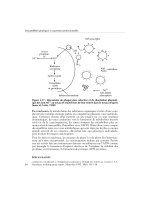
Figure 12.4. Allergic fungal sinusitis. A noncontrast axial CT image shows high
attenuation soft tissue fullness within the ethmoid and sphenoid sinuses bilaterally
with expansile bone erosion along the left laminae papyracea.
Although MRI is not a primary imaging study for the evaluation of
sinusitis, signal characteristics of sinus secretions were evaluated in chronic
sinusitis patients. Som et al. (49) reported MR signal intensity changes as
a function of protein concentration of sinus secretions. Normal sinus secre-
tions consist predominantly of water; thus it appears as low T1 and high
T2 signal intensities. As the sinus secretions become more viscous, the T1
signal intensity increases and the T2 signal intensity slowly decreases. Fur-
thermore, as sinus secretions become more desiccated and sludge-like, they
appear as low intensity in both T1 and T2 signals (50), and may become
signal void. Fungal sinusitis is also associated with signal void on MRI as
paramagnetic substance deposition such as manganese is fairly commonly
seen with fungal infection.
IV. Chronic Sinusitis: What Is the Role of Imaging in
Chronic Sinusitis? Does Imaging Change Treatment
Decision Making?
Summary of Evidence: The roles of sinus CT for chronic sinusitis patients
are to support clinical diagnosis, to evaluate the extent of disease, and to
provide detailed anatomy to assist treatment planning. The literature sug-
gests that sinus CT findings do not always correlate with patients’ clinical
symptoms. Whether patients with a normal CT but with persistent clinical
symptoms should undergo surgery remains controversial. There is not
enough evidence that sinus CT predicts clinical outcomes or that sinus CT
affects treatment decisions. Evidence for the CEA of diagnosis and treat-
ment of chronic sinusitis is lacking (insufficient evidence).
Supporting Evidence
A. The Role of Sinus Computed Tomography for Chronic Sinusitis
Despite a lack of evidence and problems related to the diagnosis of chronic
sinusitis by CT, it remains the imaging study of choice for patients with
chronic sinusitis. One of the roles of sinus CT is to determine whether a
patient is truly suffering from chronic sinusitis, as symptoms related to
chronic sinusitis are often vague and nonspecific (i.e., headache or
facial pain). Completely normal sinus CT performed when a patient is
having symptoms without prior medical treatment should suggest
other diagnoses. Sinus CT is also indicated for patients who do not respond
to medical management and to evaluate any obstructive lesions such
as a polyp, inverting papilloma, or sinonasal cancer or anatomic abnor-
malities impairing mucociliary drainage of the sinus (insufficient
evidence).
Once diagnosis of chronic sinusitis is supported clinically and radi-
ographically, an imaging evaluation for chronic sinusitis patients should
include the extent of the disease. The distribution of sinus involvement
may indicate a mucosal abnormality at the ostiomeatal complex. One
should also look for potential complications associated with sinusitis, such
as orbital cellulitis/abscess, mucocele or pyocele, epidural or brain abscess
using a soft tissue window.
226 Y. Anzai and W.E. Neighbor, Jr.
B. The Role of Sinus Computed Tomography Before and After
Endoscopic Sinus Surgery
Chronic sinusitis develops from persistent or recurrent sinus inflammation,
resulting in impaired ciliary function of the mucosa. Functional endoscopic
sinus surgery (FESS) has been developed to repair mucociliary drainage
of the sinus (51,52). Once surgery is indicated, CT is essential for providing
detailed sinus anatomy as well as the status of ostiomeatal complex prior to
FESS (insufficient evidence). Careful attention to key anatomic structures of
the ostiomeatal complex is needed. These include ethmoid infundibulum,
uncinate process, perpendicular plate and basal lamella of the middle
turbinate, ethmoid bulla, nasofrontal duct, sphenoethmoid recess, and
fovea ethmoidalis. Although certain anatomic variations such as concha
bullosa, paradoxical middle turbinate, and nasal septum deviation can
narrow the ostiomeatal complex (53,54), whether or not these anatomical
variations cause increased risk of developing chronic sinusitis is not known.
Functional endoscopic sinus surgery has been reported, primarily in the
surgical literature, to provide improved clinical outcomes for patients with
chronic sinusitis (51,55,56). However, a study evaluating the methodologic
quality of FESS investigations reports that most outcome studies of endo-
scopic sinus surgery lack a control group (57); thus the efficacy of FESS has
not been well established. Moreover, a substantial portion of patients who
had endoscopic sinus surgery have recurrent symptoms and seek further
medical care. Those patients may receive a second or third surgery and
undergo additional CT scan prior to the additional surgery.
Common CT findings following FESS include uncinectomy, partial
middle turbinectomy, and bulla ethmoidectomy. The extensive middle and
inferior turbinectomies are no longer recommended since it may cause
dryness or crusting of the nasal cavity, as well as turbulent air flow within
the nasal cavity, resulting in perception of difficulty in breathing through
the nose. One needs to look for a residual uncinate process for a patient
with persistent symptoms after sinus surgery.
C. Computed Tomography Prediction of Clinical Outcome
for Chronic Sinusitis
The value of sinus CT for predicting the clinical outcome of patients with
chronic sinusitis is highly controversial (limited evidence). Stewart et al.
(58) reported that the severity of sinus CT findings was a strong predictor
of improved clinical outcome in 57 patients. Patients with severe pretreat-
ment CT abnormality showed significantly larger improvement and lower
absolute levels of symptoms after treatment. Kennedy (52), on the other
hand, reported a strong correlation between the extent of disease on CT
and a poor surgical outcome in 120 patients with chronic sinusitis. Wang
et al. (59) also reported that in 230 consecutive patients the extent of disease
on sinus CT predicts clinical outcome of endoscopic sinus surgery for
chronic sinusitis in that the extent of disease was a consistent predictor
(p < 0.05) for bleeding, complication occurrence, medical resource utiliza-
tion, subjective sinus-specific health status, and physicians’ objective eval-
uation of surgical outcomes. Another study of endoscopic sinus surgery
indicated that advanced staging of CT and a previous history of sinus
surgery correlated with poor clinical outcome (60). Mantoni et al. (61), on
the other hand, reported that severity of sinus CT abnormality after FESS
does not correlate well with a clinical relief of patients’ symptoms.
Chapter 12 Imaging Evaluation of Sinusitis: Impact on Health Outcome 227
D. Does Sinus Computed Tomography Affect Treatment Decision
Making in Chronic Sinusitis?
Chronic sinusitis is managed either medically or surgically. Because sinus
CT has uncertain diagnostic accuracy and poor correlation with patients’
clinical symptoms for chronic sinusitis, some otolaryngologists advocate
that a treatment decision should be based solely on clinical grounds (44,46).
Surgery is indicated when the maximum medical treatment fails to resolve
the patient’s symptoms. However, there is no consensus as to what repre-
sents the maximum medical treatment. Moreover, the basis of treatment
decisions, medical versus surgical, for patients with chronic sinusitis is not
universally established. Whether or not a patient should be treated surgi-
cally, despite normal sinus CT, remains controversial (62). It is an open
question whether treatment decisions are purely based on physical exam-
ination and clinical history alone, or if sinus CT alters the treatment deci-
sions by ENT surgeons (limited evidence).
We prospectively administered questionnaires to a surgeon specializing
in endoscopic sinus surgery each time he saw a patient for suspected
sinusitis (63). After obtaining a clinical history and physical examination,
we first asked his treatment decision without a sinus CT, and then again
after reviewing the sinus CT. The abstracted clinical information of 27
patients was presented to two other otolaryngologists, and the same ques-
tionnaires were administered before and after reviewing the sinus CT.
Sinus CT altered dichotomous treatment decisions (surgical versus non-
surgical) by the surgeon in one third of patients (9/27) and there was a
tendency to offer the surgical treatment after reviewing the sinus CT
more than before. The agreement among surgeons with clinical history
and physical examination alone was poor but was much improved
after reviewing sinus CT. The results of this study indicate that sinus CT
provides pivotal objective information that affects treatment decisions
and improves the agreement of treatment plans among surgeons (limited
evidence).
E. Special Case: Cost-Effectiveness Analysis in Chronic Sinusitis
There has been no CEA for chronic sinusitis from the U.S. or Europe. Only
one recent study from Taiwan assessed cost utility analysis of endoscopic
sinus surgery. It measured the cumulative cost of treating chronic sinusi-
tis with FESS based on severity of disease. Utility assessment was per-
formed with the six-item Chronic Sinusitis Survey. The study revealed an
average cost-utility ratio of $70,221 and a high cost-utility ratio of $103,872
(after conversion to U.S. dollars at 1999 rates) for treatment of more severe
sinusitis cases due to the high cost and the limited utility gain (64). Some
patients were admitted for surgery with an average length of stay of 2.4
days (standard deviation 1.2). The cost structure in their study showed that
66% of the total cost was the operation fee. Endoscopic sinus surgery is pri-
marily performed on an outpatient basis in the U.S. Evidence is lacking in
this field, and future research is needed (insufficient evidence).
Health care costs for patients with chronic sinusitis were investigated in
health maintenance organizations (HMOs) in the state of Washington. This
study found that adult patients with chronic sinusitis have more nonurgent
outpatient visits and fill more prescriptions than adult patients without a
history of chronic sinusitis, not including endoscopic sinus surgery. The
228 Y. Anzai and W.E. Neighbor, Jr.
Chapter 12 Imaging Evaluation of Sinusitis: Impact on Health Outcome 229
Use clinical prediction rules or risk factors to differentiate bacterial and viral infection
ABX treatment Decongestant or anti-allergy Rx if h/o allergy
Good clinical response
No imaging study Screening sinus CT
Positive CT
Change ABX
Negative CT
Consider other
diagnoses
Poor response
Patients present with acute
sinusitis symptoms
Suspect bacterial sinusitis
(high probability for ABS)
Uncomplicate viral infection
(intermediate to low probability)
Good clinical response
Good clinical response
No imaging
Poor clinical response
Screening sinus CT
Positive CT Negative CT
No imaging ABX depends
on clinical exam
Poor response
Change ABX Consider other
diagnoses
Figure 12.5. Decision tree for imaging evaluation and management of acute bacterial sinusitis (ABS). ABX,
antibiotics; h/o, history of.
marginal total cost was $206 and the overall direct cost in the U.S. in 1994
was estimated to have been $4.3 billion (65).
Take-Home Figures
Decision trees for imaging evaluation and management of acute and
chronic sinusitis are shown in Figures 12.5 and 12.6.
Suggested Imaging Protocol
1. Noncontrast screening sinus CT
5-mm-thick coronal images every 10mm
140KVP, 200MA
Indications: sinusitis symptoms not responding to medical treatment
Diagnosis of sinusitis is in doubt, rule out sinusitis
Recent sinusitis, need to evaluate response to treatment
2. Noncontrast fine-cut maxillofacial CT
2.5mm thick helical
140KVP, 200MA
Indications: patients with chronic or recurrent sinusitis symptoms, need
to evaluate anatomical abnormality
Patients with chronic sinusitis failed to respond to the maximal medical
treatment; considering endoscopic sinus surgery
3. Axial fine-cut maxillofacial CT with coronal and sagittal reformat
0.625- to 1.25-mm helical scanning with coronal and sagittal reformat
140KVP, 175MA
Indications: patients require imaging-guided monitoring for endoscopic
sinus surgery for skull base lesions or complex sinus surgery
Future Research
• Randomized controlled trial of antibiotic for patients with mucosal
thickening only on CT in order to determine if this group of patients
benefits from antibiotic treatment for acute sinusitis.
• Cost-effectiveness analysis based on more realistic model assumptions
regarding types and durations of antibiotic treatment for acute sinusitis.
• Randomized controlled trial of endoscopic sinus surgery compared with
sham surgery in order to determine the efficacy of FESS for patients with
chronic sinusitis.
• Prospective outcome assessment for chronic sinusitis patients treated
medically or surgically in order to determine if CT findings predict treat-
ment response.
Summary
Acute sinusitis
• Despite inaccurate clinical diagnosis of acute or chronic sinusitis, the
initial treatment decision is based on clinical diagnosis.
• For patients present with acute sinusitis symptoms, if clinical suspicion
for acute bacterial sinusitis is high, patients should be treated with
antibiotics.
• If clinical suspicion for acute bacterial sinusitis is intermediate or low,
decongestant and conservative management is appropriate. Imaging
study is indicated when patients failed to respond to the initial treatment.
230 Y. Anzai and W.E. Neighbor, Jr.
Patients with history of chronic sinusitis presented with sinusitis symptoms
Treat with ABX and other medical management if applicable (i.e. allergy)
Good clinical response
No imaging Changes ABX or consider steroid treatment, if appropriate
Poor clinical response
Good clinical response
No imaging
Sinus CT
Screening sinus CT (if diagnosis needs to be confirmed)
Full sinus CT (if surgery is a consideration)
Normal sinus CT Localized disease on CT
Anatomic abnormality
Severe diffuse
disease on CT
Search for underlying
systemic disease
If refractory to the maximum
medical Rx, a patient
desires, consider surgery
???
Controversial
If CT correlates w
symptoms consider
surgery
Poor clinical response
Figure 12.6. Decision tree for evaluation and management of chronic sinusitis.
Chronis sinusitis
• For patients with clinical diagnosis of chronic sinusitis, imaging study
is indicated when patients failed to respond to medical management, in
order to determine if symptoms are related to sinusitis, or to evaluate
strutural abnormalities.
• Sinus CT provides objective information as to how diffuse or localized
disease is, and if symptoms are related to sinusitis, assisting treatment
decisions for patients with chronic sinusitis.
References
1. Gwaltney JM Jr, Jones JG, Kennedy DW. Ann Otol Rhinol Laryngol Suppl 1995;
167:22–30.
2. Gwaltney JM Jr, Sydnor A Jr, Sande MA. Ann Otol Rhinol Laryngol Suppl 1981;
90:68–71.
3. Collins J. National Center for Health Statistics Advanced Data 1988;1155:
1981–1916.
4. Willett LR, Carson JL, Williams JW Jr. J Gen Intern Med 1994;9:38–45.
5. Ioannidis JP, Lau J. Pediatrics 2001;108:51–58.
6. Clement PA, Bluestone CD, Gordts F, et al. Int J Pediatr Otorhinolaryngol 1999;
49:S95–100.
7. Garbutt JM, Gellman EF, Littenberg B. Qual Life Res 1999;8:225–233.
8. Turner BW, Cail WS, Hendley JO, et al. J Allergy Clin Immunol 1992;90:474–478.
9. Lau J, DZ, Engles E, et al. Diagnosis and Treatment of Acute Bacterial Rhinos-
inusitis. Rockville, MD: Agency for Health Care Policy and Research, 1999.
10. Newman LJ, Platts-Mills TA, Phillips CD, Hazen KC, Gross CW. JAMA 1994;
271:363–367.
11. Senior BA, Kennedy DW, Tanabodee J, Kroger H, Hassab M, Lanza DC. Oto-
laryngol Head Neck Surg 1999;121:66–68.
12. Amar YG, Frenkiel S, Sobol SE. J Otolaryngol 2000;29:7–12.
13. April MM, Zinreich SJ, Baroody FM, Naclerio RM. Laryngoscope 1993;103:
985–990.
14. Yang C, Talbot JM, Hwang PH. Am J Rhinol 2001;15:121–125.
15. Anderhuber W, Walch C, Braun H. Laryngorhinootologie 1997;76:315–317.
16. Lancaster J, Belloso A, Wilson CA, McCormick M. J Laryngol Otol
2000;114:630–633.
17. Schweitzer VG. Laryngoscope 1986;96:206–210.
18. Del Borgo C, Del Forno A, Ottaviani F, Fantoni M. J Chemother 1997;9:83–88.
19. Godofsky EW, Zinreich J, Armstrong M, Leslie JM, Weikel CS. Am J Med 1992;
93:163–170.
20. Collins JG. Vital Health Stat 1997;10:1–89.
21. National Center for Health Statistics: Sinusitis. NCHS, Hyattsville, MD, 2002.
22. Kaliner MA, Osguthorpe JD, Fireman P, et al. Otolaryngol Head Neck Surg 1997;
116:S1–20.
23. Gliklich RE, Metson R. Otolaryngol Head Neck Surg 1995;113:104–109.
24. Hickner JM, Bartlett JG, Besser RE, Gonzales R, Hoffman JR, Sande MA. Ann
Emerg Med 2001;37:703–710.
25. Gonzales R, Bartlett JG, Besser RE, et al. Ann Emerg Med 2001;37:690–697.
26. Low DE, Desrosiers M, McSherry J, et al. Can Med Assoc J 1997;156:S1–14.
27. Hudgins PA, Mukundan S. AJNR 1997;18:1850–1854.
28. Shapiro GG, Furukawa CT, Pierson WE, Gilbertson E, Bierman CW. J Allergy
Clin Immunol 1986;77:59–64.
29. Gwaltney JM Jr, Phillips CD, Miller RD, Riker DK. N Engl J Med 1994;330:25–30.
30. Lindbaek M, Hjortdahl P, Johnsen UL. BMJ 1996;313:325–329.
31. Engels EA, Terrin N, Barza M, Lau J. J Clin Epidemiol 2000;53:852–862.
Chapter 12 Imaging Evaluation of Sinusitis: Impact on Health Outcome 231
32. Varonen H, Makela M, Savolainen S, Laara E, Hilden J. J Clin Epidemiol 2000;
53:940–948.
33. Chen LC, Huang JL, Wang CR, Yeh KW, Lin SJ. Asian Pac J Allergy Immunol
1999;17:69–76.
34. Lindbaek M, Hjortdahl P, Johnsen UL. Fam Med 1996;28:183–188.
35. American Academy of Pediatrics. Pediatrics 2001;108:798–808.
36. Garbutt JM, Goldstein M, Gellman E, Shannon W, Littenberg B. Pediatrics 2001;
107:619–625.
37. Balk EM, Zucker DR, Engels EA, Wong JB, Williams JW Jr, Lau J. J Gen Intern
Med 2001;16:701–711.
38. Benninger MS, Sedory Holzer SE, Lau J. Otolaryngol Head Neck Surg 2000;
122:1–7.
39. Fagnan LJ. Am Fam Physician 1998;58:1795–1802, 1805–1796.
40. Gonzalez Morales JE, Leal de Hernandez L, Gonzalez Spencer D. Usefulness of
simple paranasal sinus radiographs and axial computed tomography in the
diagnosis of chronic sinusitis (in Spanish). Rev Alerg Mex 1998;45:17–21.
41. Garcia DP, Corbett ML, Eberly SM, et al. J Allergy Clin Immunol 1994;94:
523–530.
42. Burke TF, Guertler AT, Timmons JH. Acad Emerg Med 1994;1:235–239.
43. Lee HS, Majima Y, Sakakura Y, Inagaki M, Sugiyama Y, Nakamoto S. Nippon
Jibiinkoka Gakkai Kaiho 1991;94:1250–1256.
44. Stewart MG, Sicard MW, Piccirillo JF, Diaz-Marchan PJ. Am J Rhinol 1999;
13:161–167.
45. Piccirillo JF, Merritt MG Jr, Richards ML. Otolaryngol Head Neck Surg 2002;
126:41–47.
46. Bhattacharyya T, Piccirillo J, Wippold FJ. Arch Otolaryngol Head Neck Surg
1997;123:1189–1192.
47. Arango P, Kountakis SE. Laryngoscope 2001;111:1779–1782.
48. Mukherji SK, Figueroa RE, Ginsberg LE, et al. Radiology 1998;207:417–422.
49. Som P, Dillon W, Fullerton G, et al. Radiology 1989;172:515–520.
50. Som PM, Brandwein M. In: Som PM, Curtin HD, eds. Head and Neck Imaging,
3rd ed. St. Louis: Mosby, 1996;125–315.
51. Kennedy DW, Senior BA. Otolaryngol Clin North Am 1997;30:313–330.
52. Kennedy DW. Laryngoscope 1992;102:1–18.
53. Calhoun KH, Waggenspack GA, Simpson CB, Hokanson JA, Bailey BJ. Oto-
laryngol Head Neck Surg 1991;104:480–483.
54. Yousem DM, Kennedy DW, Rosenberg S. J Otolaryngol 1991;20:419–424.
55. Senior BA, Kennedy DW, Tanabodee J, Kroger H, Hassab M, Lanza D. Laryn-
goscope 1998;108:151–157.
56. Metson R, Gliklich RE. Arch Otolaryngol Head Neck Surg 1998;124:1090–1096.
57. Lieu JE, Piccirillo JF. Arch Otolaryngol Head Neck Surg 2003;129:1230–1235.
58. Stewart MG, Donovan DT, Parke RB, Bautista MH. Otolaryngol Head Neck
Surg 2000;123:81–84.
59. Wang PC, Chu CC, Liang SC, Tai CJ. Otolaryngol Head Neck Surg 2002;
126:154–159.
60. Marks SC, Shamsa F. Am J Rhinol 1997;11:187–191.
61. Mantoni M, Larsen P, Hansen H, Tos M, Berner B, Orntoft S. Eur Radiol
1996;6:920–924.
62. Kennedy DW. JAMA 2000;283:2143–2150.
63. Anzai Y, Weymuller EA, Yueh B, Maronian N, Jarvik JG. The impact of sinus
computed tomography on treatment decisions for chronic sinusitis. Arch Oto-
laryngol Head Neck Surg 2004;130(4):423–428.
64. Wang PC, Chu CC, Liang SC, Tai CJ. Otolaryngol Head Neck Surg 2004;
130:31–38.
65. Murphy MP, Fishman P, Short SO, Sullivan SD, Yueh B, Weymuller EA Jr. Oto-
laryngol Head Neck Surg 2002;127:367–376.
232 Y. Anzai and W.E. Neighbor, Jr.
13
Neuroimaging for Traumatic
Brain Injury
Karen A. Tong, Udo Oyoyo, Barbara A. Holshouser, and Stephen Ashwal
I. Which patients with head injury should undergo imaging in the
acute setting?
II. What is the sensitivity and specificity of imaging for injury requir-
ing immediate treatment/surgery?
III. What is the sensitivity and specificity of imaging for all brain
injuries?
IV. Can imaging help predict outcome after traumatic brain injury
(TBI)?
A. Imaging classification schemes
B. Normal scans
C. Brain swelling
D. Midline shift
E. Hemorrhage
F. Number/size/depth of lesions
G. Diffuse axonal injury
H. Combinations of imaging abnormalities and progressive brain
injury
I. Abnormalities of perfusion or activation
J. Measures of atrophy
K. Combinations of clinical and imaging findings
V. Is the approach to imaging children with traumatic brain injury
different from that for adults?
233
Head injury is not a homogeneous phenomenon and has a complex
clinical course. There are different mechanisms, varying severity,
diversity of injuries, secondary injuries, and effects of age or under-
lying disease.
Classifications of injury and outcomes are inconsistent. Differences in
diagnostic procedures and practice patterns prevent direct compari-
son of population-based studies.
Issues
Key Points
234 K.A. Tong et al.
There are a variety of imaging methods that measure different aspects
of injury, but there is not one all-encompassing imaging method.
Plain films have limited use for evaluating traumatic brain injury
(moderate evidence).
Computed tomography (CT) is an important part of the initial evalu-
ation and currently is the imaging modality of choice for screening of
life-threatening lesions requiring surgical intervention. It is probably
more useful for predicting short-term/crude (survival versus mortal-
ity) outcomes (moderate evidence).
Magnetic resonance imaging (MRI) is more sensitive than CT and is
useful for secondary evaluation. It is more useful for predicting long-
term outcome, although utility remains controversial (moderate
evidence). Functional MRI holds promise for predicting neuropsy-
chological outcomes (limited evidence).
Accurate prognostic information is important for determining man-
agement, but there are different needs for different populations. In
severe traumatic brain injury, information is important for acute
patient management, long-term rehabilitation, and family counseling.
In mild or moderate traumatic brain injury, patients with subtle
impairments may benefit from counseling and education.
Definition and Pathophysiology
Head trauma is difficult to study because it is a heterogeneous entity that
encompasses many different types of injuries that may occur together
(Table 13.1). Definitions of age groups, injuries and outcomes are also vari-
able. Classification of injury severity is usually defined by the Glasgow
Coma Scale (GCS) score, a scale ranging from 3 to 15, which is often
grouped into mild, moderate, or severe categories. There is inconsistency
in the timing of measurement, with some investigators using initial or field
GCS while others use postresuscitation GCS. Grouping of GCS scores also
vary. There is no universal definition of mild or minor head injury (1), as
some use GCS scores of 13 to 15 (2,3), while others use 14 to 15 (1) and
others use only 15. Variable definitions result in inconsistencies in imaging
recommendations. Moderate traumatic brain injury (TBI) is defined by a
GCS of 9 to 12. Severe TBI is defined by a GCS of 3 to 8.
Classification and measures of outcome are even more variable. The
most commonly used outcome measure is the Glasgow Outcome Scale
(GOS) (4). It is an overall measure based on degree of independence and
ability to participate in normal activities, with five categories: 5, good
recovery; 4, moderate disability; 3, severe disability; 2, vegetative state
(VS); and 1, death. The GOS is often dichotomized, although grouping is
variable. Recently modified, the extended GOS (5) has eight categories: 8,
good recovery; 7, good recovery with minor physical or mental deficits;
6, moderate disability, able to return to work with some adjustments; 5,
works at a lower level of performance; 4, severe disability, dependent on
others for some activities; 3, completely dependent on others; 2, VS; and 1,
death. Less common outcome scales include: the Differential Outcome
Scale (DOS) (6), the Rappaport Disability Rating Scale (DRS) (7), the Dis-
Chapter 13 Neuroimaging for Traumatic Brain Injury 235
ability Score (DS) (8), the FIM instrument (9), the Supervision Rating Scale
(SRS) (10), and the Functional Status Examination (FSE) (11,12).
The timing of outcome measurement also varies. Some investigators
measure outcomes at discharge and at 3, 6, or 12 months (or more) after
injury. This may be problematic because outcomes often improve with
time. However, there is moderate to strong evidence that 6 months is an
appropriate time point to measure outcomes for clinical trials (13). Neu-
ropsychological assessment is the most sensitive measure of outcome,
although this is difficult to perform in severely injured patients, resulting
in selection bias. There is a wide variety of psychometric scales for various
components of cognitive function such as intellect, orientation, attention,
language, speech, information processing, motor reaction time, memory,
learning, visuoconstructive ability, verbal fluency, mental flexibility, exec-
utive control, and personality. Currently, research has not been able to
demonstrate strong relationships between neuroimaging in the acute
period and long-term neuropsychological impairment (14,15).
Epidemiology in the United States
There is difficulty in determining the prevalence of TBI because many less
severely injured patients are not hospitalized, and cases with multiple
injuries may not be included. Estimates are often based on existing dis-
Table 13.1. Types of head injury (excluding pene-
trating/missile injuries and nonaccidental trauma)
Primary injuries
• Peripheral, nonintracranial
᭺
Scalp or soft tissue injury
᭺
Facial or calvarial fractures
• Extraaxial
᭺
Extradural or epidural hemorrhage
᭺
Subdural hemorrhage
᭺
Traumatic subdural effusion or “hygroma”
᭺
Subarachnoid hemorrhage
᭺
Intraventricular hemorrhage
• Parenchymal
᭺
Contusion
Hemorrhagic
Nonhemorrhagic
Both
᭺
Shearing injury or “diffuse axonal injury”
Hemorrhagic
Nonhemorrhagic
Both
• Vascular
᭺
Arterial dissection/laceration/occlusion
᭺
Dural venous sinus laceration/occlusion
᭺
Carotid-cavernous fistula
Secondary injuries
• Cerebral edema
• Focal infarction
• Diffuse hypoxic-ischemic injury
• Hydrocephalus
• Infection
abilities. Approximately 1.74 million/year suffer mild TBI that results in a
physician visit or temporary disability of at least 1 day (16) and more than
1 million visits per year to emergency departments are for TBI-related
injuries (17). There are more than 230,000 TBI-related hospitalizations/year
(17), perhaps up to 500,000/year (18), which account for 12% of all hospi-
tal admissions (18). Traumatic brain injury is responsible for nearly 40%
of all deaths from acute injuries (16). Between 1989 and 1998, there were
approximately 53,000 TBI-related deaths/year, for a rate of 20.6/100,000
population (17). The major causes of TBI-related deaths are firearms (40%),
motor vehicle accidents (MVAs) (34%), and falls (10%) (17). The risk of TBI
peaks between the ages of 15 and 30 (16), with the highest TBI-related death
rates occurring in American Indian/Alaska natives, males, and persons
over the age of 75 (17).
Overall Cost to Society
From 1989 to 1998 there has been an overall decline in TBI-related deaths,
probably due to multiple factors including improvements in medical care,
use of evidence-based guidelines, and injury-prevention efforts (17). An
estimated 5.3 million U.S. residents live with permanent TBI-related dis-
abilities (17). Direct costs are estimated at $4 billion/year (16). In 1995, total
direct and indirect costs of TBI were estimated at $56 billion/year (17).
There are few data on the costs of TBI related solely to imaging. There has
been one small study (limited evidence) that determined that 60% of
patients were found to have additional lesions on MRI, but because none
of these additional findings changed management, MRI resulted in a
non–value-added benefit incremental increase of $1891 per patient and a
$3152 incremental increase in charges to detect each patient with a lesion
not identified on CT (19).
Goals of Neuroimaging
• To detect the presence of injuries that may require immediate surgical
or procedural intervention.
• To detect the presence of injuries that may benefit from early medical
therapy.
• To determine the prognosis of patients to tailor rehabilitative therapy or
help with family counseling.
Methodology
A search of the Medline/PubMed electronic database (National Library of
Medicine, Bethesda, Maryland) was performed using the following key-
words: (1) head injury, head trauma, brain injury, brain trauma, traumatic brain
injury, or TBI; and (2) CT, computed tomography, computerized tomography,
MR, magnetic resonance, spectroscopy, diffusion, diffusion tensor, functional
magnetic, functional MR*, T2*, FLAIR, GRE, gradient-echo. No time limits
were applied for the searches, which were repeated several times up to
April 16, 2004. Searches were limited to the English-language literature,
abstracts, and human subjects. A search of the National Guideline Clear-
236 K.A. Tong et al.
inghouse at www.guideline.gov was also performed using the following
key words: (1) head injury, head trauma,andbrain injury; and (2) parameter
and guideline.
I. Which Patients with Head Injury Should Undergo
Imaging in the Acute Setting?
Summary of Evidence: The need for acute imaging is generally based on the
severity of injury. It is agreed that severe TBI (based on GCS score) indi-
cates the need for urgent CT imaging to determine the presence of lesions
that may require surgical intervention (strong evidence). There is greater
variability concerning recommendations for imaging of patients with mild
or moderate TBI, although there are several recent guidelines (strong evi-
dence) summarized in take-home Tables 13.2 and 13.3.
Supporting Evidence: There are several clinical prediction rules (strong evi-
dence) for evaluating mild/minor head injury in adults, based on prospec-
tive studies. The Canadian Head CT Rule (2001) (20) was developed from
prospective analysis of 3121 patients with GCS scores of 13 to 15. A CT scan
was recommended if a patient had any of the following: GCS score <15
after 2 hours; suspected open or depressed skull fracture; any sign of basal
skull fracture; episode(s) of vomiting; age greater than 65 (associated with
high risk for neurosurgical intervention); amnesia for the period occurring
30 minutes or more before impact; or an injury due to a dangerous mech-
anism, such as being struck by or ejected from a motor vehicle (associated
with a medium risk for brain injury on CT). Another guideline by Haydel
and colleagues (21) was developed after prospective analysis of 520
patients in the first phase and 909 patients in the second phase. After recur-
Chapter 13 Neuroimaging for Traumatic Brain Injury 237
Table 13.2. Suggested guidelines for acute neuroimaging in adult
patient with mild TBI (Glasgow Coma Scale score 13 to 15)
If GCS 13–15, CT recommended if patient has any one of the following:
• High risk
᭺
GCS remains <15 at 2 hours after injury
᭺
Suspected open or depressed skull fracture
᭺
Any clinical sign of basal skull fracture
᭺
Two or more episodes of vomiting
᭺
Aged 65 years or older
• Medium risk
᭺
Possible loss of consciousness
᭺
Amnesia for period before impact, of at least 30-minute time span
᭺
Dangerous mechanism (pedestrian versus motor vehicle, ejected from
motor vehicle, fall from greater than 3 feet or five stairs)
᭺
Any transient neurologic deficit
᭺
Headache, vomiting
If GCS of 15, patient can be discharged without CT scan if:
• Low risk
᭺
GCS remains 15
᭺
No loss of consciousness or amnesia
᭺
No neurologic/cognitive abnormalities
᭺
No headache, vomiting
CT, computed tomography, TBI, traumatic brain injury, GCS, Glasgow coma scale.
Source: Modified from the Canadian Head CT Rule (20), EAST guidelines (2), and the Neuro-
traumatology Committee of the World Federation of Neurosurgical Societies (1).
sive partitioning of variables in the first phase, seven variables were tested
in the second phase: headache, vomiting, age over 60 years, drug or alcohol
intoxication, short-term memory deficits, physical evidence of trauma
above the clavicles, and seizure. All patients with positive CT scans had at
least one variable, resulting in 100% sensitivity (21). An older guideline
(1995), prospectively analyzed 51 clinical variables in 540 patients in the
first phase and 10 remaining variables in 273 patients in the second phase.
The resulting sensitivity and negative predictive value were 96% and 94%,
respectively (22).
A guideline, “Practice Management Guidelines for the Management of
Mild Traumatic Brain Injury,” developed by the Eastern Association for the
Surgery of Trauma (EAST) Practice Management Guidelines Work Group
(2001) (2), was based on level II evidence from several studies (three ret-
rospective and one uncontrolled prospective). They reported that 3% to
17% of patients with mild injuries had significant CT findings, although
they noted that there was no uniform agreement as to what constitutes a
positive CT scan in different studies. They also reported that a patient with
a normal head CT had a 0% to 3% probability of neurologic deterioration.
Therefore, if a patient had a GCS of 15 and no neurologic/cognitive abnor-
malities, it was recommended that the patient be discharged. A CT scan
was recommended for all patients with transient neurologic deficits.
One guideline for severe TBI, “Management and Prognosis of Severe
Traumatic Brain Injury” (2000), was developed by the American Associa-
tion of Neurological Surgeons (AANS), and approved by the American
Society of Neuroradiology, the American Academy of Neurology, the
American College of Surgeons, the American College of Emergency Physi-
cians, the Society for Critical Care Medicine, and the American Academy
of Physical Medicine and Rehabilitation (23,24). An extensive review of the
CT literature supported the need for CT in the acute period. Computed
tomography was reported to be abnormal in 90% of patients with severe
head injury. Computed tomography is included as a necessary step in the
algorithm of initial management.
238 K.A. Tong et al.
Table 13.3. Suggested guidelines for acute neuroimaging in adult
patient with severe TBI (GCS 3–8)
• CT scan patient with severe TBI as soon as possible to determine if require
surgical intervention
• If initial scan is normal, but patient has neurologic deterioration, repeat CT
scan or consider MRI as soon as possible
• If initial scan is abnormal, but patient status is unchanged, repeat CT scan
within 24 to 36 hours to determine possible progressive hemorrhage or
edema requiring surgical intervention, particularly if initial scan showed:
᭺
Any intracranial hemorrhage
᭺
Any evidence of diffuse brain injury
• If initial scan is abnormal, repeat CT scan or consider MRI as soon as
possible if GCS worsens
• Consider MRI within first few days if:
᭺
Suspect secondary injury such as focal infarction, diffuse hypoxic-ischemic
injury or infection
TBI, traumatic brain injury; CT, computed tomography; MRI, magnetic resonance imaging;
GCS, Glasgow coma scale.
II. What Is the Sensitivity and Specificity of Imaging for
Injury Requiring Immediate Treatment/Surgery?
Summary of Evidence: Computed tomography is the mainstay of imaging
in the acute period. The majority of evidence relates to the use of CT for
detecting injuries that may require immediate treatment or surgery. Speed,
availability, and lesser expense of CT studies remain important factors for
using this modality in the acute setting. Sensitivity of detection also
increases with repeat scans in the acute period (strong evidence).
Supporting Evidence: The incidence of injury-related abnormalities on CT
is related to the severity of injury. After minor head injury, the incidence is
approximately 6% (25) and increases up to 15% in the elderly population
(26); those with GCS 13 or 14 have higher frequency of abnormalities than
those with GCS 15 (27). The incidence of CT abnormalities in moderate
head injury (with GCS of 9 to 13) has been reported to be 61% (28). The
sensitivity of CT for detecting abnormalities after severe TBI (GCS below
9) varies from 68% to 94%, while normal scans range from approximately
7% to 12% (29). Several studies have shown that the timing of CT studies
also affects the sensitivity. Oertel and colleagues (30) (strong evidence)
prospectively studied 142 patients with moderate or severe injury who had
undergone more than one CT scan within the first 24 hours, and found that
the initial CT scan did not detect the full extent of hemorrhagic injuries in
almost 50% of patients, particularly if scanned within the first 2 hours. The
likelihood of progressive hemorrhagic injury that potentially required sur-
gical intervention was greatest for parenchymal hemorrhagic contusions
(51%), followed by epidural hematoma (EDH) (22%), subarachnoid hem-
orrhage (SAH) (17%), and subdural hemorrhage (SDH) (11%). Servadei
and colleagues (31) (strong evidence) prospectively studied 897 patients
with more than one CT scan, and found that 16% of patients with diffuse
brain injury demonstrated significant evolution of injury. This was
more frequent in those with midline shift, often evolving to mass lesions.
Similar results have been seen in retrospective studies (32). Therefore, it is
useful to perform repeat CT scans in the acute period, particularly after
moderate and severe injury, although the timing has not been clearly
determined.
III. What Is the Sensitivity and Specificity of Imaging
for All Brain Injuries?
Summary of Evidence: The sensitivity and specificity of MRI for brain injury
is generally superior to CT, although most studies have been retrospective
and very few head-to-head comparisons have been performed in the recent
decade. Computed tomography is clearly superior to MRI for the detec-
tion of fractures, but MRI outperforms CT in detection of most other lesions
(limited to moderate evidence), particularly diffuse axonal injury (DAI).
However, MRI is expensive and not widely available, which also hinders
research. Because different sequences vary in the ability to detect certain
lesions, it is often difficult to compare results. Although MRI facilitates
more detailed analysis of injuries, including metabolic and physiologic
measures, further evidence-based research is needed.
Chapter 13 Neuroimaging for Traumatic Brain Injury 239
Supporting Evidence: Magnetic resonance imaging has higher sensitivity
than CT, though most comparison studies were performed in the late 1980s
and early 1990s (with older generation or lower field scanners). Orrison
and colleagues (33) (moderate evidence) retrospectively studied 107
patients with MRI and CT within 48 hours and showed that MRI had an
overall sensitivity of 97% compared to 63% for CT, even when a low-field
MRI scanner was used, with better sensitivity for contusion, shearing
injury, and subdural and epidural hematoma. Ogawa and colleagues (34)
(moderate evidence) detected more lesions with conventional MRI than
with CT, with the exception of subdural and subarachnoid hemorrhages,
in a prospective study of 155 patients, although they were studied at vari-
able time points. Other studies (moderate evidence), showed better detec-
tion of nonhemorrhagic contusions and shearing injuries (35) and of
brainstem lesions (36).
Some lesions, such as DAI, are clearly better detected with MRI, and
have been reported in up to 30% of patients with mild head injury with
normal CT (37) (limited evidence). However, sensitivity depends on the
sequence, field strength, and type of lesion. Gradient echo (GRE) sequences
are best for detecting hemorrhagic DAI, although the proportion of hem-
orrhagic versus nonhemorrhagic DAI is not truly known. An early report
(limited evidence) suggested that fewer than 20% of DAI lesions were
visibly hemorrhagic (38), but this is likely to be erroneously low, due to
poor sensitivity of the imaging methods available at that time. We have
recently studied a new susceptibility-weighted imaging (SWI) sequence
(at 1.5T) that is a modified GRE sequence, and have shown significantly
better detection of small hemorrhagic shearing lesions compared to con-
ventional GRE (39) (limited evidence). Scheid and colleagues (moderate
evidence) (40) prospectively studied 66 patients using high-field (3.0T)
MRI and found that T2*-weighted GRE sequences detected significantly
more lesions than conventional T1- or T2-weighted sequences. The fluid-
attenuated inversion recovery (FLAIR) sequence is useful for detecting
SAH, SDH, contusions, nonhemorrhagic DAI, and perisulcal lesions, but
there are few studies comparing the sensitivity of FLAIR to other
sequences. One study (limited to moderate evidence) found that FLAIR
sequences were significantly more sensitive than spin echo (SE) sequences
(p < .01) in detection of all lesions studied within 1 to 36 days (0.5T), par-
ticularly in those who had DAI-type lesions (41).
Diffusion weighted imaging (DWI) has also recently been shown to
improve the detection of nonhemorrhagic shearing lesions, although there
are only a few small studies describing sensitivity. A small study (insuf-
ficient evidence) of patients scanned within 48 hours found that DWI
identified an additional 16% of shearing lesions that were not seen on
conventional MRI. The majority of DWI-positive lesions (65%) had
decreased diffusion (42). Another descriptive study (limited evidence)
characterized several different types and patterns of DWI lesions, although
there was no comparison with other MRI sequences or analysis of diffu-
sion changes over time (43). A recent study (limited evidence) found a
strong correlation between apparent diffusion coefficient (ADC) his-
tograms and GCS score (44). There are even fewer data on the sensitivity
of diffusion tensor imaging (DTI). A few small studies (insufficient or
limited evidence) have shown decreased anisotropy in brain parenchyma
of TBI patients (45–47).
240 K.A. Tong et al.
Although CT and MRI are often limited to observing structural abnor-
malities associated with TBI, magnetic resonance spectroscopy (MRS) can
detect subtle cellular abnormalities that may more accurately estimate the
extent of brain injury, particularly DAI. However, the sensitivity and speci-
ficity of MRS are not easily addressed, as only a small number of studies
have been published. Several small studies have been performed using
single voxel spectroscopy (SVS), although measured at variable time
points. These have reported (insufficient evidence) decreased N-acetylas-
partate (NAA) in the frontoparietal white matter (WM) (48,49), gray matter
(GM) (50), or normal-appearing brain (51). Others have shown that NAA-
derived ratios were decreased in areas particularly vulnerable to DAI
(moderate evidence), such as the splenium of the corpus callosum (52,53).
There has been insufficient evidence regarding the sensitivity of multivoxel
magnetic resonance spectroscopic imaging (MRSI), although decreases in
NAA have been detected in areas of visible T2 abnormality as well as
normal-appearing regions compared to controls (54). There has been one
small study using phosphorous MRS (insufficient evidence), which found
alkaline pH, increased free intracellular magnesium, increased phospho-
creatine to inorganic phosphate ratio (PCr/Pi), and reduced inorganic
phosphate to adenosine triphosphate ratio (Pi/ATP) (55) in brains of
severely injured patients. Further research regarding the sensitivity of MRS
in TBI is warranted.
Several imaging methods permit in vivo assessment of regional metab-
olism or blood flow, which may be impaired after brain injury. These
methods include CT, MRI, and nuclear medicine imaging techniques. The
latter have been the most studied, although evidence remains limited. Most
studies consist of small sample sizes, and have been performed in the sub-
acute period. Single photon emission computed tomography (SPECT) can
measure regional cerebral blood flow (CBF) and assess localized perfusion
deficits that may correlate with cognitive deficits even in the absence of
structural abnormalities. However, SPECT has low spatial and temporal
resolution, does not permit imaging of transient cognitive events, and
interpretation is often highly subjective. The SPECT studies generally show
patchy perfusion deficits, often in areas with no visible injury on CT. One
of the largest studies, although retrospective, was performed by Abdel-
Dayem and colleagues (56) (moderate evidence), who reviewed SPECT
findings in 228 subjects with mild or moderate TBI. They found focal areas
of hypoperfusion in 77% of patients. However, there was no comparison
to CT or MRI. Stamatakis and colleagues (57) (moderate evidence) studied
61 patients with SPECT and MRI, within 2 to 18 days after injury, and
found that SPECT detected more extensive abnormality than MRI in acute
and follow-up studies. A small study (limited evidence) of patients with
persistent postconcussion syndrome after mild TBI found that SPECT
showed abnormalities in 53% of patients, whereas MRI and CT showed
abnormalities in only 9% and 4.6%, respectively (58).
Positron emission tomography (PET) can measure regional glucose and
oxygen utilization, CBF at rest, and CBF changes related to performances
of different tasks. Spatial and temporal resolution is also limited, although
better than with SPECT. However, PET is not widely available. A few PET
studies have reported various areas of decreased glucose utilization, even
without visible injury. Bergsneider and colleagues (59) (limited to moder-
ate evidence) prospectively studied 56 patients with mild to severe TBI,
Chapter 13 Neuroimaging for Traumatic Brain Injury 241
evaluated with 18F-fluorodeoxyglucose (FDG)-PET within 2 to 39 days of
injury; 14 patients had subsequent follow-up studies. The authors state in
this and previous reports that TBI patients demonstrate a triphasic pattern
of glucose metabolism changes that consist of early hyperglycolysis, fol-
lowed by metabolic depression, and subsequent metabolic recovery (after
several weeks). There are few small studies evaluating sensitivity of xenon
CT and even fewer describing the sensitivity of functional MRI (fMRI) or
MR perfusion.
IV. Can Imaging Help Predict Outcome After
Traumatic Brain Injury (TBI)?
Summary of Evidence: The study of outcome prediction after TBI is
complex. Predictor variables may not be as accurate if measured too early,
but may be less useful if measured too late. Evaluation of prognostic vari-
ables has ranged from studying individual measures to comprehensive
multimodal evaluations. Many clinical predictors have been studied
including age, gender, GCS, pupillary reactivity, intracranial pressure
(ICP), coagulopathy, hypothermia, hypoxia, hypotension, hyperglycemia,
and electrolyte imbalance, in addition to imaging findings. Thatcher and
colleagues (60) (moderate evidence) studied 162 patients and showed that
combined measures are more reliable and accurate than any single
measure. There have been relatively few comprehensive studies of long-
term prognostic indices compared to acute prognostic indices (e.g., death
versus survival).
Analysis of CT predictors of outcome have yielded variable results in
the literature. Abnormalities found on CT have been analyzed individu-
ally, collectively (in various combinations), or combined with clinical prog-
nostic variables. Various studies have shown improvement in outcome
prediction after severe TBI when adding CT information to clinical vari-
ables (moderate evidence). Computed tomography has been studied more
extensively than other imaging modalities, although it is likely that MRI
and other imaging methods will have greater value for predicting long-
term outcome. Unfortunately, available evidence is sparse.
Supporting Evidence: Early research on CT predictors was performed with
older technology that was less sensitive to the presence of injuries. Some
studies analyzed the first scans while others analyzed the worst scans.
Many studies used a crude categorization system, with limited informa-
tion regarding the degree of abnormalities. Others have attempted to assess
outcome prediction using more detailed classification schemes. Accord-
ingly, there has been variability in the reported predictors and success at
prediction.
A. Imaging Classification Schemes
Although there are a variety of classification schemes, very few have been
used to predict clinical outcomes. The most widely studied classification
scheme is based on CT findings in the Trauma Coma Databank (TCDB),
developed by Marshall and colleagues (61), based on the status of cisterns,
midline shift, and mass lesions. Categories include (a) diffuse injury I
(normal): no visible intracranial pathology; (b) diffuse injury II (small
242 K.A. Tong et al.
lesions): cisterns are present, midline shift <5mm, no lesions greater than
25cc; (c) diffuse injury III (swelling): cisterns are compressed, midline shift
<5mm, no lesions greater than 25cc; (d) diffuse injury IV (shift): midline
shift of >5mm, no lesions greater than 25cc; (e) any surgically evacuated
lesion; and (f) any nonevacuated mass lesion greater than 25cc. The TCDB
classification was developed in severely injured patients (GCS <8) and ini-
tially compared to discharge outcomes, although it has more recently been
validated using 3- and 6-month GOS (62). It is reasonably good at pre-
dicting mortality, but it may not be as applicable to mild/moderately
injured patients and has been criticized as poorly predictive of functional
recovery (63). The TCDB classification has been variously modified, often
to include the type, number (31,64), or location of lesions (65). In the AANS
guideline (24), an extensive review of the previous CT literature (strong
evidence) showed that the TCDB CT classification scheme strongly corre-
lated with outcome.
B. Normal Scans
Extensive review (strong evidence) shows that normal CT scans in severe
TBI patients are predictive of favorable outcome (61% to 78.5% positive
predictive value) (29). In a recent study (moderate evidence) normal CT
scans in moderate/severe TBI patients were associated with better neu-
ropsychological performance at 6 months (66).
C. Brain Swelling
Brain swelling is a subjective finding and more difficult to evaluate as an
outcome predictor. Partly compressed ventricles and cisterns are not as
reliably measured as obliterated ventricles and cisterns (67). Marshall and
colleagues (61) (strong evidence) studied the TCDB classification in 746
patients and reported that brain swelling on CT (categorized by diffuse
injury III) was predictive of mortality, and that survivors showed a trend
of worse GOS associated with increasing grade of diffuse injury. Com-
pressed basal cisterns have been associated with a threefold risk of raised
ICP, and a two- to threefold increase in mortality (24). However, brain
swelling on CT does not appear to correlate with neuropsychological
outcomes (14) (moderate evidence).
D. Midline Shift
Midline shift is felt to be less important than other CT parameters for pre-
dicting mortality or GOS score (24). However, some investigators have
shown that midline shift may be predictive of worse outcomes based on
rehabilitation measures such as greater need for assistance with ambula-
tion, activities of daily living (ADLs), and supervision at rehabilitation
discharge (68).
E. Hemorrhage
The presence of hemorrhage has different prognostic significance depend-
ing on the extent and location of blood. Traumatic subarachnoid hemor-
rhage is a significant independent prognostic indicator (24,69) (strong
evidence), associated with a twofold increase in mortality, and a 70% pos-
itive predictive value for unfavorable outcome (24). Mortality is higher and
Chapter 13 Neuroimaging for Traumatic Brain Injury 243
outcome is worse with acute subdural hematoma compared to extradural
hematoma (24). Hematoma volume correlates with outcome, and has 78%
to 79% positive predictive value for unfavorable outcome (24). Another
study (moderate evidence) found that patients with combined SDH and
ICH on CT had poor outcome even after surgery compared to those with
EDH or ICH alone (70). A small study (limited evidence) also found that
intraventricular hemorrhage (IVH) in all four ventricles was significantly
associated with poor outcome (71).
F. Number/Size/Depth of Lesions
Some investigators have attempted to evaluate the predictive ability of
number, size, depth, or location of lesions. Van der Naalt and colleagues
(6) (moderate evidence) studied 67 patients with mild/moderate TBI and
found outcome (1 year extended GOS or DOS) was related to number, size,
and depth of lesions on CT. Kido and colleagues (72) (moderate evidence)
found GOS was correlated with the size of intracranial lesions (indepen-
dent of compartment or brain region) on CT. A small MRI study (limited
evidence) suggested that size, depth, and multiplicity of lesions correlated
with neurobehavioral outcome (73).
Location of lesions is partly related to mechanism of injury and is asso-
ciated with different outcomes. The most available evidence is related to
brainstem injuries. Firsching and colleagues (65) (moderate evidence)
studied 102 patients in coma with MRI in the first 8 days and found that
mortality was 100% with lesions in the bilateral pons. Kampfl and col-
leagues (74) (moderate evidence) studied 80 patients and also showed that
lesion location could predict recovery from posttraumatic VS by 12
months, whereas clinical variables such as initial GCS, age, and pupillary
abnormalities were poor predictors. Logistic regression showed that
corpus callosum and dorsolateral brainstem injuries were predictive of
nonrecovery. This information is helpful in that almost half of the patients
with initial VS may recover within 1 year (74). The association between
extent or location of injuries and neuropsychological recovery has been less
well studied, with only a few studies (limited evidence) that suggest that
location of injury may be associated with specific neuropsychological
impairments (73,75).
G. Diffuse Axonal Injury
It has been repeatedly demonstrated that CT and MRI findings are poor
predictors of functional outcome of TBI patients, probably because DAI is
frequently not detected (7). Because CT clearly underestimates DAI, this
can lead to inaccurate prediction of outcome. The CT studies, many of
which were performed with older generation CT scanners, predominantly
report that DAI is associated with mortality (limited evidence) (76) or poor
outcome (moderate evidence) (77,78). It has since been shown that patients
with mild or moderate injuries can also have DAI (37) that is better
detected with newer generation CT scanners or MRI, and can therefore
have better outcomes than previously realized. Severe DAI can transform
young productive individuals into dependent patients requiring institu-
tionalized care, while milder DAI can result in neuropsychiatric problems,
cognitive deficits including memory loss, concentration difficulties, de-
creased attention span, intellectual decline, headaches, and seizures (79).
244 K.A. Tong et al.
The improved ability to detect DAI on CT even in milder injuries has also
allowed comparison with neuropsychological outcome. Wallesch and col-
leagues (80) (moderate evidence) studied 60 patients with mild or moder-
ate injuries who underwent neuropsychological assessment 18 to 45 weeks
later. Patients with DAI identified on CT had relatively transient deficits of
psychomotor speed, verbal short-term memory, and frontal lobe cognitive
functions, whereas patients with frontal contusions had persistent behav-
ior alterations.
The MRI studies also suggest an association between TBI severity and
depth of axonal injury as well as outcomes. However, most MRI studies
evaluating prognosis after DAI have consisted of small sample sizes. Small
studies (limited or moderate evidence) have demonstrated that patients in
VS are more likely to have DAI lesions in the corpus callosum and dorso-
lateral brainstem (81), compared with patients with mild TBI who were
more likely to have lesions in the subcortical white matter without involve-
ment of the corpus callosum or brainstem (77). The presence of hemorrhage
in DAI lesions may also affect prognosis, although results depend on the
MRI sequence. One study of VS (moderate evidence) found more non-
hemorrhagic DAI lesions than hemorrhagic lesions, although only T1- and
T2-weighted sequences were used (81). In contrast, another study (limited
evidence) showed that hemorrhage in DAI lesions (detected by GRE) was
associated with poor outcomes (6-month GOS), and that isolated non-
hemorrhagic DAI lesions were not associated with poor outcome (82).
There is also disagreement over whether the degree of hemorrhage corre-
lates with outcomes, although this may be partly due to differences in
outcome measures. One study (moderate evidence) found that the number
of lesions (hypointense or hyperintense) detected by T2*-weighted GRE
images correlated with duration of coma and 3-month GOS (83). However,
another study (moderate evidence) (MRI sequence not specified) found no
correlation between hemorrhagic lesion volume and neuropsychological
outcome measures obtained more than 6 months after injury (84). A recent
prospective study (moderate evidence) of 66 patients imaged with T2*-
weighted GRE at 3.0T, found no correlation between the total amount of
microhemorrhages and patient outcomes measured by GOS. However,
these patients were imaged in the chronic phase (40).
Magnetic resonance spectroscopy is able to detect abnormalities in struc-
turally normal brain that are believed to reflect DAI, and has shown
promise in predicting outcome, although there are only a few studies con-
sisting of small sample sizes. Investigators have compared MRS findings
from noncontused brain with various measures of clinical neurologic
outcome such as GOS or DRS scores and found a general trend of reduced
NAA corresponding to poor outcome (limited evidence) (50,52,53,85).
However, results are difficult to compare since varied anatomic areas were
studied and results were often acquired over a wide range of times after
injury. It is uncertain whether the timing of MRS measurement affects
outcome prediction. Subacute MRS studies have suggested that decreased
NAA correlates with poor outcomes.
There have been few acute MRS studies evaluating outcome prediction.
In a prospective MRS study (86) of 42 severely injured adults (limited to
moderate evidence), we measured quantitative metabolite changes as soon
as possible (mean of 7 days) after injury, in normal-appearing GM and
WM. In contrast to other studies, we found no correlation between NAA-
Chapter 13 Neuroimaging for Traumatic Brain Injury 245
derived metabolites and outcomes at 6 to 12 months, possibly because our
MRS studies were performed earlier. However, we found that gluta-
mine/glutamate (Glx) and Cho were significantly elevated in occipital GM
and parietal WM in patients with poor 6- to 12-month outcomes and that
these two variables predicted outcome at 6 to 12 months with 89% accu-
racy. A combination of Glx and Cho ratios with the motor component of
the GCS score provided the highest predictive accuracy (97%). It may be
that elevated Glx and Cho are more sensitive indicators of injury and pre-
dictors of poor outcome when spectroscopy is obtained early after injury.
This may be a reflection of early excitotoxic injury (i.e., elevated Glx) and
of injury associated with membrane disruption secondary to diffuse
axonal injury (i.e., increased Cho). An example of spectra from parietal and
occipital GM in a TBI patient with poor outcome is shown in case study 2,
below.
There have been no published results comparing data from MRSI to clin-
ical outcomes. Our preliminary data (limited to moderate evidence) in 42
patients with severe TBI, taken with MRSI through the corpus callosum
and surrounding GM and WM, showed significant decreases in NAA/Cre
and increases in Cho/Cre ratios in areas of visibly injured and normal-
appearing brain. Averaged ratios from all regions were able to differentiate
between patients with mild, moderate, and severe/vegetative neurologic
outcomes as measured with the GOS at 6 months compared with control
values. The results suggest that decreased NAA-derived ratios and
increased Cho/Cre ratios, detected by MRSI, are associated with worse
outcomes.
There are other MRI techniques that can detect abnormalities in visibly
normal brain, although there is little evidence regarding their role in
outcome prediction. Small studies using magnetization transfer methods
(limited evidence) have suggested that the magnetization transfer ratio
(MTR) in normal or abnormal white matter (87) or the splenium (88) may
be associated with outcomes. Diffusion weighted imaging has only
recently been studied in the setting of TBI, and the relationship between
ADC and clinical outcome has not been adequately investigated. Diffusion
tensor imaging is an even more recent development. One study (limited
evidence) compared 15 patients and 30 control subjects and found corre-
lations between cerebral fractional anisotropy score in trauma (C-FAST)
and short-term predictors such as death, length of hospital stay, or inten-
sive care unit stay (45).
H. Combinations of Imaging Abnormalities and
Progressive Brain Injury
Some studies have shown that combinations of imaging abnormalities are
predictive of outcome, although not necessarily in agreement. Fearnside
and colleagues (89) (strong evidence) prospectively studied 315 patients
and found three CT findings—cerebral edema, intraventricular blood, and
midline shift—to be highly predictive of mortality. Three other CT find-
ings—subarachnoid hemorrhage, intracerebral hematoma, and intracere-
bral contusion–were highly predictive of poor outcome in survivors (89).
In contrast, Lannoo and colleagues (90) (moderate evidence) retrospec-
tively reviewed 115 patients and found that subarachnoid, intracerebral,
and subdural hemorrhage were predictive of mortality but not signifi-
246 K.A. Tong et al.
cantly related to morbidity. Wardlaw and colleagues (63) (moderate evi-
dence) retrospectively reviewed 414 patients and developed a simple
rating system of “overall appearance” of CT findings. They reported that
massive injuries and SAH could predict poor prognosis (1-year GOS). Stein
and colleagues (32) (moderate evidence) also showed, in a retrospective
study of 337 patients, that delayed brain injury (44.5% of their population)
was a significant independent predictor of outcome.
I. Abnormalities of Perfusion or Activation
The relationship between perfusion studies and outcome has still not been
clearly demonstrated. The most extensive evidence has been with SPECT.
However results vary, possibly related to the severity of injury or timing
of studies. The largest study with patient outcomes was performed by
Jacobs and colleagues (91) (moderate to strong evidence) who prospec-
tively studied 136 patients with mild injury, within 4 weeks of injury. They
found that SPECT had a high sensitivity and negative predictive value. A
normal scan reliably excluded clinical sequelae of mild injury. A small
study (limited evidence) of patients with severe TBI and diffuse brain
injury showed that total CBF values initially increased above normal in the
first 1 to 3 days and then decreased below normal in the subacute period
of 14 to 42 days. The early CBF increase has been postulated to reflect
vasodilatation due to high tissue CO
2
and lactic acidosis. The authors
found that the initial elevation and subsequent drop in blood flow was
more marked in the poor-outcome group (92). However, another small
study (limited evidence) of patients with a spectrum of injury, studied
within 3 weeks of brain injury, found that focal zones of hyperemia in
normal-appearing brain was associated with slightly better outcomes than
in patients without hyperemia (93). The SPECT findings have also been
compared with neuropsychological outcomes, although studies have con-
sisted of small sample sizes and have found varying results (58,94).
Several limited studies show poor correlation between PET findings and
neuropsychological outcomes. Bergsneider and colleagues (59) (limited to
moderate evidence) prospectively studied 56 patients with mild to severe
TBI who underwent FDG-PET imaging within 2 to 39 days of injury;
14 patients had subsequent follow-up studies. These patients recovered
metabolically, with similar patterns of changes in glucose metabolism,
suggesting that FDG-PET cannot estimate degree of functional recovery.
Several smaller studies have found inconsistent results. Although xenon
CT has been studied in the past, there is insufficient evidence regarding
correlation with outcome.
Magnetic resonance perfusion can also provide a measure of tissue per-
fusion similar to results found using PET or SPECT methods of CBF deter-
mination. However, there have been few data in the literature regarding
its use in predicting outcome after TBI. To date there is one small study
(insufficient evidence) that showed that patients who had reduced regional
cerebral blood volume in areas of contusions had poorer outcome. A subset
of these patients who had reduced regional cerebral blood volume in
normal-appearing white matter had significantly poorer outcomes (95).
Functional MRI (fMRI) can provide noninvasive, serial mapping of brain
activation, such as with memory tasks. This form of imaging can poten-
tially assess the neurophysiologic basis of cognitive impairment, with
Chapter 13 Neuroimaging for Traumatic Brain Injury 247
better spatial and temporal resolution than SPECT or PET. However, it is
susceptible to motion artifact and requires extremely cooperative subjects,
and therefore is more successful in mildly injured than moderately or
severely injured patients. There have only been a few small studies (insuf-
ficient evidence) attempting to correlate fMRI with outcomes (96,97).
J. Measures of Atrophy
Quantification of the atrophy of various brain structures/regions (such as
the corpus callosum, hippocampus, and ventricles) has also been studied
with respect to predicting outcome, but it is time-consuming and often
requires experienced raters and specialized software. Blatter and col-
leagues (98) (moderate evidence) studied 123 patients with moderate to
severe TBI compared to 198 healthy volunteers using MRI volumetric
analysis of total brain volume, total ventricular volume, and subarachnoid
cerebrospinal fluid (CSF) volume. The TBI patients, particularly if studied
later, had the greatest decrease in brain volume, suggesting that progres-
sive brain atrophy in TBI patients occurs at a rate greater than with normal
aging. However, because atrophy takes time to develop, it cannot be used
acutely as an early predictor of outcome. Blatter and colleagues also
showed that correlations with cognitive outcomes did not become signifi-
cant until after 70 days. One study of late CT scans (moderate evidence) of
Vietnam War veterans with penetrating or closed head injuries found that
total brain volume loss and enlargement of the third ventricle were signif-
icantly related to cognitive abnormalities and return to work (99). Another
study (moderate evidence) showed that frontotemporal atrophy on late
MRI was predictive of 1-year outcome (measured by extended GOS or
DOS) (6). In an MRI study (moderate evidence) of late MRI findings and
neuropsychological outcome, hippocampal atrophy was correlated with
verbal memory function, whereas temporal horn enlargement was corre-
lated with intellectual outcome (100).
K. Combinations of Clinical and Imaging Findings
Numerous studies have attempted to analyze combinations of clinical and
imaging findings to determine the best approach to predicting outcome.
The diversity of TBI makes this a difficult but worthy task. There is agree-
ment that there is no one single variable that can predict outcome after TBI.
In fact, there is often disagreement between studies regarding the predic-
tive value of certain clinical variables, including GCS. Ideally, a combined
clinical and imaging approach to outcome prediction would likely be most
accurate. Ratanalert and colleagues (101) (moderate evidence) studied 300
patients and reported that logistic regression showed that age, status of
basal cisterns on initial CT, GCS at 24 hours, and electrolyte derangement
strongly correlated with 6-month GOS score. Ono and colleagues (64)
(moderate evidence) retrospectively studied 272 patients who were first
divided into CT categories according to the TCDB classification and found
that within certain groups additional variables such as age and GCS score
were helpful predictors of outcome. Schaan and colleagues (102) (moder-
ate evidence) studied the utility of creating a single score based on a
weighted scale of clinical variables and CT findings including pupillary
reaction, hemiparesis, brainstem signs, contusion, SDH, EDH, and cerebral
edema. In their retrospective study of 554 patients, they divided the range
248 K.A. Tong et al.