báo cáo khoa học: "A successfully treated case of herpes simplex encephalitis complicated by subarachnoid bleeding: a case report" pot
Bạn đang xem bản rút gọn của tài liệu. Xem và tải ngay bản đầy đủ của tài liệu tại đây (406.86 KB, 4 trang )
CAS E REP O R T Open Access
A successfully treated case of herpes simplex
encephalitis complicated by subarachnoid
bleeding: a case report
Yasuyo Tonomura
1
, Hiroshi Kataoka
1*
, Noritaka Yata
2
, Makoto Kawahara
1
, Kazuo Okuchi
2
, Satoshi Ueno
1
Abstract
Introduction: Histopathologically, herpes simplex virus type 1 causes hemorrhagic necrosis. Overt hemorrhage is
infrequent in herpes simplex virus encephalitis but can lead to poor outcomes. This report describes a successfully
treated case of herpes simplex virus encephalitis associated with subarachnoid bleeding in which real-time
polymerase chain reaction was useful for diagnosis.
Case presentation: A 30-year-old previously healthy Japanese woman who had fever and headache for five days
presented with disorganised speech, unusual behavior and delusional thinking. Real-time polymerase chain
reaction amplification of herpes simplex virus type 1 in cerebrospinal fluid was positive (38,000 copies/mL) and
antivirus treatment was started. During the course of her illness, the level of her consciousness decreased in
association with desaturation and tachycardia. Thrombosis of the right pulmonary artery trunk with pulmonary
embolism was evident on enhanced chest computed tomography. In addition, cranial computed tomography
revealed subarachnoid and intraventricular bleeding. Intravenous heparin (12,000 U/day) was started and the dose
was adjusted according to the activated partial thromboplastin time for about a month (maximum dose of
heparin, 20,400 U/day). After the treatments, her Glasgow coma score increased and the thrombosis of the
pulmonary artery trunk had disappeared.
Conclusions: The present case raises the question of whether anticoagulant treatment is safe in patients with
herpes simplex virus encephalitis complicated by subar achnoid bleeding.
Introduction
Herpes simplex virus type 1 (HSV) can cause fatal
sporadic encephalitis in humans. Despite treatment, the
mortality rate remains high, ranging from 20% to 30%
[1]. Histopathologically, HSV causes hemorrhagic necro-
sis [2]. Overt hemorrhage is infrequently seen in HSV
encephalitis (HSVE) but can lead to poor outcomes. We
describe a successfully treated ca se of HSVE associated
with subarachnoid bleeding in which real-time polymer-
ase chain reaction (PCR) was useful.
Case presentation
A 30-year-old previously healthy Japanese woman, who
had fever and headache for five days, presented with
disorganized speech, unusual behavior and delusional
thinking. After two days, the level of consciousness
decreased and the patient was admitted to our hospital.
She was comatose and had a fever (39.1°C). The Glas-
gow coma score (GCS) was 7: eye opening, verbal
response and motor response were 1, 2 and 4, respec-
tively. Meningismus was present. Her eyeballs deviated
to the left; the pupils were equal and normally reactive
to light. The deep tendon reflexes were normal, with no
pathological reflex. A s she had frequently experienced
generalized seizures with hypoventilation, the patient
received mechanical ventilation. Intravenous sedation
(midazolam) was started. The white cell count was
18200/μL and the C-reactive protein concentration was
elevated (13.5 mg/dL). Other blood cell counts and the
results of routine biochemical analysis were normal.
Cranial T2-weighted magnetic resonance imaging
showed bilateral regions of increased signal intensity in
* Correspondence:
1
Department of Neurology, Nara Medical University, 840 Shijo-cho, Kashihara,
Nara 634-8522, Japan
Full list of author information is available at the end of the article
Tonomura et al. Journal of Medical Case Reports 2010, 4:310
/>JOURNAL OF MEDICAL
CASE REPORTS
© 2010 Tonomura et al; licensee BioMed Central Ltd. This is an Open Access article distributed under the terms of the Creative
Commons Attribution License ( y/2.0 ), which permits unrestricted use, distribution, and
reproduction in any medium, provided the original work is properly cited.
the hippocampus and amygdaloid body, the insular,
medial temporal and medial frontal lobes (Figure 1A
and 1B). A lumbar puncture on day one showed 321
white cells/mm
3
(93% lymphocytes, 7% polyneutrophils),
1redcell/mm
3
, a protein concentration of 66 mg/dL
and a glucose concentration of 74 mg/dL. Real-time
PCR amplification of HSV-1 in cerebrospinal fluid (CSF)
was positive (38,000 copies/mL). HSV-1 immunoglobu-
lin M (IgM) and immunoglobulin G (IgG) antibodies
were not detected in the CSF. In the serum, HSV-1 IgM
antibodies were absent and the HSV-1 IgG antibody
titer was 26.3. HSVE was diagnosed.
The patient received intravenou s acyclovir (10 mg/kg/
day, 10 days), dexamethasone (16 mg/day, five days)
with tapering and immunoglobulin (5 g/d ay, three days).
Ant iconvulsant treatment with pheny toin (250 mg/day),
valproate (900 mg/day) and phenobarbital (100 mg/day)
wasalsobegun.Asshedevelopedafever(bodytem-
perature of over 40°C), her body temperature was low-
ered using a forced-air-cooling blanket. Her core
temperature was maintain ed at be tween 36°C and 37°C
for nine days.
Cranial computed tomography (CT) performed on day
five showed hemorrhagic foci in the left amygdaloid
body and low-intensity b ilateral lesions in the frontal
and temporal lobes. We performed repeated lumbar
punctures in order to evaluate the disease severity and
the responses to these treatments because a reduced
consciousness level and cranial neuroimaging abnormal-
ities persisted. CSF analysis performed on day seven
showed 188 lymphocytes/mm
3
, 38 red cells/ mm
3
, a glu-
cose concentration of 72 mg/dL and increased titers of
HSV-1 IgM and IgG antibodies (3.08 and 6.17,
respectively).
On day 11 after admission, the results of real-time
PCR for HSV-1 in CSF were negative, but CSF lympho-
cytes and red cells had increased to 189/mm
3
and 125/
mm
3
, respectively, and intracranial hemorrhage was
clearly evident (F igure 1C). The glucose concentration
in CSF was 79 mg/dL. Antiv iral treatment was switched
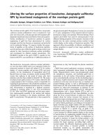
Figure 1 Cranial T2-w eighted magnetic resonance imaging (panel A and B) showed left-predominant bilateral regions of increased
signal intensity in the hippocampus and amygdaloid body, the insular, medial temporal and medial frontal lobes. Cranial computed
tomography (CT; panel C) demonstrated high intensity lesions in the left amygdaloid body. Subarachnoid and intraventricular bleeding, in
addition to low intensity lesions in the bilateral frontal and temporal lobes, was evident (panels D and E). A chest-enhanced CT demonstrated
massive thrombosis of the right pulmonary artery trunk (panel F).
Tonomura et al. Journal of Medical Case Reports 2010, 4:310
/>Page 2 of 4
from acyclovir to intravenous vidarabine (900 mg/day,
14 days). At this time, HSV-1 IgM and IgG antibodies
were 7.89 and 11.2, respectively, in the CSF and 0.56
and 76 in the serum.
On day 21, sedative medication and mechanical venti-
latory support were withdrawn and the GCS increased
to 9 (eye opening, verbal response and motor response
were 3, 2 and 4, respectively).
On day 26, the level of consciousness decreased in
association with desaturat ion and ta chycardia. Throm-
bosis of the right pulmonary artery trunk with pulmon-
ary embolism was evident on enhanced CT of the chest
(Figure 1F). A high serum D-dimer persisted (maximum
titer: 48.3 μg/mL). In addition, c ranial CT revealed
subarachnoid and intraventricular bleeding (Figure 1D
and 1E).
During her hospitalization, she did not experience any
intermittent or persistent hyperten sion. Intravenous
heparin (12,000 U/day) was star ted and the dose was
adjusted according to the activated partial thro mboplas-
tin time for about a month (maximal dose of heparin,
20,400 U/day). CSF a nalysis on day 39 showed 6 lym-
phocytes/mm
3
,52redcells/mm
3
and a gluco se concen-
tration of 78 mg/dL; the titers of HSV-1 IgM and IgG
antibodies were 1.34 and greater than 12.8, respectively.
Cranial CT on day 54 showed that the subarachnoid
and intracranial bleeding had disappeared. Enhanced CT
angiography demonstrated a n avascular area in the left
temporal lobe but no other arterial or venous abnormal-
ities, such as aneurysm formation or irregular vascular
distribution, were evident (data not shown).
Three months after admission, she responded to sim-
ple orders. Her GCS increased t o 14 (eye opening, ver-
bal response and motor response were 4, 5 and 5,
respectively) and thrombosis of the pulmonary artery
trunk had disappeared. As her consciousness level had
reduced, informed consent for the above medical treat-
ments and procedures was obtained from her family.
Discussion
PCR has become the standard diagnostic test for HSVE.
However, intrathecal antibody measurem ents are still of
value, with an estimated specificity of 80% or 95% [3].
Real-time PCR is a recent modification o f conventional
PCR for HSV. The relation between the results of PCR
and intrathecal antibody levels remains poorly under-
stood. This issue has been addressed by one study but
real-time PCR and measurement of antibody titers were
performed in many patients at different times [4].
Intrathecal viral genomes on PCR and increased
intrathecal HSV antibodies have been detected within
five days [5] and after seven days [6] from the onset of
neurologic symptoms, respectively. Our study found that
theresultsofreal-timeHSVPCRwerepositivethree
days after the onset of central nervous symptoms,
without intrathecally synthesized specific HSV antibodies.
Intracerebral hematoma is rarely associated with
HSVE [7] and only 14 cases have so far been reported.
To the best of our knowledge, this is the first report to
document a case of HSVE associated with subarachnoid
bleeding. Obvious abnormalities of major cerebral vas-
cular arteries, such as aneurysm formation and an irre-
gular distribution of the anterior, middle and posterior
cerebral arteries, were not evident which suggests that
the subarachnoid bleeding was directly attributed to
HSVE. HSV causes a necrotizing vasculopathy ascribed
to cortical and subcortical intense hemorrhagic necrosis
and perivascular cuffing in the medial temporal and
orbi tofro ntal regions [2] and CSF anal ysis often demon-
strates the presence of red cells. In gyri located near the
CSF, diffuse necrotizing angiitis of venules and capil-
laries induced by intense inflammatory necrotizing vas-
culopathy [8] can cause vessel wall necrosis and
subsequent bleeding, leading to hematogenous spread
into the CSF space. Subarachnoid bleeding in our
patient may have been caused by red-cell diapedesis
from the hemorrhagic necrotizin g amygdaloid body into
the adjacent CSF spaces, resulting in ‘ subarachnoid
bleeding with intraventricular extension’. Coagulopathy
or hepatocellular damage with a consequent insufficient
production of clotting factors can complicate severe
HSV infections [9] and may potentially cause bleeding.
Conclusions
Focal intense HSVE can increase the risk of subarach-
noid bleeding and our experience raises the ques tion of
whether anticoagulant treatment is safe for patients with
HSVE complicated by subarachnoid bleeding.
Consent
Written informed consent was obtained from the patient
for the publication of th is case report and any accompa-
nying images. A copy of the written consent is available
for review by the Editor-in-Chief of this journal.
Abbreviations
CSF: cerebrospinal fluid; CT: computed tomography; GCS: Glasgow coma
score; HSV: herpes simplex virus type 1; HSVE: HSV encephalitis; IgG:
immunoglobulin G; IgM: immunoglobulin M; PCR: polymerase chain
reaction.
Author details
1
Department of Neurology, Nara Medical University, 840 Shijo-cho, Kashihara,
Nara 634-8522, Japan.
2
Department of Emergency and Critical Care Medicine,
Nara Medical University, Kashihara, Nara, Japan.
Authors’ contributions
YT, HK, MK, NY, KO and SU reviewed the existing literature and drafted the
manuscript which was edited by HK. HK reviewed and selected radiology
images. All authors read and approved the final manuscript.
Tonomura et al. Journal of Medical Case Reports 2010, 4:310
/>Page 3 of 4
Competing interests
The authors declare that they have no competing interests.
Received: 26 March 2010 Accepted: 22 September 2010
Published: 22 September 2010
References
1. Whitley RJ, Alford CA, Hirsch MS, Schooley RT, Luby JP, Aoki FY, Hanley D,
Nahmias AJ, Soong SJ: Vidarabine versus acyclovir therapy in herpes
simplex encephalitis. N Engl J Med 1986, 314:144-149.
2. Barnes DW, Whitley RJ: CNS diseases associated with varicella zoster virus
and herpes simplex virus infection. Pathogenesis and current therapy.
Neurol Clin 1986, 4:265-283.
3. Aurelius E, Forsgren M, Skoog E: Serodiagnosis of herpes simplex
encephalitis by antibody capture enzyme-linked immunosorbent assay.
Serodiagnosis Immunotherapy Infect Dis 1989, 3:249-258.
4. Hjalmarsson A, Granath F, Forsgren M, Brytting M, Blomqvist P,
Sköldenberg B: Prognostic value of intrathecal antibody production and
DNA viral load in cerebrospinal fluid of patients with herpes simplex
encephalitis. J Neurol 2009, 256:1243-1251.
5. Aurelius E, Johansson B, Sköldenberg B, Staland A, Forsgren M: Rapid
diagnosis of herpes simplex encephalitis by nested polymerase chain
reaction assay of cerebrospinal fluid. Lancet 1991, 337:189-192.
6. Cinque P, Cleator GM, Weber T, Monteyne P, Sindic CJ, van Loon AM: The
role of laboratory investigation in the diagnosis and management of
patients with suspected herpes simplex encephalitis: a consensus report.
The EU Concerted Action on Virus Meningitis and Encephalitis. J Neurol
Neurosurg Psychiatry 1996, 61:339-345.
7. Shelley BP, Raniga SB, Al-Khabouri J: An unusual late complication of
intracerebral haematoma in herpes encephalitis after successful
acyclovir treatment. J Neurol Sci 2007, 252:177-180.
8. Menkes JH: Child Neurology Baltimore: Williams & Wilkins 1995, 528.
9. Abzug MJ, Johnson SM: Catastrophic intracranial hemorrhage
complicating perinatal viral infections. Pediatr Infect Dis J 2000, 19:556-559.
doi:10.1186/1752-1947-4-310
Cite this article as: Tonomura et al.: A successfully treated case of
herpes simplex encephalitis complicated by subarachnoid bleeding: a
case report. Journal of Medical Case Reports 2010 4:310.
Submit your next manuscript to BioMed Central
and take full advantage of:
• Convenient online submission
• Thorough peer review
• No space constraints or color figure charges
• Immediate publication on acceptance
• Inclusion in PubMed, CAS, Scopus and Google Scholar
• Research which is freely available for redistribution
Submit your manuscript at
www.biomedcentral.com/submit
Tonomura et al. Journal of Medical Case Reports 2010, 4:310
/>Page 4 of 4