báo cáo khoa học: "Long-term survival of a woman with well differentiated papillary mesothelioma of the peritoneum: a case report and review of the literature" docx
Bạn đang xem bản rút gọn của tài liệu. Xem và tải ngay bản đầy đủ của tài liệu tại đây (788.75 KB, 4 trang )
CAS E REP O R T Open Access
Long-term survival of a woman with well
differentiated papillary mesothelioma of the
peritoneum: a case report and review of the
literature
Jeffrey M Clarke
1*
, Paul Helft
2
Abstract
Introduction: Well-differentiated papillary mesothelioma of the peritoneum (WDPMP) is a rare subtype of
epitheloid mesothelioma, which is usually seen in young women. WDPMP is generally considered of low malignant
potential, however the long-term nature of the tumor remains poorly defined.
Case presentation: We describe the long-term follow-up of a 60-ye ar-old woman of West African descent who
has survived 24 years with WDPMP after receiving extensive local and systemic adjuvant chemotherapy. Her clinical
course has included three exploratory laparotomies with intraperitoneal and intravenous chemotherapy over two
decades. Her course was complicated by anthracycline-induced cardiomyopathy, for which she underwent an
orthotopic heart transplant. Our patient is alive with stable radiological evidence of peritoneal disease, and
continues to suffer from chronic abdominal pain.
Conclusion: No consensus exists regarding optimal treatment strategies for WDPMP. However, given the low
malignant potential of the tumor, careful consideration should be made before proceeding with aggressive
interventions. Further, long-term follow-up reports are required to fully characterize this tumor.
Introduction
Mesothelioma is an uncommon neoplasm which origi-
nates from the mesothelial lining of the pleura, pericar-
dium, peritoneum, and tunica vaginalis [1,2]. Malignant
peritoneal mesothelioma (MPM) makes up approxi-
mately 10% to 20% of all cases of mesothelio ma [2].
MPM is an aggressive tumor typically associated with
asbestos exposure and afflicts mainly men in the fifth to
sixth decades of life [2,3]. In contrast, well-differentiated
papillary mesothelioma of the peritoneum (WDPMP) is a
rare subtype of epitheloid mesothelioma, which is usually
seen in young women [1,4,5]. WDPMP is generally con-
sidered of low malignant potential and falls within a clini-
cohistological spectrum of papillary peritoneal tumors in
women ranging from mesothelial hyperplasia to papillary
carcinoma [1,5]. While the histological features of
WDPMP have b een described in many cases with short-
term clinical follow-up, the long-term nature of the
tumor remains poorly defined. We present a case
describing long-term survival and follow-up of woman
with WDPMP who received extensive intraperitoneal and
systemic chemotherapy.
Case presentation
We report the case of a 60-year-old woman of West
African descent, with no history of asbestos e xposure,
who originally presented 24 years ago to another institu-
tion with acute abdominal pain. At that time, she under-
went an exploratory laparotomy and was found to have
nodules diffusely covering the peritoneum. A total
abdominal hysterectomy and bilateral salpingo-oophor-
ectomy were performed for suspected ovarian carci-
noma, and biopsies were taken of the peritoneal
nodules. The pathology from this original surgery was
interpreted as low-grade papillary mesothelioma. She
then received six adjuvant cycles of intravenous
* Correspondence:
1
Department of Medicine, Duke University Medical Center, Durham, NC, USA
Full list of author information is available at the end of the article
Clarke and Helft Journal of Medical Case Reports 2010, 4:346
/>JOURNAL OF MEDICAL
CASE REPORTS
© 2010 Clarke and Helft; licensee BioMed Central Ltd. This is an Open Access article distributed under the terms of the Creative
Commons Attribution License (http://cr eativecommons.org/licenses/by/2 .0), which permits unrestricted use, distribution, and
reproduction in any mediu m, provided the original work is properly cited.
cyclophosphamide, doxorubicin and cisplatin. She
underwent a second-look laparotomy six months later,
and still had gross disease visible in the peritoneum.
Post-operatively she received three additional cycles of
intraperitoneal cisplatin and intravenous sodium thiosul-
fate. She subsequently received maintenance therapy
with alternating courses of tamoxifen and megace alter-
nating every two weeks.
She presented four years later with obstructive gastro-
intestinal symptoms and was again fo und on laparotomy
to have diffuse peritoneal studding. Pathology from this
surgery was interpreted again to be papillary mesothe-
lioma. As a re sult, she began six cycles of carboplatin
and cyclophosphamide chemotherapy for suspected pro-
gressive disease. Several months later, she presented
with complaints of shortness of breath, orthopnea, and
worsening lower extremity edema. A multi-gated acqui-
sition scan (MUGA) revealed an ejection fraction of 14%
and enlarged cardiac silhouette on chest X-ray, and she
was clinically diagnosed as having anthracycline-induced
cardiomyopathy. Medical therapy was initiated at that
time for congestive heart failure.
Two years later, she was found on a computed topo-
graphy (CT) scan to have an interval increase in locu-
lated subhepatic fluid collection and a lobular soft tissue
mass in the right subphrenic region. S he then received
three cycles of VP-16 and ifosfamide. She remained well
until 2000, when she underwent an orthotopic heart
transplant. Upon subsequent reimaging of her abdomen
the next year, she was found to have continued slow
progression of the tumor and was started on single-
agent paclitaxel followed by cyclophosphamide for two
months. She was then ref erred to our institution in late
2001 with stable disease on abdominal CT and a pre-
sumed diagnosis of malignant perito neal mesothelioma
refractory to therapy. Over the following year, she was
maintained on combination capecitabine and gemcita-
bine therapy and had stable disease as assessed by CT
scans. However, in early 2003 she was found to have
declining renal function and was forced to stop
chemotherapy.
She was observed c losely until 2004 and had little
change in her overall tumor burden, but had recurrent
ascites requiring drainage by paracentesis on multiple
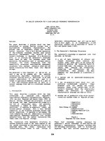
occasions. Because of doubts about the true nature of
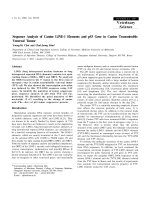
her peritoneal tumor, a further biopsy of her tumor was
performed in 2004, with th e final interpretation demon-
strating a low- grade papillary mesothelioma of the peri-
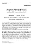
toneum (see Figure 1). She has been observed closely
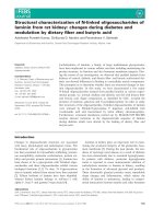
since that t ime with periodic abdominal imaging show-
ing a right side subphrenic m ass, loculated subhepatic
fluid collection, scattered soft tissue densities with calci-
fication, and extensive anterior wall and peritoneal adhe-
sive disease without obstruction (see Figure 2). She
continues to have chronic renal insufficiency and suffers
from severe chronic abdominal pain and cramping, but
has stable radiological evidence of disease.
Discussion
To the best of our knowledge, fewer than 60 cases of
WDPMP have been described in the literature. The
reported age at diagnosis has ranged from two to 74
years old [1,4]. Of 39 case reports we reviewed, the
mean age at presentation was 44 years (median 43
years). In all, 28 patients were women and 11 patients
were men [1,2,4,6-12]. Symptoms at presentation
included acute and ch ronic abdominal pain, ascites,
pleural effusion, bloating, weight loss, dyspareunia, and
menorrhagia [1,4]. However, the diagnosis of WDPMP
was frequently made incidentally during surgery [4].
Only six of the patients were reported to have possible
asbestos exposure, but no definitive causation has ever
been described [8,10]. Follow-up time was recorded for
37 of the 45 patient case reports we reviewed and ran-
ged from six weeks to 29 years (median 36 months,
mean 51 months) [1,2,4,6-12].
The reported cases of WDPMP retain several uniting
histomorphological features. Coarse papillary architec-
ture with fibrovascular cores is the most commonly
seen appearance, with occasional areas of tubulopapil-
lary pattern [1,4,7-10,12]. The papillae are lined by a
simple uniform cuboidal epithelium, with little to no
nuclear atypia or mitoses. The amount of fibrosis pre-
sent can be variable and psammoma bodies can also be
found [4,8]. Areas of invasion are typically not seen
[4,5,8]. Microscopic analysis of cytology from ascite s can
show spheroid tumor cell clusters [12]. Classically,
WDPMP exists within a spectrum of primary papillary
peritoneal tumors described in women, which ranges
Figure 1 Well di fferentiated papillary mesothelioma of the
peritoneum (40× magnification). Multiple coarse papillae are
present with varying fibrovascular cores and minimal cellular atypia.
Clarke and Helft Journal of Medical Case Reports 2010, 4:346
/>Page 2 of 4
from mesothelial hyperplasia to the more aggressive aty-
pical diffuse mesothelioma and papillary carcinoma
[1,5]. As suggested in several previous reports, the
tumor must be distinguished from its benign and malig-
nant counterparts based on degree of cellular differen-
tiation and atypia [1,4,5,8,12].
The case presented above is unusual in two respects.
She maintained follow-up and remains alive with disease
after 24 years from her initial diagnosis. This is the sec-
ond longest time of follow-up reported for WDPMP.
The longest follow-up was a 41-year-old woman
observed for 29 years who eventually died of a pancrea-
tic carcinoma [4]. While many reported cases portray
WDPMP as a clinically benign tumor, several case
reports have described more aggressive behavior with
long-term follow-up. In one case, a patient died five
years following diagnosis. He was found at autopsy to
have extensive retroperitoneal, anterior abdominal wall,
diaphragmatic, and pericardial invasion, culminating in a
large embolism of tumor cells to the pulmonary artery
[7]. A second case describes a patient who died of dif-
fuse malignant mesothelioma approximately nine years
after the diagnosis of WDPMP, suggesting a malignant
transformation at some point in the clinical course [9].
To better understand and characterize the malignant
potential of this tumor, additional case reports with
long-term follow-up are required.
Secondly, she received e xtensive chemotherapy w ith
substantial associ ated morbidity, we believe on the basis
of the fact that her tumor was thought originally to be
an ovarian-derived tumor or primary peritoneal carci-
noma, and was later thought to b e a malignant
peritoneal mesothelioma. Only after a repeat b iopsy 19
year s after her original diagnosis was the probable iden-
tity of her tumor finally understood.
Management of pat ients with WDPMP remains con-
troversial. The majority of patients undergo initial
exploratory laparotomy for diagnostic and cytoreductive
purposes [1,4,8]. However this approach is contentious,
given the low malignant potential of the tumor. Some
authors recommend close observation or s erial biopsy
for surveillance [1]. Adjuvant treatment for WDPMP
also remains poorly defined and was described in only
14 of the cases that we reviewed [2,4,8,10-12]. In the lar-
gest series, three patients received a combination of che-
motherapy and radiation therapy, one of these with
intravenous thiotepa, and two additional patients
received radiation therapy alone [4]. Of the patients who
received adjuvant radiation therapy, two patients had
died of radiation enteritis and intestinal obstruction at
two-year and seven-year follow-up, respectively [4].
Intraperitoneal administration of chemotherapy has
been described in several case reports. One patient with
simultaneous involvement of the pleural and peritoneal
surfaces with ascites and pleural effusion was treated
succ essfully with intraperitoneal, intrapl eural, and intra-
venous carboplatin [12]. Our patient remained disease
free at four years following presentation. Four patients
have received intraperitoneal hyperthermic chemoperfu-
sion (IPHP) therapy [2,6,10]. Two of these patients
received cisplatin and doxorubicin following optimal
debulking. One patient was alive with disease at 15
months, while the other patient suffered a post-operative
colobronchial fistula requiring partial colectomy and was
Figure 2 Cr oss-secti onal computed tomography (CT) images displayi ng a right-sided, perihepatic soft tissue mass containing
calcifications and subhepatic loculated fluid collection.
Clarke and Helft Journal of Medical Case Reports 2010, 4:346
/>Page 3 of 4
alive 40 months later [2,6]. Another patient received
IPHP with cisplatin and mitomycin C after suboptimal
debulking and died of disease progression 13 months
later [2]. A third patient with concurrent rectal carci-
noma underwent a low anterior resection with omen-
tectomy and peritonectomy and subsequent IPHP with
mitomycin-C and 5-fluorouracil [10]. Our patient had
no evidence of disease at six months follow-up.
Five patients whose care reports we reviewed received
intravenous chemotherapy alone, two of these with
unspecified regimens [4,8,11]. Two patients rec eived cis-
platin and doxorubicin. One of these patients had no
evidence of disease at three years, and the second
patient died of disease three years later [8]. One case
described an 11-year-old girl who was treated with com-
bination cisplatin, cyclophosphamide and maintenance
lupron [11]. She had stable diffuse peritoneal nodules at
nine months. Considerable variability exists in the litera-
ture regarding the chemotherape utic management of
this tumor.
Conclusion
Clearly, no consensus has been reached regarding opti-
mal treatment strategies for WDPMP. It is difficult to
determine the effect of systemic or intraperitoneal che-
motherapy on the tumor progression of our patient due
to imprecise past medical records. However, one must
question the necessity of extensive chemotherapeutic
and surgical therapies for a tumor with presumed low
malignant potenti al, given the inherent risks of such
interventions. Furthermore, accurate pathological diag-
nosis must be initially obtained in order to prevent over-
treatment of WDPMP. Additional information obtained
from other case reports describing the long-term beha-
vior of this tumor should also help to elucidate the pre-
cise roles for observation and therapeutic intervention.
Consent
Written informed consent was obtained from the patient
for publication of this case report and any accompany-
ing images. A copy of the written c onsent is available
for review by the journal’s Editor-in-Chief.
Abbreviations
(MPM): Malignant peritoneal mesothelioma; (WDPMP): Well-differentiated
papillary mesothelioma of the peritoneum; (MUGA): Multi-gated acquisition
scan; (CT): Computed topography; (IPHP): Intraperitoneal hyperthermic
chemoperfusion;
Author details
1
Department of Medicine, Duke University Medical Center, Durham, NC, USA.
2
Department of Medicine, Section of Hematology/Oncology, Indiana
University Melvin and Bren Simon Cancer Center, Indiana University School
of Medicine, Indianapolis, IN, USA.
Authors’ contributions
JC researched and composed the literature review and the patient history.
PH was a major contributor to the patient history and to critical revision of
the manuscript. Both authors read and approved the final manuscript.
Competing interests
The authors declare that they have no competing interests.
Received: 9 October 2009 Accepted: 29 October 2010
Published: 29 October 2010
References
1. Hoekstra A, Riben M, Frumovitz M, Liu J, Ramirez P: Well differentiated
papillary mesothelioma of the peritoneum: a pathological analysis and
review of the literature. Gynecologic Oncology 2005, 98:161-167.
2. Deraco M, Casali P, Inglese M, Baratti D, Pennacchioli E, Bertulli R,
Kusamura S: Peritoneal mesothelioma treated by induction
chemotherapy, cytoreductive surgery, and intraperitoneal hyperthermic
perfusion. J Surg Oncol 2003, 83:147-153.
3. Bani-Hani K, Gharaibeh K: Malignant peritoneal mesothelioma. J Surg
Oncol 2005, 91:17-25.
4. Daya D, McCaughey W: Well differentiated papillary mesothelioma of the
peritoneum: A clinicopathologic study of 22 cases. Cancer 1990,
65:292-296.
5. Foyle A, Al-Jabi M, McCaughey W: Papillary peritoneal tumors in women.
Am J Surg Pathol 1981, 5:241-249.
6. Laterza B, Baratti D, Cozzi G, Kusamura S, Oliva GD, Gavazzi C, Fumagalli L,
Sironi A, Sabia D, Deraco M: Colobronchial fistula: an unusual
complication after peritonectomy and hyperthermic intra-peritoneal
chemotherapy. In Vivo 2009, 23:151-154.
7. Burrig K, Pfitzer P, Hort W: Well differentiated papillary mesothelioma of
the peritoneum: a borderline mesothelioma. Virchow Arch A Pathol Anat
Histopathol 1990, 417:443-447.
8. Butnor K, Sporn T, Hammar S, Roggli V: Well differentiated papillary
mesothelioma. Am J Surg Pathol 2001, 25:1304-1309.
9. Hejmadi R, Ganesan R, Kamal N: Malignant transformation of a well-
differentiated peritoneal paillary mesothelioma. Acta Cytol 2003,
47:517-518.
10. Jatzko G, Jester J: Simultaneous occurrence of a rectal carcinoma and a
well differentiated papillary mesothelioma of the peritoneum. Int J
Colorectal Dis 1997, 12(6):326-328.
11. Lovell F, Cranston P: Well differentiated papillary mesothelioma of the
peritoneum. Am J Roentgenol 1990, 155:1245-1246.
12. Shukunami K, Hirabuki S, Kaneshima M, Kamitani N, Kotsuji F: Well
differentiated paillary mesothelioma involving the peritoneal and pleural
cavities: successful treatment by local and systemic administration of
carboplatin. Tumori 2000, 86:419-421.
doi:10.1186/1752-1947-4-346
Cite this article as: Clarke and Helft: Long-term survival of a woman
with well differentiated papillary mesothelioma of the peritoneum: a
case report and review of the literature. Journal of Medical Case Reports
2010 4:346.
Submit your next manuscript to BioMed Central
and take full advantage of:
• Convenient online submission
• Thorough peer review
• No space constraints or color figure charges
• Immediate publication on acceptance
• Inclusion in PubMed, CAS, Scopus and Google Scholar
• Research which is freely available for redistribution
Submit your manuscript at
www.biomedcentral.com/submit
Clarke and Helft Journal of Medical Case Reports 2010, 4:346
/>Page 4 of 4