báo cáo khoa học: " Retrorectal endometrioid cyst: a case report" pot
Bạn đang xem bản rút gọn của tài liệu. Xem và tải ngay bản đầy đủ của tài liệu tại đây (278.27 KB, 2 trang )
CAS E REP O R T Open Access
Retrorectal endometrioid cyst: a case report
Iraklis E Katsoulis
*
, Ioannis E Katsoulis
Abstract
Introduction: Developmental cysts are the most common retrorectal cystic lesions in adults, whereas reports of
endometrioid cysts in this anatomic location are extremely rare.
Case presentation: A 21-year-old nulliparous Greek woman presented with chronic noncyclic pelvic pain, and a
retrorectal cyst was diagnosed. The lesion was resected through a laparotomy and, on histologic examination, was
found to be an endometrioid cyst. Th e treatment was completed with a six-month course of a gonadotropin-
releasing hormone analogue. One yea r after surgery, the woman remained free of sy mptoms, and pelvic imaging
showed no recurrence of the lesion. Reviewing the literature, we found only three previous reports of an
endometrioid cyst in this anatomic location.
Conclusion: In women of reproductive age, endometriosis must be included in the differential diagnosis of
retrorectal cysts.
Introduction
Endometriosis is the presence of endometrioti c tissue in
anatomic regions outside the uterus [1]. The most-
common sites are the ovaries and the fallopian tubes,
the uterosacral ligaments, and the lateral pelvic perito-
neum. Endometriosis can less commonly be found in
laparotomy scars, the vagina, and the rectovaginal
septum, and also can involve the wall of the colon and
the rectum. This is a report of a rare retrorectal endo-
metrioid cyst that was not contiguous to the rectal wall.
Developmental cysts are the most common retrorectal
cysti c lesions in adults, whereas reports of endometrioid
cysts in this anatomic location are extremely rare [2-4].
Case presentation
A 21-year-old nulliparous Greek woman complained of
chronic noncyclic pelvic pain. Abdominal and vaginal
examinations were unremarkable, whereas on rectal
examination, a soft extraluminal mass was found poster-
iorly and left laterally.
The rectal mucosa was n ormal on rigid rectosigmoi-
doscopy. A pelvic ultrasound scan revealed a cystic
lesion posterior to the middle rectum, and blood tests
showed a mo derately elevated CA 19-9 (79IU/ml),
whereas all other tumour markers were normal. Com-
puted tomography (CT) of the whole abdomen excluded
other intra-abdominal pathology and provided further
information regarding the anatomic relations of the
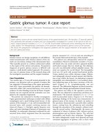
lesion . The cyst lay posterior and left lateral to the mid-
dle rectum above the level of the pelvic floor and was
contiguous neither to the rectal wall nor to the sacrum
(Figure 1). Its maximal diameter was about 7 cm.
After administration of preoperative antibiotic prophy-
laxis, a laparotomy was undertaken through an infra-
umbilical midline incision. Moderate bilateral ovarian
endometriosis and minor endometrio sis of the pelvic
peritoneum were found; these were ablated with surgical
diathermy. Subsequently, the pelvic peritoneum was
opened, and the retrorectal space was carefully dissected
to avoid injury of the pelvic nervous plexuses and the
hypogastric nerves. The retrorectal cystic lesion was
removed intact, and on histologic examination was
found to be a suppurated endometrioid cyst.
The pati ent made an uneventful recovery and was dis-
charged o n the third postoperative day. The treatment
was completed with a six-mont h course of a gonadotro-
pin-releasing hormone (GnRH) analogue. One year
postoperatively, she remained free of symptoms, and fol-
low-up pelvic imaging showed no recurrence of
endometriosis.
Discussion
Developmental cysts are the most common retrorectal
cysti c lesions in adults, occurring mostly in middle-aged
* Correspondence:
White Cross Hospital, 1 Sisini Str, 11528 Athens, Greece
Katsoulis and Katsoulis Journal of Medical Case Reports 2010, 4:389
/>JOURNAL OF MEDICAL
CASE REPORTS
© 2010 Katsoulis and Katsoulis; licensee BioMed Central Ltd. This is an Open Access article distributed under the terms of the Creative
Commons Attribution License ( /licenses/by/2.0), which permits unrestricted use, distribution, and
reproduction in any medium, provided the original work is pr operly cited.
women. They are classified as epidermoid cysts, dermoid
cysts, enteric cysts (tailgut cysts or hamartomas and cys-
tic rectal duplication), and neuroenteric cysts, accordin g
to their origin and histopathologic features [5,6]. The
diagnosis of retrorectal cysts can be accomplished with
greater tha n 90% accuracy with computed tomography
(CT) and magnetic resonance imaging (MRI) if the rec-
tum is contrasted [3,6]. Such lesions warrant surgical
excision to establish the diagnosis and to avoid compli-
cations. MRI has been suggested to increase the accu-
racy of preoperative localization and to enable surgical
planning [6]. Transrectal ultrasound, if available, can
also be useful in defining the depth of infiltration in
cases of rectal involvement [3].
The operative approach can be perineal, abdominal, or
combined, dep ending on the positio n of the lesion and
its anatomic relations with surrounding structures. Ret-
rorectal cysts have been also managed by using a laparo-
scopic approach [7]. In our patient, the information
provided by the CT regarding the size and the anatomic
relations of the cyst was considered sufficient, and
therefore a pelvic MRI was not performed. We opted to
approach the lesion through a laparotomy, aiming to
explore her pelvis thoroughly in view of her persistent
pelvic pain and elevated CA 19-9 levels.
We found foci o f endometriosis on both ovaries and
the pelvic peritoneum. A complete resection of the lesion
was achieved, and histology made the diagnosis of a sup-
purated endometrioid cyst. In cases of low perirectal
lesions, in which endometriosis is suspected, an alterna-
tive strategy can be transperineal excision combined with
a laparoscopy for assessment of the intra-abdominal
organs. We thought, however, that because the cyst lay
posterior and left lateral to the middle rectum, a
transperineal approach would neither be sufficient nor
warrant the preservation of surrounding structures.
It is not uncommon for endometriosis to involve the
rectal wall, requiring an anterior resection of the rectum
[8]. Conversely, the presentation of an endometrioid
cyst that occupies the retrorectal space, without being
contiguous to either the rectal wall or the sacrum, is a
rare entity. Reviewing the literature, we found only three
previous reports of an endometrioid cyst in this ana-
tomic location [2-4].
Conclusion
In women of reproductive age, endometriosis must be
included in the differential diagnosis of retrorectal cysts.
Consent
Written informed consent was obtained from the patient
for publication of this case report and accompanying
images. A copy of the written consent is available for
review by the Editor-in-Chief of this journal.
Authors’ contributions
Both authors contributed equally to the writing and read and approved the
final manuscript.
Competing interests
The authors declare that they have no competing interests.
Received: 27 March 2010 Accepted: 30 November 2010
Published: 30 November 2010
References
1. Farquhar C: Endometriosis. BMJ 2007, 334:249-253.
2. Rieger N, Munday D: Retrorectal endometrial cyst. Arch Gynecol Obstet
2004, 270:67-68.
3. Stroh C, Manger T: Ultrasound diagnosis of rare retrorectal tumours.
Zentralbl Chir 2003, 128:1075-1079.
4. Singer MA, Cintron JR, Martz JE, Schoetz DJ, Abcarian H: Retrorectal cyst: a
rare tumor frequently misdiagnosed. J Am Coll Surg 2003, 196:880-886.
5. Dahan H, Arrivé L, Wendum D, le Pointe HD, Tubiana JM: Retrorectal
developmental cysts in adults: clinical and radiologic-histopathologic
review: differential diagnosis, and treatment. Radiographics 2001,
21:575-584.
6. Woodfield JC, Chalmers AG, Phillips N, Sagar PM: Algorithms for the
surgical management of retrorectal tumours. Br J Surg 2008, 95:214-221.
7. Gunkova P, Martinek L, Dostalik J, Gunka I, Vavra P, Mazur M”: Laparoscopic
approach to retrorectal cyst. World J Gastroenterol 2008, 14:6581-6583.
8. Brouwer R, Woods RJ: Rectal endometriosis: results of radical excision
and review of published work. A N Z J Surg 2007, 77:562-571.
doi:10.1186/1752-1947-4-389
Cite this article as: Katsoulis and Katsoulis: Retrorectal endometrioid cyst:
a case report. Journal of Medical Case Reports 2010 4:389.
Figure 1 Computed to mography, showing the cystic lesion
posterior and left lateral to the middle rectum.
Katsoulis and Katsoulis Journal of Medical Case Reports 2010, 4:389
/>Page 2 of 2