báo cáo khoa học: "Successful interdisciplinary management of the misdeployment of two self-expanding stents into the internal carotid artery: a case report" pps
Bạn đang xem bản rút gọn của tài liệu. Xem và tải ngay bản đầy đủ của tài liệu tại đây (872.91 KB, 4 trang )
CAS E REP O R T Open Access
Successful interdisciplinary management of the
misdeployment of two self-expanding stents into
the internal carotid artery: a case report
Dominik Jost
1*
, Helfried Meissner
1
, Henning von Loewensprung
2
, Thomas Guethe
3
, Thomas Hupp
1,4
,
Hans Henkes
5,6
Abstract
Introduction: With the widespread use of carotid artery stenting, previously unknown technical mistakes of this
treatment modality are now being encountered. There are multiple strategies for the treatment of in-stent
restenosis. With regard to surgical management, endarterectomy and patch plasty are favored. To the best of our
knowledge, this report is the first description of a complete stent removal by the eversion technique.
Case presentation: We report the case of a 63-year-old Caucasian man with misde ployment of two stents into his
stenotic proximal internal carotid artery, resulting in a high-grade mechanical obstruction of the internal carotid
artery lumen. With the contralateral internal carotid artery already occluded and associated stenoses of both
proximal and distal vertebral arteries, an interdisciplinary therapeutic concept was applied. Bilateral balloon
angioplasty and stenting of the proximal and distal stenotic vertebral arteries were carried out to provide sufficient
posterior collateral blood flow, followed by successful surgical stentectomy and carotid endarterectomy using the
eversion technique. Duplex scanning and neurological assessments were normal over a 12-month follow-up
period.
Conclusions: Interdisciplinary treatment is a recommended option to prot ect patients from further impairment.
Further evaluation in larger studies is highly recommended.
Introduction
Stroke is the most common cause of disability. Preven-
tion of stroke by carotid endarterectomy (CEA) or caro-
tid artery st enting (CAS) is widely accepted, and they
are basically equivalent treatment modalities [1,2]. The
endovascular treatment of internal carotid stenoses is an
appropriate treatment method not just for patients at
high surgical risk. It is not unexpected that procedural
safety and complication rates of CAS are closely related
to the operator’ s skill and the institutional experience
with the technique. This can be expressed in terms of
caseload or patient enrollment numbers into clinical
trials [3]. With the widespread and unregulated use of
CAS, however, complication rates could increase and
their management is sometimes a challenge for vascular
specialists. Apart from the inherent risks of stenting
procedures (for example, stent thrombosis, distal emboli,
hyperperfusion, hemorrhage and so on), a variety of
technical failures have also been observed. They include,
among others, sizing issues with overdilation or under-
dilatation, distal wire injury, and disconnection of pro-
tection filters, with their respective clinical sequelae.
Here, we report the case of a patient in whom a proxi-
mal internal carotid artery (ICA) stenosis was stented in
another hospital. At presentation to our institution, t he
apparent misdeployment of t he two stents in a partly
overlapping position was found (’hugging’ stents). Well
co-ordinated endovascular and surgical management
saved our patient from further impairment.
Case presentation
A 63-year-old Caucasian man initially presented with an
asymptomatic 55% stenosis of the right proximal ICA
(Figure 1A) to another institution. The left ICA was
* Correspondence:
1
Department of Vascular Surgery, Klinikum Stuttgart, Stuttgart, Germany
Full list of author information is available at the end of the article
Jost et al. Journal of Medical Case Reports 2010, 4:397
/>JOURNAL OF MEDICAL
CASE REPORTS
© 2010 Jost et al; licensee BioMed Central Ltd. This is an Open Access article distribute d under the terms of the Creative Commons
Attribution License ( which permits unrestricted use, distribution, and reproduction in
any medium, provided the original work is properly cited.
found to be occluded; the vessel s of the posterior circu-
lation were not examined. Our patient underwent a n
endovascular procedure at the other institution, which
included the deployment of two 7 mm/40 mm Walls-
tents (Wallstent, Boston Scientific Corporation, Natick,
MA, USA) (Figure 1B). The reason why two stents were
inserted without balloon angioplasty remains unex-
plained. Then, three months later, our patient was
referred to our hospital with clinical signs and symp-
tom s of a transient left hemispheric ischemia, including
global aphasia, right hemiparesis, and paresthesia of the
right upper extremity. His cardiovascular risk factors
included arterial hypertension, hyperlipidemia and non-
insulin dependent diabetes mellitus. Our patient was a
non-smoker. Our patient’s history included severe cor-
onary heart disease, cardiac insufficiency (New York
Heart Association stage II) and recurrent atrial fibrilla-
tion, which altogether resulted in an American Society
of Anesthesiologists (ASA) physical status of IV.
Magnetic resonance imaging and angiography includ-
ing diffusion-weighted imaging did not reveal any
ischemic lesions of the left hemisphere. Digital sub-
tracted angiography (DSA) of the supra-aortic and intra-
cranial vessels confirmed the occlusion of the left ICA
and revealed a highly effective mechanical obstruction of
the right ICA caused by two stents deployed in an over-
lappi ng side-by-side position (Figure 2A). Further at her-
osclerotic lesions included significant stenoses of both
proximal vertebral arteries (V1), a stenosis of the entire
left V4 segment and a focal stenosis at the junction of
the right V4 segment with the basilar artery. The ante-
rior communicating artery and the right posterior
communicating artery were widely patent and the left
external carotid artery contributed to the supply of the
left hemisphere via the ophthalmic artery. When giving
his informed consent, our patient w as informed of a
peri- procedural and post-procedural mortality and mor-
bidi ty rate of between 3% and 5%, and an increased risk
of peri-operative bleeding due to the antiplatelet medi-
cation. Our patient accepted this, and the off-label use
of both the Coroflex (Coroflex Please, B Braun Melsun-
gen AG, Melsungen, Germany) and Enterprise (Codman
Enterprise, Raynham, MA, USA) stents.
The first step of the treatment strategy focused on the
posterior circulation stenoses in order to improve the
potential collateral supply during subsequent stentect-
omy and CEA. Dual platelet antiaggregation with acetyl-
salicylic acid and clopidogrel was initiated. Under
general anesthesia the stenoses of both vertebral artery
origins were treated with short drug-eluting stents (Cor-
oflex), followed by the stent percutaneous transluminal
angioplasty of the left and right V4 stenosis using a
combination of moderately undersized balloon dilatation
and deployment of oversized self-expanding stents
(Enterprise).
The surgical stent removal from the right ICA com-
bined with CEA completed the treatment. Stentecto my
and CEA were carried out under regional anesthesia.
A
B
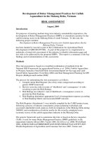
Figure 1 A) Digital subtracted angiography (DSA) of the right
common carotid artery reveals a mid-grade proximal internal
carotid artery stenosis. B) In another institution two Wallstents
were deployed without balloon angioplasty, resulting in a significant
residual stenosis of the ICA. Due to monoplane imaging in
apparently only one projection, the incondite position of the stents
remained unrecognized.
A B
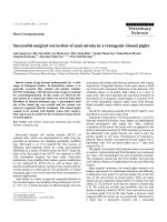
Figure 2 A) During the diagnostic investigation for transient
left hemispheric ischemic signs and symptoms, misdeployment
of the two stents became apparent. Instead of being inserted in
a coaxial way, the distal end of the lower and the proximal end of
the upper stent were found side by side. B) Both stents and the
proximal internal carotid artery stenosis were removed surgically
with excellent reconstruction of the proximal carotid artery lumen
after six months.
Jost et al. Journal of Medical Case Reports 2010, 4:397
/>Page 2 of 4
As routinely, we operated using ultrasound-guided
regional anesthesia (Micr oMaxx, Sonosite GmbH, Erlan-
gen, Germany) of the cervical plexus using 20 cc of lido-
caine 1% (Xylocain, Braun Melsungen AG, Me lsungen,
Germany) and 50 cc of ropivacaine 0.375% (Naropin,
Astra Zeneca GmbH, Wedel, Germany). The dispensa-
tion of analgosedation allowed our patient to be awake
throughout the operation, while neurological function
was monitored by assessing the level of consciousness
and our patient’ s motor function on the left side. At
first the proximal ICA was dissected circumfe rentially
beyond the level of the carotid bifurcation after unevent-
ful clamping. The removal of the proximal stent from
the common carotid artery was then possible without
any neurological deficit, and we decided to proceed with
the eversion technique. The simultaneous removal of
the two ‘hugging’ stents together with the atherosclero-
tic plaque in the proximal ICA was possible. The ICA
and common carotid artery (CCA) were reanastomosed
using a 6/0 polypropylene suture in a continuous fash-
ion. Intra-operative angiographic assessment was per-
formed to ensure patency and in order to con trol the
distal end of the plaque removal. Our patient made an
uneventful recovery with no additional neurological
deficit.
Histologi cal examination revealed a thickened layer of
arterial neointima. Duplex scanning was within normal
limits after five days and three months, and was con-
firmed by DSA after six months (Figure 2B). Follow-up
duplex scanning surveillance and neurological assess-
ments were unremarkable after 12 months.
Discussion
With experienced staff at dedicated centers, CEA and
CAS are considered equally sa fe and efficient methods
for the treatment of proximal carotid artery stenoses.
However, carotid artery angiography and stenting
requires pro per training [4]. In institutions with a suffi-
ciently large caseload, low complication rates of CAS
can be achieved [5]. However, t he technical risks and
the clinical sequelae of CAS procedures performed by
inexperienced operators have also been noted [6].
Our case report deserves some discussion. Whether an
asymptomatic mid-grade ICA stenoses with contralateral
ICA occlusion should be treated is the subject of an
ongoing debate [7,8]. The deployment of a self-expand-
ing stent without balloon angioplasty has been proposed
by others [9], but would certainly not be our preferred
technique. The deployment of two stents is hard to jus-
tify, and in this particular case is likely just a technical
mistake. Unfortunately, the reason why two stents were
inserted in this way remains unexplained by t he opera-
tor at t he other hospital. The non-coaxial deployment,
which apparently remained unrecognized by the
operator, made the si tuation even wors e. The significant
residual stenosis (Figure 1B) can be interpreted as a fail-
ure to improve the cerebral perfusion and should have
prompted immediate action. The omission of the exami-
nation of the posterior circulation vessels, which would
have shown the complexity of the cerebral blood supply,
is also an area for criticism.
Our subsequent efforts applied generally accepted
endovascular and surgical methods, and started with the
deployment of a short drug-eluting stent into the proxi-
mal vertebral artery stenoses [10]. For treatment of the
basilar artery stenosis, a combination of undersized bal-
loon dilatation followed by the deployment o f a moder-
ately oversized self-e xpanding stent was used [11]. The
following surgical stentectomy and CEA was not carried
out in a standard fashion.
At present, surgical experience with complications
after stenting is limited [12]. In the case of our patient,
with a symptomatic occlusion of the left ICA and a
high-grade mechanical obstruction of the right proximal
ICA, we ‘prepared’ him for the operative stentectomy
with temporary clamping of t he right ICA b y improving
the collateral supply using endovascular means. With
regard to the anesthesiological and surgical methods,
local anesthesia during CEA with the eversion technique
offers the possibility of continuous neurological assess-
ment; an inherent advantage over general anesthesia.
The eversion e ndarterectomy enabled the simultaneous
removal of the two stents and the underlying carotid
plaque. As the proximal part of the two ‘hugging ’ stents
could be removed easily, we decided not to use the stan-
dard endarterectomy with patch plasty. Thus, a biologi-
cal carotid reconstruct ion without extraneous tissue was
possible in this re-do procedure. To the best of our
knowledge this is the first description of a stentectomy
by carotid endarterectomy with the eversion technique
[13]. A single suture for reanastomosis of ICA and CCA
aft er plaque eversion reduces the risk of bleeding under
high antiplatelet medication with acetylsalicylic acid and
clopidogrel in comparison to the standard patch plasty.
Thestrategyofremovalofthetwo‘ hugging’ stents
with prior hemodynamic ‘preparation’ led to a good
clinical outcome for our patient, without any further
neurological deficit during follow-up of 12 months.
Conclusion
Post-procedural complication management in vascular
medici ne is a continuous task requiring interdisciplinary
co-operation. The increasing numbers of stent proce-
dures will increase the related surgical expertise. Recom-
mendations for re-do procedures required by local
complications of carotid stenting are: (1) the earlier the
better, (2) biological reconstruction with the eversion
technique is beneficial, and (3) institutions which offer
Jost et al. Journal of Medical Case Reports 2010, 4:397
/>Page 3 of 4
the full range of therapeutic options on site are
advantageous.
Further evaluation in larger studies is highly
recommended.
Consent
Written informed consent was obtained from the patient
for publication of this case report and any accompany-
ing images. A copy of the written consent is available
for review by the Editor-in-Chief of this journal.
Author details
1
Department of Vascular Surgery, Klinikum Stuttgart, Stuttgart, Germany.
2
Department of Anaesthesiology and Intensive Care, Klinikum Stuttgart,
Stuttgart, Germany.
3
Department of Cardiovascular Disease, Klinikum
Stuttgart, Stuttgart, Germany.
4
Faculty of Medicine University of Tuebingen,
Tuebingen, Germany.
5
Department of Neuroradiology, Klinikum Stuttgart,
Stuttgart, Germany.
6
Faculty of Medicine University of Duisburg-Essen, Essen,
Germany.
Authors’ contributions
DJ, HvL and TH performed the surgical procedure and drafted the case
report. TG and HH performed the endovascular procedure. HM participated
in the diagnostic and therapeutic decisions and was responsible for follow-
up examinations. TH and HH made major contributions to writing the
manuscript. All authors read and approved the final manuscript.
Competing interests
The authors declare that they have no competing interests.
Received: 13 May 2010 Accepted: 9 December 2010
Published: 9 December 2010
References
1. Coward LJ, Featherstone RL, Brown MM: Safety an efficacy of
endovascular treatment of carotid artery stenosis compared carotid
endarterectomy: a cochrane systematic review of the randomized
evidence. Stroke 2005, 36:905-911.
2. van der Vaart MG, Meerwaldt R, Reijnen MM, Tio RA, Zeebregts CJ:
Endarterectomy or carotid artery stenting: the quest continues. Am J
Surg 2008, 195:259-269.
3. Fiehler J, Jansen O, Berger J, Eckstein HH, Ringleb PA, Stingele R:
Differences in complication rates among the centres in the SPACE study.
Neuroradiology 2008, 50:1049-1053.
4. Connors JJ, Sacks D, Furlan AJ, Selman WR, Russell EJ, Stieg PE, Hadley MN,
NeuroVascular Coalition Writing Group, Wojak JC, Koroshetz WJ, Heros RC,
Strother CM, Duckwiler GR, Durham JD, Tom-sick TO, Rosenwasser RH,
McDougall CG, Haughton VM, Derdeyn CP, Wechsler LR, Hudgins PA,
Alberts MJ, Raabe RD, Gomez CR, Cawley CM, Krol KL, Futrell N, Hauser RA,
Frank JI: Training, competency, and credentialing standards for
diagnostic cervicocerebral angiography, carotid stenting, and
cerebrovascular intervention: a joint statement from the American
Academy of Neurology, the American Association of Neurological
Surgeons, the American Society of Interventional and Therapeutic
Neuroradiology, the American Society of Neuroradiology, the Congress
of Neurological Surgeons, the AANS/CNS Cerebrovascular Section, and
the Society of Interventional Radiology. J Vasc Interv Radiol 2009, 20:
S292-301.
5. Ecker RD, Sauvageau E, Levy EI, Hopkins LN: Complications of carotid
artery stenting at a high-volume teaching center: experience of
University at Buffalo endovascular fellows from 2004 to 2006.
Neurosurgery 2008, 62:812-816.
6. Mas JL, Trinquart L, Leys D, Albucher JF, Rousseau H, Viguier A, Bossavy JP,
Denis B, Piquet P, Garnier P, Viader F, Touzé E, Julia P, Giroud M, Krause D,
Hosseini H, Becquemin JP, Hinzelin G, Houdart E, Hénon H, Neau JP,
Bracard S, Onnient Y, Padovani R, Chatellier G, EVA-3S investigators:
Endarterectomy Versus Angioplasty in Patients with Symptomatic Severe
Carotid Stenosis (EVA-3S) trial: results up to 4 years from a randomised,
multicentre trial. Lancet Neurol 2008, 7:885-892.
7. Wholey MH, Barbato JE, Al-Khoury GE: Treatment of asymptomatic carotid
disease with stenting: pro. Semin Vasc Surg 2008, 21:95-99.
8. Naylor AR, Bell PR: Treatment of asymptomatic carotid disease with
stenting: con. Semin Vasc Surg 2008, 21:100-107.
9. Bussière M, Pelz DM, Kalapos P, Lee D, Gulka I, Leung A, Lownie SP: Results
using a self-expanding stent alone in the treatment of severe
symptomatic carotid bifurcation stenosis. J Neurosurg 2008, 109:454-460.
10. Vajda Z, Miloslavski E, Güthe T, Fischer S, Albes G, Heuschmid A, Henkes H:
Treatment of stenoses of vertebral artery origin using short drug-eluting
coronary stents: improved follow-up results. AJNR Am J Neuroradiol 2009,
30:1653-1656.
11. Bose A, Hartmann M, Henkes H, Liu HM, Teng MM, Szikora I, Berlis A, Reul J,
Yu SC, Forsting M, Lui M, Lim W, Sit SP: A novel, self-expanding, nitinol
stent in medically refractory intracranial atherosclerotic stenoses: the
Wingspan study. Stroke 2007, 38:1531-1537.
12. Jimenez JC, Moore WS, Lawrence PF, Quinones-Baldrich WJ: Technical
strategies for recurrent carotid stenosis following angioplasty and
stenting. Ann Vasc Surg 2008, 22:179-184.
13. King BN, Scher LA, Lipsitz EC: Refractory in-stent restenosis following
carotid artery stenting. Vasc Endovasc Surg 2009, 43:306-311.
doi:10.1186/1752-1947-4-397
Cite this article as: Jost et al.: Successful interdisciplinary management
of the misdeployment of two self-expanding stents into the internal
carotid artery: a case report. Journal of Medical Case Reports 2010 4:397.
Submit your next manuscript to BioMed Central
and take full advantage of:
• Convenient online submission
• Thorough peer review
• No space constraints or color figure charges
• Immediate publication on acceptance
• Inclusion in PubMed, CAS, Scopus and Google Scholar
• Research which is freely available for redistribution
Submit your manuscript at
www.biomedcentral.com/submit
Jost et al. Journal of Medical Case Reports 2010, 4:397
/>Page 4 of 4