báo cáo khoa học: "Benign perimesencephalic hemorrhage occurring after previous aneurysmal subarachnoid hemorrhage: a case report" pot
Bạn đang xem bản rút gọn của tài liệu. Xem và tải ngay bản đầy đủ của tài liệu tại đây (1.11 MB, 4 trang )
CAS E REP O R T Open Access
Benign perimesencephalic hemorrhage occurring
after previous aneurysmal subarachnoid
hemorrhage: a case report
Richard H Singleton, Dean B Kostov, Hilal A Kanaan, Michael B Horowitz
*
Abstract
Introduction: Both aneurysmal subarachnoid hemorrhage and benign perimesencephalic hemorrhage are well-
described causes of spontaneous subarachnoid hemorrhage that arise as a result of different pathologic processes.
To the best of the authors’ knowledge, ther e have been no reports of both vascular pathologies occurring in the
same individual.
Case presentation: A 51-year-old Caucasian woman with a history of aneurysmal subarachnoid hemorrhage
presented five years after her initial treatment with ictal headache, meningismus, nausea and emesis similar to her
previous bleeding event. Computed tomographic imaging revealed perimesencephalic bleeding remote from her
previously coiled anterior communicating artery aneurysm. Both immediate and delayed diagnostic angiography
revealed no residual fil ling of the previously coiled aneurysm and no other vascular anomalies, consistent with
benign perimesencephalic hemorrhage. The patient had an uneventful hospital course and was discharged to
home in good condition.
Conclusions: This report for the first time identifies benign perimesencephalic hemorrhage occurring in the setting
of previous aneurysmal subarachnoid hemorrhage. The presence of a previously treated aneurysm can complicate
the process of diagnosing benign perimesencephalic hemorrhage. Fortunately, in this case, the previously treated
anterior communicating artery aneurysm was remote from the perimesencephalic hemorrhage and could be ruled
out as a source. The patient’s prior aneurysmal subarachnoid hemorrhage did not worsen the anticipated good
outcome associated with benign perimesencephalic hemorrhage.
Introduction
Spontaneous subarachnoid hemorrhage (SAH) is a sig-
nificant clinical problem that occurs most commonly as
a result of aneurysm rupture. In approximately 15% of
cases, however, no aneurysm can be identified by cere-
bral angiography. Although in a minority of cases occult
aneurysms are eventually identified, non-aneurysmal
SAH represents an interesting clinical problem that can
occur as a result of many different pathologies, includ-
ing vasculitis, arterial dissection, intra-cranial or cervical
arteriovenous malformation or fistula, clotting diatheses,
antiplatelet and/or anticoagulant medication, pituitary
apoplexy and tumors [1]. Benign perimesencephalic
hemorrhage (BPH) is another described type of non-
aneurysmal SAH and is thought to account for approxi-
mately one- to two-thirds of non-aneurysmal SAH and
5-10% of SAH as a whole [1,2]. The presenting symp-
toms of both aneurysmal SAH and BPH overlap and
include sudden onset “th underclap” headache, nausea,
emesis a nd meningismus. The diagnosis of BPH can be
made on the basis of the appearance of hemorrhage lim-
ited to the prepontine and/or perimesencephalic cisterns
on computed tomography (CT) scans in the absence of
an aneurysm on cerebral angiography [1].
Despite the fact that aneury smal SAH and BPH are
the respective leading diagnoses in spontaneous SAH
with and without an identifiable point of origin [1], to
the best of the authors’ knowledge, no cases of both vas-
cular pathologies occurring in the same i ndividual have
bee n previously reported. Herein we present the case of
* Correspondence:
Department of Neurological Surgery, University of Pittsburgh Medical Center,
Suite B-400, 200 Lothrop Street, Pittsburgh, PA 15213, USA
Singleton et al. Journal of Medical Case Reports 2010, 4:405
/>JOURNAL OF MEDICAL
CASE REPORTS
© 2010 Singleton et al; licensee BioMed Central Ltd. This is an Open Access article distributed under the terms of the Creative
Commons Attribution License ( which permits unrestri cted use, distribution, and
reproduction in any medium, provided the original work is properly cited.
a patient with aneurysmal SAH followed five years later
by BPH.
Case Presentation
The patient was a Caucasian, non-smoking 51-year-old
woman with insulin-dependent diabetes and hyperten-
sion who init ially presented at the age of 46 with acute-
onset ictal headache, meningismus and emesis (Hunt/
Hess grade I). A non-contrasted head CT scan revealed
SAH in a n aneurysmal pattern (Figure 1A). She under-
went cerebral angiography, which revealed a 6 mm ante-
rior communicating artery (Acomm) aneurysm (Figure
2A) that was treated with endovascular coiling in the
samesetting.Attheendoftheprocedure,a0.5mm
residual was noted at the base of the aneurysm that
incorporated the anterior cerebral arteries and was not
treated (not shown). Follow-up angiography eight
months later showed complete obliteration (Figure 2B).
The patient’s hospital course was complicated by vasos-
pasm, which was treated with hypervolemia, hyperten-
sion and intra-arterial nicardipine, as well as cerebral
salt wasting, which was treated with sodium and volume
supplementation. She was ultimately discharged to
home and returned to work six weeks later with no resi-
dual neurologic deficits. After her eight-month posthe-
morrhage angiogram, she was lost to neurosurgical
follow-up. Of note, the patient developed peritoneal dia-
lysis-dependent renal f ailure three yea rs later. A renal
ultrasound did not demonstrate evidence of polycystic
kidney disease (not shown).
Prior to the patient’s current admission (five years
aft er her aneurysmal SAH), the patient had experienced
three days of intermittent nausea and vomiting. On
the day of admission, she developed an ac ute-onset
severe headache with worsened nausea and emesis. On
admission, the patien t’s condition was Hunt/Hess grade
II with an initial non-contrasted h ead CT scan demon-
strating perimesencephalic SAH in the prepontine, inter-
peduncular, ambient and crural cisterns (Figure 1B and
1C). Of note, no hemorrhage w as noted adjacent to
the previously treated Acomm aneurysm (Figure 1C,
arrowhead). Although there was no evidence of throm-
bocytopenia or other coagulopathy on her admission
laboratory testing, she was t aking clopidogrel and
received a pool of platelets. A toxicology screen revealed
no evidence of sympathomimetic use. Diagnostic cere-
bral angiography did not reveal any new aneurysms or
vasculopathy and showed the previo usly treated aneur-
ysm to be stable with no residual (Figure 2C). The diag-
nostic angiogram also demonstrated patent cerebral
venous sinuses without evidence of thrombosis or steno-
sis. Given the lack of vasculitic changes on the angio-
gram, further workup for vasculitis was not performed.
The patient had a follow-up angiogram eight days later
that again failed to show any source for the hemorrhage,
consistent with BPH (not shown). The patient had an
uneventful hospit al cours e and was dischar ged to home
in good condition on post-bleed day 10. Follow-up mag-
netic resonance imaging and angiography performed six
months later demonstra ted no vascular abnormalities
(not shown).
Discussion
Despite the recent identification of BPH as a distinct
vascular pathology [2], it is now purported to be a pri-
mary etiology of non-aneurysmal SAH [1,3]. In con-
trasttoaneurysmalSAH,BPH,forwhichsome
authors have proposed the term pre-truncal non-
aneurysmal hemorrhage [4], is thought to arise from
multiple poss ible non-arterial sources [2,5]. Previous
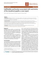
Figure 1 The patient’s non-contrasted h ead computed tomography scan (A) at the time of her initial aneurysmal subarachnoid
hemorrhage admission and (B and C) subsequent non-aneurysmal subarachnoid hemorrhage revealing perimesencephalic
hemorrhage. The superior aspect of the coil mass from her previously treated anterior communicating artery aneurysm, remote from the new
non-aneurysmal subarachnoid hemorrhage, is noted (C, arrowhead).
Singleton et al. Journal of Medical Case Reports 2010, 4:405
/>Page 2 of 4
studies have reported that patients with BPH have nor-
mal life expectancies and are not at risk for re-bleeding
[1]. Other studies have noted some, albeit reduced,
post-hemorrhage complications compared to aneurys-
mal SAH, including vasospasm, post-hemorrhagic
hydrocephalus and death [6].
The patient in this case had BPH that was both tem-
porally and spatially remote from her previous aneurys-
mal SAH. Other than her general risk factors for SAH,
which include female sex, hypertension and previous
ruptured aneurysm, the literature offers little insight
regarding a probable underlying pathology that could
account for both of these hemorrha ges. There have
been previous reports of BPH occurring in individuals
with various vascular pathologies, including ischemic
stroke [5] and venous stenosis or thro mbosis [7,8], but
none that the authors know of in the setting of a pre-
viously ruptured intra-cranial aneurysm. Rebleeding
from BPH has been reported only once, although it
occurred after early anti-coagulation [9].
The location of the patient’s aneurysmal rupture was
fortuitous as it related to her subsequent BPH. It was
evident that the location of her perimesencephalic
hemorrhage did not extend to the region of her pre-
viously coiled Acomm aneurysm and most likely a rose
from a separate process. This permitted the diagnosis of
BPH to be made and a less aggressi ve treatment course
to be pursued. Her treatment would have been signifi-
cantly more complicated had her aneurysm been in the
posterior circulation within the region of her BPH. In
this setting, a diagnosis of BPH would have been diffi-
cult to justify, and she possibly would have undergone
attempts at either recoiling or even open clipping of a
suspected unsecured aneurysm.
Multiple previous case series have attested to the rela-
tively benign course of perimesencephalic hemorrhage
[2,3,6,10]. It would not be unreasonable, however, to
posit that the currently presented patient may have
fared worse than expected, given her previous aneurys-
mal SAH. Fortunately, this was not the case. There is
no evidence that the first bleeding event rendered her
more susceptible to a second, less severe event. It is
unknown what effect BPH occurring shortly after aneur-
ysmal SAH or in someone with a poorer grade injury
would have on neurologic outcome.
Conclusions
This work represents the first report of both aneurysmal
SAH and non-aneurysmal BPH occurring in the same
individual. The diagnosis of BPH may be complicated by
previous aneurysmal SAH. The expected good prognosis
associated with BPH does not appear to be altered by a
previous episode of aneurysmal SAH. To those in the
fields of neurology and neurosurgery, this case serves as
an important reminder that in patients with a history of
previous aneurysmal SAH, subsequent episodes of SAH
need to be fully investigated because they may be attri-
butable to an entirely different pathology.
Consent
Written informed consent was obtained from the patient
for publication of this case report and accompanying
images. A copy of the written consent is available for
review by the Editor-in-Chief of this journal.
Authors’ contributions
RS was responsible for the initial care of the patient, along with the
conception and writing of the manuscript. DK and HK were responsible for
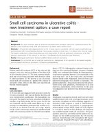
Figure 2 (A) The patient’s initial diagnostic angiogram at the time of aneurysmal subarachnoid hemorrhage.A6mmanterior
communicating artery aneurysm is shown (arrow). The aneurysm was treated in the same setting with endovascular coiling. A follow-up
diagnostic angiogram obtained eight months after aneurysmal subarachnoid hemorrhage shows complete obliteration (B, arrow). At the time of
her perimesencephalic hemorrhage five years later, a diagnostic angiogram reveals the previously coiled anterior communicating artery
aneurysm (C, arrowhead); no residual filling of the aneurysm was noted, and no other vascular abnormalities were seen.
Singleton et al. Journal of Medical Case Reports 2010, 4:405
/>Page 3 of 4
patient management and workup for benign perimesencephalic
hemorrhage. MH was responsible for the patient’s original aneurysmal
management and supervised her case on her subsequent admission. All
authors read and approved the final manuscript.
Competing interests
The authors declare that they have no competing interests.
Received: 2 June 2010 Accepted: 14 December 2010
Published: 14 December 2010
References
1. Van Gijn J, Rinkel GJ: Subarachnoid haemorrhage: diagnosis, causes and
management. Brain 2001, 124:249-278.
2. Van Gijn J, van Dongen KJ, Vermeulen M, Hijdra A: Perimesencephalic
hemorrhage: a nonaneurysmal and benign form of subarachnoid
hemorrhage. Neurology 1985, 35:493-497.
3. Flaherty ML, Haverbusch M, Kissela B, Kleindorfer D, Schneider A, Sekar P,
Moomaw CJ, Sauerbeck L, Broderick JP, Woo D: Perimesencephalic
subarachnoid hemorrhage: incidence, risk factors, and outcome. J Stroke
Cerebrovasc Dis 2005, 14:267-271.
4. Schievink WI, Wijdicks EF: Pretruncal subarachnoid hemorrhage: an
anatomically correct description of the perimesencephalic subarachnoid
hemorrhage. Stroke 1997, 28:2572.
5. Lansberg MG: Concurrent presentation of perimesencephalic
subarachnoid hemorrhage and ischemic stroke. J Stroke Cerebrovasc Dis
2008, 17:248-250.
6. Andaluz N, Zuccarello M: Yield of further diagnostic work-up of
cryptogenic subarachnoid hemorrhage based on bleeding patterns on
computed tomographic scans. Neurosurgery 2008, 62:1040-1047.
7. Mathews MS, Brown D, Brant-Zawadzki M: Perimesencephalic
nonaneurysmal hemorrhage associated with vein of Galen stenosis.
Neurology 2008, 70:2410-2411.
8. Shad A, Rourke TJ, Hamidian Jahromi A, Green AL: Straight sinus stenosis
as a proposed cause of perimesencephalic non-aneurysmal
haemorrhage. J Clin Neurosci 2008, 15:839-841.
9. Van der Worp HB, Fonville S, Ramos LM, Rinkel GJ: Recurrent
perimesencephalic subarachnoid hemorrhage during antithrombotic
therapy. Neurocrit Care 2009, 10:209-212.
10. Greebe P, Rinkel GJ: Life expectancy after perimesencephalic
subarachnoid hemorrhage. Stroke 2007, 38:1222-1224.
doi:10.1186/1752-1947-4-405
Cite this article as: Singleton et al.: Benign perimesencephalic
hemorrhage occurring after previous aneurysmal subarachnoid
hemorrhage: a case report. Journal of Medical Case Reports 2010 4:405.
Submit your next manuscript to BioMed Central
and take full advantage of:
• Convenient online submission
• Thorough peer review
• No space constraints or color figure charges
• Immediate publication on acceptance
• Inclusion in PubMed, CAS, Scopus and Google Scholar
• Research which is freely available for redistribution
Submit your manuscript at
www.biomedcentral.com/submit
Singleton et al. Journal of Medical Case Reports 2010, 4:405
/>Page 4 of 4