Báo cáo khoa học: "Synchronous association of rectal adenocarcinoma and three ileal carcinoids: a case report" pdf
Bạn đang xem bản rút gọn của tài liệu. Xem và tải ngay bản đầy đủ của tài liệu tại đây (613.64 KB, 3 trang )
BioMed Central
Page 1 of 3
(page number not for citation purposes)
World Journal of Surgical Oncology
Open Access
Case report
Synchronous association of rectal adenocarcinoma and three ileal
carcinoids: a case report
Seamus M McHugh*, Jill O'Donnell and Peter Gillen
Address: Department of Surgery, Our Lady of Lourdes Hospital, Drogheda, Ireland, UK
Email: Seamus M McHugh* - ; Jill O'Donnell - ; Peter Gillen -
* Corresponding author
Abstract
Background: Synchronous midgut carcinoids with gastrointestinal adenocarcinoma are a rare but
recognised association.
Case presentation: The patient, a 74 year old woman, underwent anterior resection for a low
rectal adenocarcinoma. Intra-operatively 3 serosal deposits of tumour were noted in the distal
ileum. Histology revealed these to be ileal carcinoids.
Conclusion: During resection of a gastrointestinal tumour, a thorough inspection of the
abdominal cavity should be undertaken to investigate the possibility of metastatic secondaries or a
synchronous tumour as is reported in this case.
Background
The natural history of midgut carcinoid tumours is to
progress slowly, arising from neuro-endocrine cells that
line the tract. They often present with metastasis at diag-
nosis and occur most frequently in the ileum (52%) and
the appendix (22%) [1]. The incidence of ileal carcinoids
appears to be increasing [2]. The concept of an association
with a synchronously occurring non-carcinoid neoplasia
was first broached in 1949 [3], and several reviews since
have stressed this connection [4-7]. We present the case of
a 74 year old woman who underwent elective anterior
resection for a high rectal adenocarcinoma with the inci-
dental discovery of 3 ileal carcinoids during her surgery.
Case presentation
The patient was referred to by her G.P. to surgical outpa-
tients with a 9 month history of diarrhoea with tenesmus.
Physical exam was normal, including a soft non-tender
abdomen. Serum full blood count, urea & electrolytes,
liver function tests and coagulation screen were all within
normal limits. Elective colonoscopy revealed a large vil-
lous tumour in the lower rectum, which proved to be a
moderately differentiated adenocarcinoma histologically.
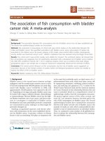
Staging abdomino-pelvic CT & MRI scans confirmed a 6.5
× 4 cm T3N1 irregular low rectal mass lesion extending
inferiorly with involvement of the muscularis and serosa
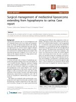
(figure 1), but no distal metastasis or small bowel pathol-
ogy (figure 2).
Neo adjuvant chemo-radiotherapy was given to down-
stage the tumour and repeat MRI scan showed marked
reduction of the mass lesion with no evidence of lym-
phadenopathy.
At laparotomy a mobile tumour was noted in the upper
rectum following mobilisation. In addition three serosal
deposits of tumour were noted in the distal ileum. An
anterior resection was performed and 25 cm of ileum was
also resected
Published: 19 February 2009
World Journal of Surgical Oncology 2009, 7:21 doi:10.1186/1477-7819-7-21
Received: 29 October 2008
Accepted: 19 February 2009
This article is available from: />© 2009 McHugh et al; licensee BioMed Central Ltd.
This is an Open Access article distributed under the terms of the Creative Commons Attribution License ( />),
which permits unrestricted use, distribution, and reproduction in any medium, provided the original work is properly cited.
World Journal of Surgical Oncology 2009, 7:21 />Page 2 of 3
(page number not for citation purposes)
The patient made an uncomplicated recovery and was dis-
charged on 15 post operative day.
Two nodules of carcinoid were confirmed in the small
bowel segment using immuno-staining. They both
invaded to serosal level. In the third, tumour was lost on
deeper sectioning but it's H&E appearance supported car-
cinoid. Two tiny mesenteric nodes taken were clear. The
12 cm segment of rectum showed radiation change but no
residual primary mucosal lesion, demonstrating complete
pathological response with 12 negative lymph nodes.
Discussion
In 1888, Otto Lubarsch, a pathologist based in Berlin first
described carcinoid lesions in detail. He reported the
autopsy findings of a patient with multiple carcinoid
tumours involving the ileum. Carcinoid syndrome and it's
classic associated symptoms was reported two years later
by Ransom. He described a patient with diarrhoea and
wheezing secondary to an ileal carcinoid which had a dis-
tant metastasis to the liver [8].
The annual age-adjusted incidence of small intestine car-
cinoids is reported as increasing, from 1.09 per 100,000 in
1973 to 5.25 per 100,000 in 2004 [9]. The incidental find-
ing of malignant carcinoid at autopsy is reported at 21 per
million [10]. Surgical resection is the preferred treatment
option. With regards neoadjuvant therapy, combined
associations (including either 5-fluorouracil and/or strep-
tozotocin) rarely exceed a 20% response rate [11].
Five year survival for resected isolated carcinoid is deter-
mined by site, with appendiceal carcinoids having a better
5 year survival prognosis (> 95%) than small intestine car-
cinoids (70–80%) [12]. Recent European guidelines for
surveillance of midgut carcinoids post resection with cur-
ative intent suggest follow up every 6–12 months, with
the exception of grade 3 tumours which should be fol-
lowed every 3 months. Minimal examinations include
measurement of chromogranin A (a neuroendocrine
secretory protein located in the secretory vesicles of neu-
rons and neuroendocrine cells) and 5-Hydroxyindoleace-
tic acid (5-HIAA is the main metabolite of serotonin) in
24 hour urine and with three-phasic CT scan [13]. Follow
up should be life-long.
Synchronous carcinoids with non-carcinoid neoplasms in
the G.I. tract were first noted by Pearson and Fitzgerald in
1949 [2]. A study by Gerstle et al in 1995 reported on 69
patients with carcinoids of the gastrointestinal tract were
discovered, of whom 29 (42 percent) had second synchro-
nous tumours [14]. To our knowledge this is the first
report with as many as 3 midgut carcinoids discovered
with a synchronous adenocarcinoma. This further
cements the association elucidated by Gerstle et al.
Hypotheses put forward to explain this association
include the secretion of active neuroendocrine peptides
such as gastrin and cholecystokinin [15]. Both have been
previously implicated as directly regulating growth in
colorectal carcinoma [16]. Non-neuroendocrine peptides
regulating cell growth and differentiation have been dem-
onstrated in gastrointestinal carcinoid tumours and may
also play a role in carcinogenesis [17].
Pre-operative CT Abdomen-Pelvis image showing polypoid rectal lesion extending into the lumenFigure 1
Pre-operative CT Abdomen-Pelvis image showing
polypoid rectal lesion extending into the lumen.
Pre-operative CT Abdomen Pelvis image showing normal appearing ileum at ileo-caecal junctionFigure 2
Pre-operative CT Abdomen Pelvis image showing
normal appearing ileum at ileo-caecal junction.
Publish with BioMed Central and every
scientist can read your work free of charge
"BioMed Central will be the most significant development for
disseminating the results of biomedical research in our lifetime."
Sir Paul Nurse, Cancer Research UK
Your research papers will be:
available free of charge to the entire biomedical community
peer reviewed and published immediately upon acceptance
cited in PubMed and archived on PubMed Central
yours — you keep the copyright
Submit your manuscript here:
/>BioMedcentral
World Journal of Surgical Oncology 2009, 7:21 />Page 3 of 3
(page number not for citation purposes)
Conclusion
In the case of a synchronous carcinoid with adenocarci-
noma, management is directed towards the carcinoma,
since the finding of carcinoid is incidental and so it is usu-
ally at an early stage. During resection of the colorectal
tumour, a thorough inspection of the abdominal cavity
should be undertaken to investigate the possibility of met-
astatic secondaries or a synchronous tumour as is reported
in this case. Because of their slow growing natural history,
the discovery of an asymptomatic gastrointestinal carci-
noid during the operative treatment of another malig-
nancy usually requires resection alone without additional
treatment and will have little effect on the prognosis of the
individual [18].
Consent
Written consent was obtained from the patient for publi-
cation of this case report
Competing interests
The authors declare that they have no competing interests.
Authors' contributions
SM was involved in data acquisition and interpretation,
writing initial drafts and subsequent revisions and under-
took review of literature on topic. JOD made a substantial
contribution regarding conception of report, was the edi-
tor of multiple drafts of case report, made many sugges-
tions regarding format of report for inclusion of
intellectual content and undertook review of literature on
topic. PG made a substantial contribution regarding con-
cept of case report, was the supervisor of work done and
drafts of case report prepared by first and second authors.
All authors read and approved final approval of version to
be published given.
References
1. Jenson RT: Carcinoid and pancreatic endocrine tumors:
recent advances in molecular pathogenesis, localization, and
treatment. Curr Opin Oncol 2000, 12:368-77.
2. Maggard M, O'Connell JB, Ko CY: Updated population-based
review of carcinoid tumors. Ann Surg 2004, 240(1):117-22.
3. Pearson CM, Fitzgerald PJ: Carcinoid tumours: A re-emphasis of
their malignant nature: Review of 140 cases. Cancer 1949,
2:1005-26.
4. Lotliker U, Fogler R, Novetsky AD, Yoon NY: Concurrent colonic
carcinoma and small bowel carcinoid tumour: Case reports
and review of the literature. Dis Colon Rectum 1982, 25:375-82.
5. Brown NK, Smith MP: Neoplastic diathesis of patients with car-
cinoid: Report of a case with four other neoplasms. Cancer
1973, 32:216-22.
6. Khubchandani M, Alford JE: Primary carcinoid and carcinoma of
the rectum occurring simultaneously: Report of a case. Dis
Colon Rectum 1974, 17:117-22.
7. Sparta D, Massimilliano B, Lleshi A, Rossela F, Santo B, Salvatore B:
Right colon cancer and incidental ileal carcinoid: case report.
Acta Chirurgica Mediterranea 2005, 21:15.
8. Sweeney JF, Rosemurgy AS: Carcinoid tumors of the gut. Cancer
Control 1997, 4(1):18-24.
9. Yao JC, Hassan M, Phan A, Dagohoy C, Leary C, Mares JE, Abdalla EK,
Fleming JB, Vauthey JN, Rashid A, Evans DB: One hundred years
after "carcinoid": epidemiology of and prognostic factors for
neuroendocrine tumors in 35,825 cases in the United States.
J Clin Oncol 2008, 26(18):3063-72.
10. Berge T, Linell F: Carcinoid tumours. Frequency in a defined
population during a 12-year period. Acta Pathol Mircobio Immunol
Scand [A] 1976, 84(4):322-330.
11. O'Toole D, Hentic O, Corcos O, Ruszniewski P: Chemotherapy
for Gastro-Enteropancreatic Tumours. Neuroendocrinology
2004, 80 Suppl 1:79-84.
12. Sutton R, Doran HE, Williams EM, Vora J, Vinjamuri S, Evans J, Camp-
bell F, Raraty MG, Ghaneh P, Hartley M, Poston GJ, Neoptolemos JP:
Surgery for midgut carcinoid. Endocrine-Related Cancer
2003:469-481.
13. Eriksson B, Klöppel G, Krenning E, Ahlman H, Plöckinger U, Wieden-
mann B, Arnold R, Auernhammer C, Körner M, Rindi G, Wildi S, Fra-
scati Consensus Conference participants: Consensus guidelines
for the management of patients with digestive neuroendo-
crine tumors – Well-differentiated Jejunal-ileal tumor/carci-
noma. Neuroendocrinology 2008, 87:8-19.
14. Gerstle JT, Kauffman GL Jr, Koltun WA: The incidence, manage-
ment, and outcome of patients with gastrointestinal carci-
noids and second primary malignancy. J Am Coll Surg 1995,
180:427-32.
15. Zucker KA, Longo WE, Modlin IM, Bilchik AJ, Adrian TE: Malignant
diathesis from jejuno-ileal carcinoids. Am J Gastroenterol 1989,
84:182-186.
16. Reubi JC, Schaer JC, Waser B: Cholecystokinin (CCK)-A and
CCK-B/gastrin receptors in human tumors. Cancer Res 1997,
57:1377-1386.
17. Habal N, Sims C, Bilchik AJ: Gastrointestinal carcinoid tumors
and second primary malignancies. J Surg Oncol 2000,
75:310-316.
18. McCabe HL: Adenocarcinoma of the gastro-oesophageal junc-
tion with a synchronous carcinoid of the duodenum. Postgrad
Medical Journal, BMJ 2001, 77:255-256.