báo cáo khoa học: " Spontaneous biloma managed with endoscopic retrograde cholangiopancreatography and percutaneous drainage: a case report" pdf
Bạn đang xem bản rút gọn của tài liệu. Xem và tải ngay bản đầy đủ của tài liệu tại đây (1006.07 KB, 3 trang )
CAS E REP O R T Open Access
Spontaneous biloma managed with endoscopic
retrograde cholangiopancreatography and
percutaneous drainage: a case report
Gurhan Bas
1
, Ismail Okan
1
, Mustafa Sahin
1
, Ramazan Eryılmaz
2
, Arda Isık
1*
Abstract
Introduction: Spontaneous biloma formation is a very rare condition, which mandates immediate treatment.
Case presentation: An 80-year-old Caucasian man was referred to our department with a diagnosis of intra-
abdominal collection located in his right upper quadrant. Further radiological examination demonstrated multiple
calculi in his gallbladder and common bile duct. Our patient underwent endoscopic retrograde
cholangiopancreatography and the stones in the common bile duct were extracted. Percutaneous drainage of the
abdominal collection revealed a spontaneous biloma formation. Continuous drainage of bile persisted for one
week, so endoscopic retrograde cholangiopancreatography was repeated and a 10Fr stent was placed;
subsequently the biliary leak ceased and our patient was discharged. A control abdominal computed tomography
did not show any residual fluid collection.
Conclusion: Spontaneous biloma formation is a very ra re incidence; awareness is necessary for prompt recognition
and treatment.
Introduction
A biloma is defined as an encapsulated collectio n of bile
outside the biliary tree [1]. It is mainly caused by iatro-
genic injury (surgery, percutaneous trans-hepatic inter-
ventions) or abdominal trauma [1,2]. Spontaneous
rupture of the biliary tree is a very rare condition [3].
We report here the case of a patient with spontaneous
biloma formation developed secondary to cholecysto-
choledocholithiasis, and managed with percutaneous
drainage and endoscopic biliary decompression.
Case report
An 80-year-old Caucasian man was referred to our
depar tment with the diagnosis of right upper abdominal
encapsulated fluid collection. Two weeks before, he was
admitted to the emergency room in a state hospital with
abdominal pain and nausea. Subsequent analysis, includ-
ing abdominal ultrasonography (US) and computed
tomography (CT), showed a large fluid collection in his
right upper abdominal cavity, and gallbladder stones. He
had no past history of abdominal surgery or trauma. On
admission, his vital signs and physical examination were
normal, except asymmetry and slight tenderness in his
right upper quadrant with a palpable mass. Complete
blood count and blood biochemistry results were evalu-
ated. Abnormal laboratory findings included (normal
range in parenthesis): albumin, 2.3 g/dL (3.5-5.0 g/dL);
erythrocyte sedimentation rate (ESR), 82 mm/h; C-reac-
tive protein (CRP), 5.2 mg/dL (0.00-0.80 mg/dL); and
calcium levels, 7.7 mg/dL (8.6-1.2 mg/dL). His viral
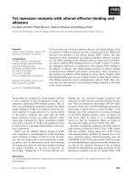
hepatitis marker tests were all negat ive. A repeat CT
revealed a large right hepatic subcapsular collection with
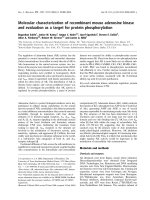
a size of 18.9 cm (Figure 1). Abdominal magnetic reso-
nance imaging (MRI) demonstrated multiple common
bile duct (CBD) stones with an enlarged biliary tree, and
a large subcapsular f luid collection extending around
the lower margin of his right hepatic lobe (Figure 2)
without a ny direct communication with the biliary sys-
tem. Nine days after our patient’s admission, endoscopic
retrograde cholang iopancreatog raphy (ERC P) and endo-
scopic sphincterotomy with stone extraction were per-
formed. Two days later, a percutaneous drainage of fluid
* Correspondence:
1
Department of Surgery, Vakif Gureba Training and Research Hospital,
Istanbul
Full list of author information is available at the end of the article
Bas et al. Journal of Medical Case Reports 2011, 5:3
/>JOURNAL OF MEDICAL
CASE REPORTS
© 2011 Bas et al; licensee BioMe d Ce ntral Ltd. This is an Open Access article distributed under the terms of the Creative Commons
Attribution License ( which permits unrestricted use, distribution, and re production in
any medium, provided the original work is properly cited.
under US guidance was performed and 800 ml of bile-
stained fluid was aspirated. Drain fluid revealed a total
bilirubin level of 22.3 mg/dL and a direct bilirubin level
of 18.9 mg/dL. Direct microbiological examinatio n with
gram staining showed a Gram-negative bacillus. Since a
residual collection was detected with US after one week,
an 8Fr p igtail catheter was in troduced percutaneously.
However, daily 50-100 ml drainage continued over
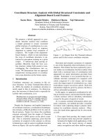
seven days, and so a repeat ERCP was performed. It
showed extravasation of contrast material from a distal
biliary radicle in his right hepatic lobe and communica-
tion with the biloma (Figure 3). After the insertion of a
10Fr stent to his CBD, the drainage decreased dramati-
cally and ceased. The percutaneous catheter was
removed after five days and our patient was discharged
two days later. The 10Fr stent at his CBD was removed
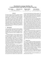
two months after his discharge. Control CT scans taken
two months (Figure 4) and one year after discharge
were normal.
Discussion
Biloma formation is encountered mainly after surgical or
interventional procedures and trauma involving the bili-
ary system [2]. However, there are few reported cases of
spontaneous biloma in the literature. The most frequent
Figure 1 Initial abdominal CT demonstrating a large right
hepatic subcapsular collection.
Figure 2 Abdominal MRI showing CBD stones.
Figure 3 The ERCP findings reveal relation of the biloma with
the intrahepatic biliary tree.
Figure 4 Abdominal CT showing complete resolution of the
biloma after management.
Bas et al. Journal of Medical Case Reports 2011, 5:3
/>Page 2 of 3
cause of spontaneous biloma is choledocholithiasis [4,5].
Less commonly reported causes include biliary tree
malignancy, acute cholecystitis, hepatic infarction and
abscess, obstructive jaundice and tuberculosis [3-5].
Although the pathophysiology of spontaneous biloma
remains to be elucidated [5], one suggested contributing
factor is an intraductal pressure increase due to obstruc-
tive lesions or infarcti ons on any part of the biliary tree
[4]. Bilomas are generally localized in the right upper
quadrant of the abdomen, neighboring the right hepatic
lobe [4]. The clinical presentation of biloma varies
greatly from nonspecific abdominal pain to biliary sepsis
[6]. Encapsulation of bile within the omentum and
mesentery [2] prevents g eneralized peritonitis in most
cases. Abdominal US is the first modality to e valuate
the nature of a biloma and the underlying pathology.
However, an abdominal CT can define the disease, the
causeandtherelationswiththe adjacent structures
more accurately [3]. Differential diagnosis should
include hematoma, seroma, liver abscess, cysts, pseudo-
cysts, and lymphocele [5]. Percutaneous aspiration
under radiologic guidance can also aid in diagnosis and
treatment. Biochemical and microbiological analysis of
the fluid helps differentiation from pyogenic abscesses
or other causes [7]. An MRI may be of value to evaluate
theetiologysinceitcanbeusedsafelyforthepatholo-
gies of the biliary system [8]. ERCP is also used for diag-
nostic and therapeutic purposes. Management of the
biloma in a p atient includes appropriate measures such
as intravenous hydration and initiation of a ntibiotic
treatment if sepsis is present. Although some bilomas,
especially those that are small in size and asymptomatic,
can be followed without intervention [3], most require
treatment. Percutaneous [9] and endoscopic modalities
provideadequatedrainageandmaybetherapeuticin
most cases [6]. These treatments are preferable to sur-
gery as the first step in treatment [ 4,5,10]. ERCP is indi-
cated particularly in treat ment failure, such as persistent
bile leakage despite percutaneous catheterizatio n. Sur-
gery always remains an option in emergency and persis-
tent cases. In our patient , the biloma was located in the
right upper quadrant and was detected with abdominal
US. Because an MRI demonstrated CBD stones, ERCP
was preferred for the first modality for diagnosis and
treatment. Although it did not show the communication
between the biliary tree and the collection and proved
biloma, his CBD was clea red from stones. Repeat ERCP
with stenting was necessary because the drainage didn’t
stop. In ERCP, the communication b etween the biliary
tree and biloma was shown clearly, probably due to the
decompression of the biloma by percutaneous drainage.
The drainage ceased after five days. During our one year
follow-up, there has been no recurrence by clinical or
radiological means.
Conclusion
Percutaneous treatment should be considered as the
first-line option for patients with symptomatic sponta-
neous biloma. In cases of persistent bile leaks, ERCP
and endoscopic sphincterotomy with or without stent
placement should be performed.
Consent
Written informed consent was obtained from the patient
for publication of this case report and any accompany-
ing images. A copy of the written consent is available
for review by the Editor-in-Chief of this journal.
Author details
1
Department of Surgery, Vakif Gureba Training and Research Hospital,
Istanbul.
2
Department of Surgery, Antalya Training and Research Hospital,
Antalya, Istanbul.
Authors’ contributions
GB, IO, MS and RE analyzed and interpreted the patient data. AI was a major
contributor in writing the manuscript. All authors read and approved the
final manuscript.
Competing interests
The authors declare that they have no competing interests.
Received: 10 March 2010 Accepted: 6 January 2011
Published: 6 January 2011
References
1. Gould L, Patel A: Ultrasound detection of extrahepatic encapsulated bile:
“biloma”. AJR Am J Roentgenol 1979, 132(6):1014-1015.
2. Vazquez JL, Thorsen MK, Dodds WJ, Quiroz FA, Martinez ML, Lawson TL,
Stewart ET, Foley WD: Evaluation and treatment of intraabdominal
bilomas. AJR Am J Roentgenol 1985, 144(5):933-938.
3. Lee JH, Suh JI: A Case of infected Biloma due to spontaneous
intrahepatic biliary rupture. Korean J Intern Med 2007, 22(3):220-224.
4. Fujiwara H, Yamamoto M, Takahashi M, Ishida H, Ohashi O, Onoyama H,
Takeyama Y, Kuroda Y: Spontaneous rupture of an intrahepatic bile duct
with biloma treated by percutaneous drainage and endoscopic
sphincterotomy. Am J Gastroenterol 1998, 93(11):2282-2284.
5. Akhtar MA, Bandyopadhyay D, Montgomery HD, Mahomed A:
Spontaneous idiopathic subcapsular biloma. J Hepatobiliary Pancreat Surg
2007, 14(6):579-581.
6. Binmoeller KF, Katon RM, Shneidman R: Endoscopic management of
postoperative biliary leaks: review of 77 cases and report of two cases
with biloma formation. Am J Gastroenterol 1991, 86(2):227-231.
7. Middleton JP, Wolper JC: Hepatic biloma complicating sickle cell disease.
A case report and a review of the literature. Gastroenterology 1984,
86(4):743-744.
8. Hekimoglu K, Ustundag Y, Dusak A, Erdem Z, Karademir B, Aydemir S,
Gundogdu S: MRCP vs. ERCP in the evaluation of biliary pathologies:
review of current literature. J Dig Dis 2008, 9(3):162-169.
9. Chang ML, Lin DY: Symptomless cyst formation at the location of a
biloma resolved with a single aspiration: case report. Chang Gung Med J
2000, 23(12):794-798.
10. Kuligowska E, Schlesinger A, Miller KB, Lee VW, Grosso D: Bilomas: a new
approach to the diagnosis and treatment. Gastrointest Radiol 1983,
8(3):237-243.
doi:10.1186/1752-1947-5-3
Cite this article as: Bas et al.: Spontaneous biloma managed with
endoscopic retrograde cholangiopancreatography and percutaneous
drainage: a case report. Journal of Medical Case Reports 2011 5:3.
Bas et al. Journal of Medical Case Reports 2011, 5:3
/>Page 3 of 3