báo cáo khoa học: "Eosinophilic pneumonia associated with daptomycin: a case report and a review of the literature" pptx
Bạn đang xem bản rút gọn của tài liệu. Xem và tải ngay bản đầy đủ của tài liệu tại đây (321.15 KB, 5 trang )
CAS E REP O R T Open Access
Eosinophilic pneumonia associated with
daptomycin: a case report and a review
of the literature
Andreas S Kalogeropoulos
1*
, Sotirios Tsiodras
2
, Dionysios Loverdos
1
, Panagiotis Fanourgiakis
1
,
Athanasios Skoutelis
1
Abstract
Introduction: Although several studies did not demonstrate that daptomycin may cause significantly higher rates
of pulmonary adverse effects when compared with vancomycin or penicillinase-resistant penici llins, there have
been a few case reports of severe pulmonary complications associated with daptomycin administration.
Case presentation: A rare case of eosinophilic pneumonia occurring 10 days after daptomycin administration in a
78-year-old Caucasian man with possible infectious endocarditis is described. He developed new onset fever, up to
38.5°C, with bilateral pulmonary crackles on physical examination and with no signs of severe respiratory failure.
A chest computed tomography-scan showed bilateral nodular consolidations with air bronchograms and pleural
effusions. Immediate discontinuation of daptomycin was followed by vigorous improvement of clinical signs and
symptoms with progressive resolution of pulmonary consolidations a month later.
Conclusion: Physicians should be aware of this rare but serious complication during daptomycin treatment, and
prompt discontinuation of the offending agent, with or without additional supportive treatment, must occur
immediately.
Introduction
Eosinophilic pneumonia (EP) belongs to a heteroge-
neous group of lung diseases characterized by pulmon-
ary infiltrates and increased numbers of eosinophils in
lung tissue or broncho-alveolar lavage (BAL) fluid,
with or without increased levels of eosinophils in the
peripheral blood [1]. Acute EP due to drugs or toxins
has similar clinical, radiographic and histopathologic
manifestations to idiopathic acute or chronic EP, mak-
ing the distinction of these entities difficult. The most
common drugs a ssociated with EP are antibiotics and
anti-inflammatory drugs [2]. A complete and updated
list of drugs suspected of causing lung disease can be
found on a website maintained by the Groupe Etude
de la Pathologie Pulmonaire Iatrogene at http://www.
pneumotox.com.
Daptomycin, an antimicrobial agent of the cyclic
lipopeptide group of ant ibiotics, has an outstanding cov-
erage for Gram-positive bacteria and is licensed for the
treatment of bacteraemia and right-sided endocarditis
due to methicillin-susceptible and methicillin-resistant
Staphylococcus aureus [3]. It is also effective for vanco-
mycin-resistant enterococci [3]. Although daptomycin
has a favorable adverse effect summary, and even
though several retrospective studies did not show signif-
icantly increased incidence of pulmonary adverse drug
reactions when compared to other anti-microbial agents
[4-7], recently published case reports pointed out
serious respiratory complications associated with
daptomycin [8-11].
We present a case of pulmonary infilt rates and
broncho-alveolar lavage eosinophilia occurring during
treatment with daptomycin in a patient with possible
infectious endocarditis (IE). In this particular case, and
in contrast to previously published reports, our patient
did not develop severe respiratory failure, and direct dis-
continuation of daptomycin without the systemic
* Correspondence:
1
5th Department of Internal Medicine and Infectious Diseases,
“EVANGELISMOS” General Hospital, 45-47 Ipsilantou Street, 106 76 Kolonaki,
Athens, Greece
Full list of author information is available at the end of the article
Kalogeropoulos et al. Journal of Medical Case Reports 2011, 5:13
/>JOURNAL OF MEDICAL
CASE REPORTS
© 2011 Kalogeropoulos et al; licensee BioMed Central Ltd. This is an Open Access article distributed under the terms of the Creative
Commons Attribution License (ht tp://creativecomm ons.org/licenses/by/2.0), which permits unrestricted use, distribution, and
reproduction in any medium, provided the original work is properly cited.
administration of corticosteroids was associated with the
progressive and complete resolution of clinical manifes-
tations and laboratory disturbances.
Case presentation
A 78-year-old Caucasian man, with a history of coronary
artery disease, presented with symptoms of acute con-
gestive heart failure (CHF) including dyspnea at rest,
orthopnea and paroxysmal nocturnal dyspnea.
The patient had a history of a transurethral prostatect-
omy (TURP) one month before admission. A week after
the TURP, he developed a fever of 38.5°C that was con-
sidered a ma nifestation of a post-o perative urinary tract
infection and was treated empirically with oral ciproflox-
acin 500 mg twic e daily. The fever did not respond and
treatment changed to oral amoxicilli n/clavulanic 1 g
twice daily and intramuscular netilmicin, 300 mg once
daily. The fever resolved and no other clinical manifes-
tations developed until the day of admission to our
hospital. Regarding his past m edical history, he was a
non-smoker, he had no known allergies and he did not
mention any recent travels.
On admission, he was afebrile. His blood pressure was
120/55 mmHg, his heart rate 105/minute a nd his SaO
2
was 92% on ambient air. The remaining physical exami-
nation revealed decreased breath sounds at both lung
bases and inspiratory crackles at the lower pulmonary
fields bilaterally, a 4/6 diastolic heart murmur at the
lowe r left parasternal area and a 4/6 systolic heart mur-
mur at the right upper parasternal area. Laboratory stu-
dies revealed a leukocyte count 8350/μL, hematocrit
36.8%, platelet count 270,000/μL and C-reactive protein
(CRP) 1.0 mg/dL (normal range <0.5 mg/dL). A chest
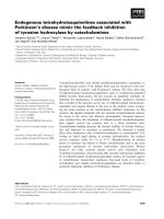
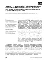
radiogram showed bilateral perihilar alveolar edema
with a “butterfly” appearance and bilateral pleural effu-
sions (Figure 1a). A transesophageal, two-dimensional
Doppler echocardiogram showed a tricuspid aortic valve
with a mobile vegetation of 9 mm in length on the right
cusp and the presence of severe aortic valve regurgita-
tion with possible perforation of the left cusp. Moderate
mitral valve regurgitation was present as well, whereas
the ejection fraction was 55%.
Following emergent treatment of CHF, all symptoms
and p hysical signs were completely resolved. Addition-
ally, a new chest radiogram showed signific ant improve-
ment of the aforementioned radiographic finding s
(Figure 1b). Six sets of bloo d cultures from three sepa-
rate body sites, drawn over 24 hours, were negative for
a common bacterial pathogen. Considering the patient’ s
previous history of TURP, and the previous admission
of an antimicrobial regimen, empirical treatment for IE
due t o possible resistant enterococci was initiated
including ampicillin 12 g daily, gentamicin 80 mg thrice
daily and daptomycin (8 mg/kg) once daily. The patient
responded positively to the empirical treatment until
day 10, when he developed a new onset fever up to
38.5°C, a ccompanied by chills and dia phoresis. Physical
examination revealed new onset crackles, predominantly
at the left upper and medial pulmonary fields. The
patient a lso showed significant hypoxemia with arterial
blood gases analyses revealing a pH of 7.44, an oxygen
saturation of 88%, a partial pressure of oxygen of
58 mmHg and a partial pressure of carbon dioxide of
38 mmHg, while breathing on ambient air. Laboratory
studies revealed a leukocyte count of 9970/μL, with
78.3% neutrophils and 2.3% eosinophils. The erythrocyte
sedimentation rate was 79 mm/h and CRP was 16.1
mg/dL. A chest x-ray w as immediately pe rformed
demonstrating bilateral non-cavitating, reticulo-nodular
infiltrates. All blood cultures were negative. The patient
was treated with supplemental oxygen to maintain an
oxygen saturation >92% and an additional empirical
antimicrobial regimen for suspected health care acquired
pneumonia (HCAP) was initiated (intravenous moxiflox-
acin 400 mg once daily and meropenem 3 g thrice
daily). Inhaled corticosteroids and bronchodilators were
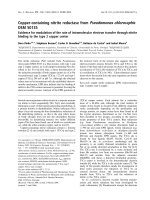
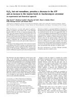
also administrated. The high resolution chest computed
tomography (chest HRCT) disclosed patchy areas of
consolidation with ground-glass peripheral opacities and
bilateral pleural effusions (Figure 2a). Urine examination
for S. pneumoniae and Legionella antigen was negative.
Serology for Chlamydia pneumoniae, Mycoplasma pneu-
moniae, Bartonella spp, Coxiella burnetii , Brucella and
Cytomegalovirus was negative. The se rological screening
was negative for a uto-immune markers (anti -nuclear
antibodies, cytoplasmic and perinuclear anti-neutrophil
cytoplasmic antibodies and anti- double-stranded DNA
antibodies) as well. Despite the treatment, there was no
clinical improvement. A thoraco centesis with a collec-
tion of pleuritic fluid for analysis was performed and the
latter revealed a transudate with 6700 nucleated cells
(70% lymphocytes, 15% eosinophils, 15% neutrophils). In
addition, cultures for acid fast-bacilli and adenosine dea-
minase activity test of the pleurit ic fluid were negative.
To further investigat e the nature of th e aforementio ned
clinical syndrome a bronchoscopy with BAL was carried
out, which disclosed 480 nucleated cells/μL (55% macro-
phages, 27.5% eosinophils, 12.2% neutrophils and 5.3%
lymphocytes). Additional cultures for acid fast-bacilli,
fungal and parasitic infections were also ne gative. Given
the above findings the diagnosis of EP was made,
daptomycin (as a probable cause of EP) was replaced by
linezolid and moxifloxacin with meropenem were dis-
continued. Twenty-four hours after the daptomycin
withdrawal the f ever resolved completely. During the
following seven days a significant improvement of the
clinical and radiographic findings occurred, whereas
CRP was within the normal range. One month later,
Kalogeropoulos et al. Journal of Medical Case Reports 2011, 5:13
/>Page 2 of 5
a follow up chest-HRCT was normal (Figure 2b). The
Naranjo causality scale yielded a score of 7 suggesting a
probable adverse reaction due to daptomycin [12].
Discussion
Eosinophilic pneumonia is a rationally uncommon entity
and has been associated with several medications and
chemicals, with antibiotics and non steroidal anti-
inflammatory drugs being th e most co mmon eliciting
factor [9]. The pathophysiology of EP is thought to
involve the triggering of immune response due to an
offending agent (for example, a drug or an infecting
pathogen), principally expressed through antigen presen-
tation by alveolar macrophages. This process may conse-
quently provoke the recruitment of T-helper 2 (Th2)
lymphocytes that sequenti ally release interleukin-5.
Further eosinophil migration into the alveoli is facili-
tated through various mechanisms. Initially, interleukin-
5 may promote significant eosinophil production and
resettlement in t he pulmonary alveoli. In addition,
Figure 1 Chest Radiograms. a. Chest X-ray demonstrating bilateral perihilar alveolar edema with a “butterfly” appearance and bilateral pleural
effusions. b. Chest X-ray after pharmaceutical treatment for the congestive heart failure symptoms. Most of the initially appeared radiographic
findings have been almost completely resolved.
Figure 2 Chest HRCT-scans. a. Chest HRCT-scan demonstrating bilateral irregular ly shaped nodular consolidations with air bronchograms and
bilateral pleural effusions. b. Chest HRCT-scan, one month after daptomycin discontinuation, demonstrating complete resolve of nodular
consolidations and bilateral effusions.
Kalogeropoulos et al. Journal of Medical Case Reports 2011, 5:13
/>Page 3 of 5
alveolar macrophages can excrete eotaxin, a cytokine
that selectively recruits eosinophils by inducing their
chemotaxis, which in turn may promote further eosino-
phil localization into the lungs [1].
Drug-induced EP can appear either as an acute or as a
chronic syndrome that may occur within days or weeks
after starting the offending agent. Diagnosis usually
requires synthesis of information including clinical his-
tory, laboratory data and radiologic findings [13].
Patients with EP normally have cough and dyspnea for
several days or weeks and may have a rash and/or fever.
In acute patterns of EP patients may appear to have
symptoms of sever e dyspnea and hypoxemia resembling
acute lung injury (PaO
2
/FiO
2
<300 mmHg) or acute
respiratory distress syndrome (PaO
2
/FiO
2
<200 mmHg).
It typically appears a s area s of conso lidation and
ground-glass opacity on CT imaging, usually involving
the peripheral pulmonary parenchyma. In addition, it
may or may not be associated with peripheral blood
eosinophilia, however pulmonary eosinophilic infiltrates
or BAL eosinophilia are the corner stone for the diagno-
sis of EP [8]. A lung biopsy can verify the diagnosis but
is not always a requisite given a typical c linical appear-
ance and consistent laboratory and radiographic find-
ings. In addition, according to the criteria of Solomon
and Schwarz, the diagnosis of drug induced EP requires
further evidence of pneumonitis with the aforemen-
tioned features, throughout treatment, with a drug that
has the potential to provoke this syndrome. Infectious
causes of eosinophilia, such as fungal or parasitic infec-
tions, need to be excluded, whereas clinical improve-
ment should ensue drug cessation and symptoms should
reappear after a rechallenge [2]. In our case, most of the
criteria for the diagnosis of eosinophilic pneumonia
were ful filled. In particular, the patient developed fever
and an abrupt abatement of respiratory function with
hypoxemia during the treatment with an offending
agent like daptomycin. However, the aforementioned
syndrome did not progress to a severe respiratory failure
and the patient did not require mechanical or non-
mechanical ventilation. The arterial blood gases analysis
revealed an acute lung injury with a PaO
2
/FiO
2
ratio
being 276. Moreover, in imaging studies with chest-
HCRT he developed t he characteristic pattern of bilat-
eral peripheral consolidations and ground-glass opacities
that we usually find in cases with eosinophilic pneumo-
nia. Finally, BAL fluid examination revealed significant
eosinophilia, a condition that is fundamental for the
diagnosis of EP, whereas parasitic and fungal infections
were excluded. However, before the accomplishment of
the b ronchoscopy procedure, we considered it essential
to carry out a serological screening for autoimmune
markers and a thoracocentesis, in order to examine the
pleuritic fluid. Indeed, pleuritic fluid analysis revealed a
transudate while cultures for acid fast bacilli and ADA
test were negative; findings th at were in consi sten t with
the possibility of tuberculosis as a cause of the clinical
syndrome in our case. In addition, serological screening
for autoimmune markers was also performed with the
intention of excluding diseases of autoimmune origin,
such as sm all vessel vasculitis, or systemic lupus erythe-
matosus, conditions that may both provoke significant
pulmonary lesions and non-infectious endocarditis
[14,15].
Patients with idiopathic EP often require systemi c cor-
ticosteroids treatment whereas those with drug induced
EP demonstrate significant improvement, only with
offending agent withdrawal. However , cases with persis-
tent symptoms may need treatment with systemic corti-
costeroids and additional respiratory support with
supplemental oxygen and assisted ventilation [16].
The E P described in our case is most likely attributa-
ble to an adverse drug reaction due t o daptomycin
administration. The patient had no history of c hronic
primary lung disease and he developed s ignificant pul-
monary abnormalities early after daptomycin initiation
and had a remarkable improvement soon after daptomy-
cin discontinuation. To the best of our knowledge, only
five cases of EP associated with daptomycin have been
reported thus far, but they should be considered in indi-
viduals who receive the drug and develop new pulmon-
ary infiltrates [8-11]. In the majority o f these reports
(80%) [8,10,11] patients developed severe respirato ry
failur e requiring systemic corticosteroids administration,
intubation and assisted ventilation or supplemental
oxygen and bimodal intermittent airway pressure sup-
port. In fact, in two of these cases persistent complete
recovery did not occur and patients became chronically
steroid dependent. Unfortunately, we are not able to
make any comments regarding the association of the
severity of the symptoms and the daptomycin dosage,
since the daptomycin dosage regimen was not referred.
In our patient, daptomycin was administered in high
doses (8 mg/kg) in view of the fact that in previous stu-
dies higher doses of daptomycin were more effective
and well tolerated when compared to other antimicro-
bial agents [17]. Additionally, our patient did not receive
any systemic corticosteroid treatment since clinical
presentation was not associated with severe respiratory
failure and the patient exhibited significant clinical
improvement after daptomycin discontinuation.
Daptomycin’s toxicity m echanism remains uncertain
and further in vitro and in vivo studies are necessary
in order to elucidate its toxicity biochemical pathways.
The primary mechanism of action involves calcium-
dependent transitions, which are responsible for confor-
mational changes of the daptomycin molecule that allow
interactions with cytoplasmic membrane, enhancing
Kalogeropoulos et al. Journal of Medical Case Reports 2011, 5:13
/>Page 4 of 5
daptomycin-c ytoplasmic membrane binding capacity
and cytoplasmic membrane permeability. The latter may
induce significant leakage of intracellular ions, such as
potassium. It has been recently demonstrated that syn-
thetic surfactant binds to daptomycin and diminishes its
antibacterial activity [ 18]. Therefore, we may assume
that the administration of daptomycin for long periods
of time could lead to increased accumulation of the
drug near the alveolar epithelial surface, which subse-
quently may cause severe epithelial injury and organized
pneumonia. Furthermore interaction of daptomycin with
pulmonary surfactant may result in the deterioration of
lipid integrity in the alveolar space, which in turn may
trigger and conserve an inflammatory process [9].
Conclusion
Daptomycin is a relatively new drug extensively used in
tertiary health care units and in intensive care practice
with excellent results regarding its antimicrobial activity.
Although extremely rare, daptomycin-i nduced EP must
be considered for patients who receive the drug and
develop new unexplained pulmonary infiltrates. Signifi-
cant morbidity and mortality may occur if this condition
remains unrecognized and not properly treated in a
timely fashion. Finally, further investigation through
experimental and clinical studies needs to be completed
in order to elucidate the exact mechanism behind this
rare yet grave adverse drug reaction.
Consent
Written informed consent was obtained from the patient
for publicatio n of this case report and any accompany-
ing images. A copy of the written consent is available
for review by the Editor-in-Chief of this journal.
Author details
1
5th Department of Internal Medicine and Infectious Diseases,
“EVANGELISMOS” General Hospital, 45-47 Ipsilantou Street, 106 76 Kolonaki,
Athens, Greece.
2
4th Academic Department of Internal Medicine and
Infectious Diseases, University of Athens Medical School, Attikon University
Hospital, Athens, Greece.
Authors’ contributions
All authors are aware of and approved the manuscript being submitted to
this journal. AK has made substantial contributions in drafting and revising
the manuscript. ST, DL, PF and AS have been involved in revising the
manuscript critically for important intellectual content. AS has given final
approval of the version to be published.
Competing interests
The authors declare that they have no competing interests.
Received: 6 May 2010 Accepted: 17 January 2011
Published: 17 January 2011
References
1. Allen JN: Drug-induced eosinophilic lung disease. Clin Chest Med 2004,
25:77-88.
2. Solomon J, Schwarz M: Drug-, toxin-, and radiation therapy-induced
eosinophilic pneumonia. Semin Respir Crit Care Med 2006, 27:192-197.
3. Kosmidis C, Levine DP: Daptomycin: pharmacology and clinical use. Expert
Opin Pharmacother 2010, 11:615-625.
4. Arbeit RD, Maki D, Tally FP, Campanaro E, Eisenstein BI: The safety and
efficacy of daptomycin for the treatment of complicated skin and skin-
structure infections. Clin Infect Dis 2004, 38:1673-1681.
5. Figueroa DA, Mangini E, Amodio-Groton M, Vardianos B, Melchert A,
Fana C, Wehbeh W, Urban CM, Segal-Maurer S: Safety of high-dose
intravenous daptomycin treatment: three-year cumulative experience in
a clinical program. Clin Infect Dis 2009, 49:177-180.
6. Mohr JF, Friedrich LV, Yankelev S, Lamp KC: Daptomycin for the treatment
of enterococcal bacteraemia: results from the Cubicin Outcomes
Registry and Experience (CORE). Int J Antimicrob Agents 2009, 33:543-548.
7. Fowler VG Jr, Boucher HW, Corey GR, Abrutyn E, Karchmer AW, Rupp ME,
Levine DP, Chambers HF, Tally FP, Vigliani GA, Campion M, Abrutyn E,
Levine DP, Price CS, Rehm SJ, Corey GR, Karchmer AW, S. aureus
Endocarditis and Bacteraemia Study Group.l: Daptomycin versus standard
therapy for bacteremia and endocarditis caused by Staphylococcus
aureus. N Engl J Med 2006, 355:653-665.
8. Hayes D Jr, Anstead MI, Kuhn RJ: Eosinophilic pneumonia induced by
daptomycin. J Infect 2007, 54:e211-213.
9. Cobb E, Kimbrough RC, Nugent KM, Phy MP: Organizing pneumonia and
pulmonary eosinophilic infiltration associated with daptomycin. Ann
Pharmacother 2007, 41:696-701.
10. Shinde A, Seifi A, DelRe S, Moustafa Hussein WH, Ohebsion J: Daptomycin-
induced pulmonary infiltrates with eosinophilia. J Infect 2009, 58:173-174.
11. Lal Y, Assimacopoulos AP: Two cases of daptomycin-induced eosinophilic
pneumonia and chronic pneumonitis. Clin Infect Dis 2010, 50:737-740.
12. Naranjo CA, Busto U, Sellers EM, Sandor P, Ruiz I, Roberts EA, Janecek E,
Domecq C, Greenblatt DJ: A method for estimating the probability of
adverse drug reactions. Clin Pharmacol Ther 1981, 30:239-245.
13. Camus PH, Foucher P, Bonniaud PH, Ask K: Drug-induced infiltrative lung
disease. Eur Respir J Suppl 2001, 32:93s-100s.
14. Chirinos JA, Corrales-Medina VF, Garcia S, Lichtstein DM, Bisno AL, Chakko S:
Endocarditis associated with antineutrophil cytoplasmic antibodies: a
case report and review of the literature. Clin Rheumatol 2007, 26
:590-595.
15. Bouma W, Klinkenberg TJ, van der Horst IC, Wijdh-den Hamer IJ,
Erasmus ME, Bijl M, Suurmeijer AJ, Zijlstra F, Mariani MA: Mitral valve
surgery for mitral regurgitation caused by Libman-Sacks endocarditis: a
report of four cases and a systematic review of the literature.
J Cardiothorac Surg 2010, 5:13.
16. Philit F, Etienne-Mastroianni B, Parrot A, Guerin C, Robert D, Cordier JF:
Idiopathic acute eosinophilic pneumonia: a study of 22 patients. Am J
Respir Crit Care Med 2002, 166:1235-1239.
17. Moise PA, Hershberger E, Amodio-Groton MI, Lamp KC: Safety and clinical
outcomes when utilizing high-dose (>or = 8 mg/kg) daptomycin
therapy. Ann Pharmacother 2009, 43:1211-1219.
18. Silverman JA, Mortin LI, Vanpraagh AD, Li T, Alder J: Inhibition of
daptomycin by pulmonary surfactant: in vitro modeling and clinical
impact. J Infect Dis 2005, 191:2149-2152.
doi:10.1186/1752-1947-5-13
Cite this article as: Kalogeropoulos et al.: Eosinophilic pneumonia
associated with daptomycin: a case report and a review
of the literature. Journal of Medical Case Reports 2011 5:13.
Kalogeropoulos et al. Journal of Medical Case Reports 2011, 5:13
/>Page 5 of 5